1
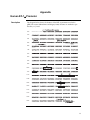
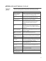
pEF6/His A, B, and C Catalog no. V963-20 Rev. date: 28 October 2009 Manual part no. 25-0240 MAN0000082 Corporate Headquarters Invitrogen Corporation 1600 Faraday Avenue Carlsbad, CA 92008 T: 1 760 603 7200 F: 1 760 602 6500 E: [email protected] For country-specific contact information visit our web site at www.invitrogen.com User Manual ii Table of Contents Contents and Storage ...............................................................................................................................................iv Introduction ......................................................................................................................1 Product Overview ..................................................................................................................................................... 1 Methods ............................................................................................................................2 Cloning into pEF6/His A, B, and C ........................................................................................................................ 2 Transfection................................................................................................................................................................ 7 Creating Stable Cell Lines ........................................................................................................................................ 9 Appendix.........................................................................................................................13 Human EF-1α Promoter ......................................................................................................................................... 13 pEF6/His A, B, and C Vectors............................................................................................................................... 14 pEF6/His/lacZ......................................................................................................................................................... 16 Accessory Products ................................................................................................................................................. 17 Technical Support.................................................................................................................................................... 18 Purchaser Notification ............................................................................................................................................ 19 References................................................................................................................................................................. 21 iii Contents and Storage Shipping and Storage pEF6/His vectors are shipped on wet ice. Upon receipt, store vectors at –20ºC. Contents 20 μg each of pEF6/His A, B, and C are supplied at 0.5 μg/μL in 10 mM Tris-HCl, 1 mM EDTA, pH 8.0 in a total volume of 40 μL. 20 μg of pEF6/His/lacZ is supplied at 0.5 μg/μL in 10 mM Tris-HCl, 1 mM EDTA, pH 8.0 in a total volume of 40 μL. iv Introduction Product Overview Description of the System pEF6/His A, B, and C are 5.8 kb vectors designed for high-level expression and purification of recombinant proteins in mammalian cell lines. Refer to page 15 for specific features of the vectors that allow for purification and detection of expressed proteins. High-level stable and transient expression can be carried out in most mammalian cells. The vectors contain the following elements: • The human elongation factor 1 α (hEF-1α) subunit promoter provides highlevel expression in a wide range of mammalian cells (Kim et al., 1990; Mizushima and Nagata, 1990; Uetsuki et al., 1989). See page 13 for more information. • Three reading frames to facilitate in-frame cloning with an N-terminal tag encoding the Xpress™ epitope and a polyhistidine (6xHis) metal-binding tag. • Blasticidin resistance gene (bsd) for selection of stable cell lines (Kimura et al., 1994). • Episomal replication in cell lines that are latently infected with SV40 or that express the SV40 large T antigen (e.g., COS7). The control plasmid, pEF6/His/lacZ, is included for use as a positive control for transfection, expression, and detection in the cell line of choice. Experimental Outline Use the following outline to clone and express your gene of interest in pEF6/His. • Consult the multiple cloning sites depicted on pages 3–5 to determine which vector (A, B, or C) should be used to clone your gene in-frame with the N-terminal Xpress™ epitope and polyhistidine tag. • Ligate your insert into the appropriate vector and transform into E. coli. Select transformants with 50 to 100 μg/mL ampicillin (or 50 μg/mL blasticidin). • Analyze your transformants for the presence of insert by restriction digestion. • Select a transformant with the correct restriction pattern and use sequencing to confirm that your gene is cloned in-frame with the N-terminal peptide. • Transfect your construct into the cell line of choice using your own method of transfection. Generate a stable cell line, if desired. • Test expression of your recombinant gene by western blot analysis or other functional assay. For ordering information about an antibody against the Xpress™ epitope, see page 17. • To purify your recombinant protein, you may use a metal-chelating resin such as ProBond™. ProBond™ resin is available separately (see page 17 for ordering information). 1 Methods Cloning into pEF6/His A, B, and C Before Starting Diagrams are provided on pages 3–5 to help you ligate your gene of interest inframe with the N-terminal peptide. General considerations for ligation and transformation are listed below. General Molecular Biology Techniques For help with DNA ligations, E. coli transformations, restriction enzyme analysis, purification of single-stranded DNA, DNA sequencing, and DNA biochemistry, see Molecular Cloning: A Laboratory Manual (Sambrook et al., 1989) or Current Protocols in Molecular Biology (Ausubel et al., 1994) (See References, page 21). Transformation Method You may use any method of your choice for transformation. Chemical transformation is the most convenient for most researchers. Electroporation is the most efficient and the method of choice for large plasmids. Maintaining pEF6/His To propagate and maintain pEF6/His A,B, and C, use the supplied 0.5 μg/μl stock solution in TE, pH 8.0 to transform a recA (recombination deficient), endA (endonuclease A deficient) E. coli strain, such as TOP10F’, INVαF´, or equivalent (see page 17 for ordering information). Select transformants on LB plates containing 50–100 μg/mL ampicillin or 50 μg/mL blasticidin. Be sure to prepare a glycerol stock of each plasmid for long-term storage (see page 6). The pEF6/His vectors are fusion vectors. To ensure proper expression of your recombinant protein, you must clone your gene in-frame with the ATG at base pairs 1716–1718. This will create a fusion with the N-terminal polyhistidine tag, Xpress™ epitope, and the enterokinase cleavage site. The vector is supplied in three reading frames to facilitate cloning. See the diagrams on pages 3–5 to develop a cloning strategy. If you wish to clone as close as possible to the enterokinase cleavage site, follow the guidelines below: • Digest pEF6/His A, B, or C with Kpn I. • Create blunt ends with T4 DNA polymerase and dNTPs. See Ausubel, et al., 1994 for a detailed protocol. • Clone your blunt-ended insert in-frame with the lysine codon (AAG) of the enterokinase recognition sequence. Following enterokinase cleavage, no vector-encoded amino acid residues will be present in your protein. Continued on next page 2 Cloning into pEF6/His A, B, and C, Continued If you will be recombining your entry clone with a destination vector for mammalian expression, your insert should contain a Kozak consensus sequence with an ATG initiation codon for proper initiation of translation (Kozak, 1987; Kozak, 1990; Kozak, 1991). An example of a Kozak consensus sequence is provided below. Other sequences are possible, but the G or A at position –3 and the G at position +4 (shown in bold) illustrates the most commonly occurring sequence with strong consensus. Replacing one of the two bases at these positions provides moderate consensus, while having neither results in weak consensus. The ATG initiation codon is shown underlined. Kozak Sequence for Mammalian Expression (G/A)NNATGG Multiple Cloning Site of pEF6/His A Below is the multiple cloning site for pEF6/His A. Restriction sites are labeled to indicate the cleavage site. The boxed nucleotide indicates the variable region. Note that there is a stop codon after the Xba I site. The multiple cloning site has been confirmed by sequencing and functional testing. The sequence may be downloaded from www.invitrogen.com or requested from Technical Support (see page 18). For more information on the hEF-1α promoter, see page 13. 3´ end of hEF-1a Intron 1 1579 GTTTGGATCT TGGTTCATTC TCAAGCCTCA GACAGTGGTT CAAAGTTTTT TTCTTCCATT TCAGGTGTCG TGAGGAATTA 1659 GCTTGGTACT AATACGACTC ACTATAGGGA GACCCAAGCT GGCTAGTTAA GCTTACC ATG GGG GGT TCT CAT CAT Met Gly Gly Ser His His 5´ end of hEF-1a Exon 2 T7 promoter/priming site Xpress epitope Polyhistidine Region 1734 CAT CAT CAT CAT GGT ATG GCT AGC ATG ACT GGT GGA CAG CAA ATG GGT CGG GAT CTG TAC GAC GAT His His His His Gly Met Ala Ser Met Thr Gly Gly Gln Gln Met Gly Arg Asp Leu Tyr Asp Asp 1800 GAC GAT AAG GTA CCT AGG ATC CAG TGT GGT GGA ATT CTG CAG ATA TCC AGC ACA GTG GCG GCC GCT Asp Asp Lys Val Pro Arg Ile Gln Cys Gly Gly Ile Leu Gln Ile Ser Ser Thr Val Ala Ala Ala Asp718 I Kpn I EK Recognition site Xba I BamH I EK Cleavage site BstX I* EcoR I Pme I EcoR V BstX I* Not I BGH Reverse priming site 1866 CGA GTC TAG AGG GCC CGT TTA AAC CCG CTG ATC AGC CTC GAC TGT GCC TTC TAG TTGCCAGCC Arg Val *** Arg Ala Arg Leu Asn Pro Leu Ile Ser Leu Asp Cys Ala Phe *** 1929 ATCTGTTGTT TGCCCCTCCC CCGTGCCTTC CTTGACCCTG GAAGGTGCCA CTCCCACTGT CCTTTCCTAA TAAAATGAGG 2009 AAATTGCATC GCATTGTCTG AGTAGGTGTC ATTCTATTCT GGGGGGTGGG GTGGGGCAGG ACAGCAAGGG GGAGGATTGG BGH polyadenylation signal *There are two BstX I sites in the multiple cloning site. Continued on next page 3 Cloning into pEF6/His A, B, and C, Continued Multiple Cloning Site of pEF6/His B Below is the multiple cloning site for pEF6/His B. Restriction sites are labeled to indicate the cleavage site. The boxed nucleotides indicate the variable region. The multiple cloning site has been confirmed by sequencing and functional testing. The sequence may be downloaded from www.invitrogen.com or requested from Technical Support (see page 18). For more information on the hEF-1α promoter, see page 13. 3´ end of hEF-1a Intron 1 1579 GTTTGGATCT TGGTTCATTC TCAAGCCTCA GACAGTGGTT CAAAGTTTTT TTCTTCCATT TCAGGTGTCG TGAGGAATTA 1659 GCTTGGTACT AATACGACTC ACTATAGGGA GACCCAAGCT GGCTAGTTAA GCTTACC ATG GGG GGT TCT CAT CAT Met Gly Gly Ser His His 1734 CAT CAT CAT CAT GGT ATG GCT AGC ATG ACT GGT GGA CAG CAA ATG GGT CGG GAT CTG TAC GAC GAT His His His His Gly Met Ala Ser Met Thr Gly Gly Gln Gln Met Gly Arg Asp Leu Tyr Asp Asp 1800 GAC GAT AAG GTA CCT AAG GAT CCA GTG TGG TGG AAT TCT GCA GAT ATC CAG CAC AGT GGC GGC CGC Asp Asp Lys Val Pro Lys Asp Pro Val Trp Trp Asn Ser Ala Asp Ile Gln His Ser Gly Gly Arg 5´ end of hEF-1a Exon 2 T7 promoter/priming site Xpress epitope Polyhistidine Region Asp718 I Kpn I Bsu36 I BamH I EK Recognition site Xba I EK Cleavage site BstX I* EcoR I Pme I EcoR V BstX I* Not I BGH Reverse priming site 1866 TCG AGT CTA GAG GGC CCG TTT AAA CCC GCT GAT CAG CCT CGA CTG TGC CTT CTA GTT GCC AGC CAT Ser Ser Leu Glu Gly Pro Phe Lys Pro Ala Asp Gln Pro Arg Leu Cys Leu Leu Val Ala Ser His 1932 CTG TTG TTT GCC CCT CCC CCG TGC CTT CCT TGA CCCT GGAAGGTGCC ACTCCCACTG TCCTTTCCTA Leu Leu Phe Ala Pro Pro Pro Cys Leu Pro *** 1999 ATAAAATGAG GAAATTGCAT CGCATTGTCT GAGTAGGTGT CATTCTATTC TGGGGGGTGG GGTGGGGCAG GACAGCAAGG 2079 GGGAGGATTG GGAAGACAAT AGCAGGCATG CTGGGGATGC GGTGGGCTCT ATGGCTTCTG AGGCGGAAAG AACCAGCTGG BGH polyadenylation signal *There are two BstX I sites in the multiple cloning site. Continued on next page 4 Cloning into pEF6/His A, B, and C, Continued Multiple Cloning Site of pEF6/His C Below is the multiple cloning site for pEF6/His C. Restriction sites are labeled to indicate the cleavage site. Note that there is a stop codon within the Pme I site. The multiple cloning site has been confirmed by sequencing and functional testing. The sequence may be downloaded from www.invitrogen.com or requested from Technical Support (see page 18). For more information on the hEF-1α promoter, see page 13. 3´ end of hEF-1a Intron 1 1579 GTTTGGATCT TGGTTCATTC TCAAGCCTCA GACAGTGGTT CAAAGTTTTT TTCTTCCATT TCAGGTGTCG TGAGGAATTA 1659 GCTTGGTACT AATACGACTC ACTATAGGGA GACCCAAGCT GGCTAGTTAA GCTTACC ATG GGG GGT TCT CAT CAT Met Gly Gly Ser His His 1734 CAT CAT CAT CAT GGT ATG GCT AGC ATG ACT GGT GGA CAG CAA ATG GGT CGG GAT CTG TAC GAC GAT His His His His Gly Met Ala Ser Met Thr Gly Gly Gln Gln Met Gly Arg Asp Leu Tyr Asp Asp 1800 GAC GAT AAG GTA CCA GGA TCC AGT GTG GTG GAA TTC TGC AGA TAT CCA GCA CAG TGG CGG CCG CTC Asp Asp Lys Val Pro Gly Ser Ser Val Val Glu Phe Cys Arg Tyr Pro Ala Gln Trp Arg Pro Leu 5´ end of hEF-1a Exon 2 T7 promoter/priming site Xpress epitope Polyhistidine Region Asp718 I Kpn I BamH I EK Recognition site Xba I EK Cleavage site BstX I* EcoR I Pme I EcoR V BstX I* Not I BGH Reverse priming site 1866 GAG TCT AGA GGG CCC GTT TAA AC CCGCTGATCA GCCTCGACTG TGCCTTCTAG TTGCCAGCCA TCTGTTGTTT Glu Ser Arg Gly Pro Val *** 1939 GCCCCTCCCC CGTGCCTTCC TTGACCCTGG AAGGTGCCAC TCCCACTGTC CTTTCCTAAT AAAATGAGGA AATTGCATCG 2019 CATTGTCTGA GTAGGTGTCA TTCTATTCTG GGGGGTGGGG TGGGGCAGGA CAGCAAGGGG GAGGATTGGG AAGACAATAG BGH polyadenylation signal *There are two BstX I sites in the multiple cloning site. Continued on next page 5 Cloning into pEF6/His A, B, and C, Continued MEND ION AT RECOM E. coli Transformation Transform the ligation mixtures into a competent recA, endA E. coli strain (e.g., TOP10F´, INVαF´) and select on LB plates containing 50–100 μg/mL ampicillin or 50 μg/mL blasticidin. Select 10–20 clones and analyze for the presence and orientation of your insert. Sequence your construct with the T7 Forward and BGH Reverse primers (see page 17 for ordering information) to confirm that your gene is fused in-frame with the Xpress™ epitope and the N-terminal polyhistidine tag. Refer to the diagrams on pages 3–5 for the sequences and locations of the priming sites. For your convenience, Invitrogen offers a custom primer service. For more information, visit www.invitrogen.com or call Technical Support (see page 18). Primer T7 Forward BGH Reverse Preparing a Glycerol Stock Sequence 5’-TAATACGACTCACTATAGGG-3’ 5’-TAGAAGGCACAGTCGAGG-3’ After identifying the correct clone, purify the colony and make a glycerol stock for long-term storage. It is also a good idea to keep a DNA stock of your plasmid at −20°C. 1. Streak the original colony on an LB plate containing 50 μg/mL ampicillin or 50 μg/mL blasticidin. Incubate the plate at 37°C overnight. 2. Isolate a single colony and inoculate into 1–2 mL of LB containing 50 μg/mL ampicillin (or 50 μg/mL blasticidin). 3. Grow the culture to mid-log phase (OD600 = 0.5–0.7). 4. Mix 0.85 mL of culture with 0.15 mL of sterile glycerol and transfer to a cryovial. 5. Store at −80°C. Applying Selective Take some (if not all) of the following precautions to prevent your clone from being “overrun” by background contaminants: Pressure • Use carbenicillin instead of ampicillin. Carbenicillin is more stable than ampicillin, and allows for a longer period of selective pressure. • Increase the antibiotic concentration. More antibiotic means that your clones will not be overwhelmed by β-lactamase buildup. • Periodically refresh plate media. If you suspect that tubes/plates may be beginning to fail, spin them down, remove the old media, and replenish the wells with fresh LB media plus glycerol and antibiotic. Streak clones on selective (preferably carbenicillin) LB agar plates. After about 12 hours, isolate colonies for downstream usage. This will isolate your desired clones from potential background contaminants. 6 Transfection Introduction After confirming that your construct is in the correct orientation and fused inframe with the N-terminal peptide, transfect your cell line of choice. Include the positive control vector and a mock transfection to evaluate your results. Plasmid Preparation Plasmid DNA for transfection into eukaryotic cells must be clean and free from phenol and sodium chloride. Contaminants will kill the cells and salt will interfere with lipid complexing, decreasing transfection efficiency. Isolate DNA using the PureLink™ HiPure Miniprep Kit, the PureLink™ HiPure Midiprep Kit (see page 17 for ordering information), or CsCl gradient centrifugation. Methods of Transfection For established cell lines (e.g., HeLa), consult original references or the supplier of your cell line for the optimal method of transfection. Precisely follow the protocol for your cell line. Pay particular attention to medium requirements, when to pass the cells, and at what dilution to split the cells. Further information is provided in Current Protocols in Molecular Biology (Reference section, page 21). Methods for transfection include calcium phosphate (Chen and Okayama, 1987; Wigler et al., 1977), lipid-mediated (Felgner et al., 1989; Felgner and Ringold, 1989) and electroporation (Chu et al., 1987; Shigekawa and Dower, 1988). For high efficiency transfection in a broad range of mammalian cells, use Lipofectamine™ 2000 Reagent available from Invitrogen (see page 17). Positive Control pEF6/His/lacZ is provided as a positive control vector for mammalian transfection and expression (see page 16). It may be used to optimize transfection conditions for your cell line. The gene encoding β-galactosidase is expressed in mammalian cells under the control of the hEF-1α promoter. A successful transfection will result in β-galactosidase expression that can be easily assayed. Assay for β-galactosidase Activity You may assay for β-galactosidase expression by activity assay using cell-free lysates (Miller, 1972) or by staining the cells for activity. Invitrogen offers the β-Gal Assay Kit and the β-Gal Staining Kit (see page 17 for ordering information) for fast and easy detection of β-galactosidase expression. The N-terminal peptide containing the Xpress epitope and the polyhistidine tag will add approximately 3.4 kDa to your protein. Purification You will need 5 × 10⁶ to 1 × 10⁷ of transfected cells for purification of your protein on a 2 mL ProBond™ column (or other metal-chelating column). Refer to the manufacturer's instructions before attempting to purify your fusion protein. To prepare cells for lysis, see the protocol on page 12. Continued on next page 7 Transfection, Continued Detecting Fusion Proteins The Anti-Xpress™ Antibody is available from Invitrogen and can be used to detect expression of your fusion protein from pEF6/His (see page 17). To detect the fusion protein by western blot, prepare a cell lysate from transfected cells. We recommend that you perform a time course to optimize expression of the fusion protein (e.g., 24, 48, 72 hours, etc. after transfection). To lyse cells: 6 1. Wash cell monolayers (~10 cells) once with phosphate-buffered saline (PBS). 2. Scrape cells into 1 mL PBS and pellet the cells at 1,500 × g for 5 minutes. 3. Resuspend in 50 μL Cell Lysis Buffer (see below) or other suitable lysis buffer. 4. Incubate cell suspension at 37°C for 10 minutes to lyse the cells. 5. Centrifuge at 10,000 × g for 10 minutes to pellet nuclei and transfer the post-nuclear lysate to a new tube. Assay the lysate for protein concentration. Note: Do not use protein assays utilizing Coomassie Blue or other dyes. NP-40 interferes with the binding of the dye with the protein. Cell Lysis Buffer 6. Add SDS-PAGE sample buffer to a final concentration of 1X and boil the sample for 5 minutes. 7. Load 20 μg of lysate onto an SDS-PAGE gel and electrophorese. Use the appropriate percentage of acrylamide to resolve your fusion protein. 50 mM Tris, pH 7.8 150 mM NaCl 1% Nonidet P-40 1. Prepare the solution from the following common stock solutions. For 100 mL, combine: Stock Solution Volume 1 M Tris base 5 mL 5 M NaCl 3 mL Nonidet P-40 1 mL 2. Bring the volume to 90 mL with deionized water and adjust the pH to 7.8 with HCl. 3. Bring the volume to 100 mL. Store at room temperature. Note: Protease inhibitors may be added at the following concentrations: 1 mM PMSF 1 μg/mL Pepstatin 1 μg/mL Leupeptin 8 Creating Stable Cell Lines Introduction The pEF6/His vectors contain the blasticidin resistance gene for selection of stable cell lines using blasticidin. Test the sensitivity of your mammalian host cell to blasticidin as natural resistance varies among cell lines. General information and guidelines are provided below for your convenience. Blasticidin Blasticidin S HCl is a nucleoside antibiotic isolated from Streptomyces griseochromogenes which inhibits protein synthesis in both prokaryotic and eukaryotic cells (Takeuchi et al., 1958; Yamaguchi et al., 1965). Resistance is conferred by expression of either one of two blasticidin S deaminase genes: bsd from Aspergillus terreus (Kimura et al., 1994) or bsr from Bacillus cereus (Izumi et al., 1991). These deaminases convert blasticidin S to a non-toxic deaminohydroxy derivative (Izumi et al., 1991) . Molecular Weight, Formula, and Structure The formula for blasticidin is C17H26N8O5-HCl, and the molecular weight is 458.9. The diagram below shows the structure of blasticidin. NH2 N N HOOC Handling Blasticidin NH N NH O -HCl CH3 H2N O NH2 O Always wear gloves, mask, goggles, and protective clothing (e.g., a laboratory coat) when handling blasticidin. Weigh out blasticidin and prepare solutions in a hood. Continued on next page 9 Creating Stable Cell Lines, Continued Blasticidin may be obtained from Invitrogen in 50 mg aliquots (see page 17 for ordering information). Blasticidin is soluble in water and is generally used to prepare stock solutions of 5 to 10 mg/mL. Preparing and Storing Stock Solutions Possible Sites for Linearization Enzyme • Dissolve blasticidin in sterile water and filter-sterilize the solution. • Aliquot in small volumes suitable for one time use (see last point below) and freeze at −20°C for long-term storage or store at 4°C for short term storage. • Aqueous stock solutions are stable for 1–2 weeks at 4°C and 6–8 weeks at −20°C. • The pH of the aqueous solution should not exceed 7 to prevent inactivation of blasticidin. • Do not subject stock solutions to freeze/thaw cycles (do not store in a frostfree freezer). • Upon thawing, use what you need and discard the unused portion. To obtain stable transfectants, you may choose to linearize your vector before transfection. While linearizing your vector may not improve the efficiency of transfection, it increases the likelihood that the vector will not integrate in a way that disrupts the gene of interest. The table below lists unique sites that may be used to linearize your construct prior to transformation. Other restriction sites are possible. Note that the cleavage site is indicated for versions A, B, and C of pEF6/His. Be sure that your insert does not contain the restriction enzyme site you wish to use to linearize your vector. Restriction Site (bp) (A, B, C) Location Supplier Ssp I 4 Upstream of hEF-1α promoter Invitrogen* Aat II 122 Upstream of hEF-1α promoter Many Mlu I 351 Upstream of hEF-1α promoter Invitrogen* Bst1107 I 3751 (A), 3752 (B), 3750 (C) End of SV40 poly A Roche Sap I 4014 (A), 4015 (B), 4013 (C) Backbone New England Biolabs Eam1105 I 5023 (A), 5024 (B), 5022 (C) Ampicillin gene Roche Fsp I 5245 (A), 5246 (B), 5244 (C) Ampicillin gene New England Biolabs Sca I 5503 (A), 5504 (B), 5502 (C) Ampicillin gene Invitrogen* *see page 17 for ordering information Continued on next page 10 Creating Stable Cell Lines, Continued Selection in Mammalian Cell Lines Selecting Stable Integrants To generate a stable cell line expressing your protein, you need to determine the minimum concentration of blasticidin required to kill your untransfected host cell line. Typically, concentrations between 2 and 10 μg/mL blasticidin are sufficient to kill the untransfected host cell line. Test a range of concentrations (see below) to determine the minimum concentration necessary for your cell line. 1. Seed cells (2 × 10⁵ cells/60 mm plate) for each time point and allow cells to adhere overnight. 2. The next day, substitute culture medium with medium containing varying concentrations of blasticidin (e.g. 0, 1, 3, 5, 7.5, and 10 μg/mL). 3. Replenish the selective medium every 3–4 days. Cells sensitive to blasticidin will round up and detach from the plate. Dead cells will accumulate in the medium. 4. Count the number of viable cells at regular intervals to determine the appropriate concentration of blasticidin that prevents growth. Once the appropriate blasticidin concentration is determined, generate a stable cell line with your construct. Colonies can generally be identified in 7 to 10 days with complete selection and expansion in 2 weeks. 1. Transfect your cells using the appropriate protocol for your cell line. Include a sample of untransfected cells as a negative control. 2. After transfection, wash the cells once with 1X PBS and add fresh medium to the cells. 3. 48 hours after transfection, split the cells into fresh medium containing blasticidin at the appropriate concentration for your cell line. Split the cells such that they are no more than 25% confluent. 4. Replenish selective medium every 3–4 days until blasticidin-resistant colonies are detected. 5. Pick and expand colonies. Continued on next page 11 Creating Stable Cell Lines, Continued Preparing Cells for Use the procedure below to prepare cells for lysis prior to purifying your protein on ProBond™. You will need 5 × 10⁶ to 1 × 10⁷ cells for purification of Lysis your protein on a 2 mL ProBond™ column (see the ProBond™ Protein Purification manual). Lysing Cells 1. Seed cells in five T-75 flasks or 2 to 3 T-175 flasks. 2. Grow the cells in selective medium until they are 80–90% confluent. 3. Harvest the cells by treating with trypsin-EDTA for 2 to 5 minutes or by scraping the cells in PBS. 4. Inactivate the trypsin by diluting with fresh medium (if necessary) and transfer the cells to a sterile microcentrifuge tube. 5. Centrifuge the cells at 250 × g for 5 minutes. Resuspend the cell pellet in PBS. 6. Centrifuge the cells at 250 × g for 5 minutes. You may lyse the cells immediately or freeze in liquid nitrogen and store at −70°C until needed. If you are using ProBond™ resin, refer to the ProBond™ Protein Purification manual for details about sample preparation for chromatography. If you are using another metal-chelating resin, refer to the manufacturer's instructions for recommendations on sample preparation. 12 Appendix Human EF-1α Promoter Description The diagram below shows the features of the hEF-1α promoter used in the pEF6/His vectors (Mizushima and Nagata, 1990). Features are marked as per Uetsuki, et al., 1989. 459 AAGGAGTGGG AATTG 519 579 639 699 759 819 879 939 999 1059 1119 1179 1239 1299 1359 1419 1479 1539 1599 13 pEF6/His A, B, and C Vectors Xpress Epitope P BGH pA f1 or i bsd pUC EM-7 n pEF6/His A,B,C 5.8 kb ri 40 o SV A m p i c i ll i Comments for pEF6/His C 5822 nucelotides -1a EF EK Recognition Site Asp718 I Kpn I Bsu36 I* BamH I BstX I EcoR I EcoR V BstX I Not I Xba I** Pme I ATG 6xHis A T7 The figure below summarizes the features of the pEF6/His vectors. The sequences for pEF6/His A, B, and C can be downloaded from www.invitrogen.com or requested from Technical Support (see page 18). p 40 SV Map of pEF6/His EF-1a promoter: bases 474-1651 T7 promoter/priming site: bases 1668-1687 ATG initiation codon: bases 1716-1718 Polyhistidine (6xHis) region: bases 1728-1745 Xpress epitope: bases 1785-1808 Enterokinase recognition site: bases 1794-1808 Multiple cloning site: bases 1808-1887 BGH reverse priming site: bases 1900-1917 BGH polyadenylation sequence: bases 1903-2130 f1 origin of replication: bases 2176-2604 SV40 promoter and origin: bases 2631-2939 EM-7 promoter: bases 2986-3041 Blasticidin resistance gene (ORF): bases 3060-3458 SV40 polyadenylation sequence: bases 3616-3746 pUC origin: bases 4130-4803 Ampicillin resistance gene (ORF): bases 4948-5808 (complementary) * Bsu36 I is unique to version B. **There is a stop codon following the Xba I site in version A. There is a stop codon in the Pme I site in version C. Continued on next page 14 pEF6/His A, B, and C Vectors, Continued Features of pEF6/His pEF6/His A (5,823 bp), pEF6/His B (5,824 bp), and pEF6/His C (5,822 bp) contain the following elements. All features have been functionally tested. Feature Benefit Human elongation factor 1α (hEF-1α) promoter Permits overexpression of your recombinant protein in a broad range of mammalian cell types (Goldman et al., 1996; Mizushima and Nagata, 1990). T7 promoter/priming site Allows for in vitro transcription in the sense orientation and sequencing through the insert. N-terminal polyhistidine tag Permits purification of your recombinant protein on metal-chelating resin such as ProBond™. Xpress™ epitope tag Allows for the detection of an 8 amino acid epitope (Asp-Leu-Tyr-Asp-Asp-Asp-Asp-Lys) on the recombinant protein with the Anti-Xpress™ Antibody. Enterokinase cleavage site Allows for the removal of the N-terminal polyhistidine tag from the recombinant protein using an enterokinase such as EKMax™ Enterokinase (see page 17). Multiple cloning site in three reading frames Allows insertion of your gene and facilitates cloning in-frame with the polyhistidine N-terminal tag and the Xpress™ epitope. BGH reverse priming site Permits sequencing through the insert. Bovine growth hormone Efficient transcription termination and (BGH) polyadenylation polyadenylation of mRNA (Goodwin and Rottman, signal 1992). f1 origin Allows for the rescue of single-stranded DNA. SV40 early promoter and origin Allows efficient, high-level expression of the blasticidin resistance gene and episomal replication in cells expressing SV40 large T antigen. EM-7 promoter Allows for the expression of the blasticidin resistance gene in E. coli. Blasticidin resistance gene (bsd) Selection of transformants in E. coli and stable transfectants in mammalian cells (Kimura et al., 1994). SV40 polyadenylation signal Efficient transcription termination and polyadenylation of mRNA. pUC origin High-copy number replication and growth in E. coli. Ampicillin resistance gene (β-lactamase) Selection of transformants in E. coli. 15 pEF6/His/lacZ Map of Control Vector The figure below summarizes the features of the pEF6/His/lacZ vector. The sequence for pEF6/His/lacZ can be downloaded from www.invitrogen.com or requested from Technical Support (see page 18). T7 ATG 6xHis Xpress EK Recognition Epitope Site a F-1 PE f1 or i A 40 p SV EF-1a promoter: bases 471-1653 T7 promoter/priming site: bases 1670-1689 pUC ATG initiation codon: bases 1718-1720 Polyhistidine (6xHis) region: bases 1730-1747 Xpress epitope: bases 1787-1810 Enterokinase recognition site: bases 1796-1810 lacZ portion of fusion: bases 1839-4888 BGH reverse priming site: bases 4966-4983 BGH polyadenylation sequence: bases 4969-5196 f1 origin of replication: bases 5242-5670 SV40 promoter and origin: bases 5697-6006 EM-7 promoter: bases 6054-6109 Blasticidin resistance gene (ORF): bases 6128-6526 SV40 polyadenylation sequence: bases 6684-6814 pUC origin: bases 7197-7870 Ampicillin resistance gene (ORF): bases 8015-8875 (complementary) 16 bs d n 8.9 kb EM-7 pEF6/His/ lacZ ori 40 SV A m p i c i ll i Comments for pEF6/His/lacZ 8889 nucelotides BGH pA lacZ EcoR I Not I Xba I Pme I pEF6/His/lacZ is a 8,889 bp control vector containing the gene for β-galactosidase. pcDNA6/His/lacZ was digested with Sca I and Kpn I to remove the CMV promoter. A 2.1 kb Sca I-Kpn I fragment containing the EF-1α promoter was ligated into pcDNA6/ His/lacZ to create pEF6/His/lacZ. Asp718 I Kpn I Description Accessory Products Additional Products The following additional products may be used with the pEF6/His vectors. For more information, visit www.invitrogen.com or contact Technical Support (see page 18). Item ™ Electrocomp Kit (TOP10F´) Cat. no. 2 × 20 reactions C665-11 6 × 20 reactions C665-24 One Shot® TOP10F´ Chemically Competent E. coli 20 × 50 μL C3030-03 One Shot® INVαF’ Chemically Competent E. coli 20 × 50 μL C2020-03 40 × 50 μL C2020-06 Ampicillin 200 mg 11593-027 Blasticidin 50 mg R210-01 Carbenicillin 5g 10177-012 T7 promoter primer 2 μg N560-02 BGH Reverse primer 2 μg N575-02 ™ 100 preps K2100-03 ™ PureLink HiPure Plasmid Midiprep Kit 25 preps K2100-04 Lipofectamine™ 2000 Reagent 1.5 mL 11668-019 β–Gal Assay Kit 1 kit K1455-01 β–Gal Staining Kit 1 kit K1465-01 Anti-Xpress™ Antibody 50 μL R910-25 PureLink HiPure Plasmid Miniprep Kit ™ EKMax Enterokinase 250 units E180-01 ™ 6 purifications K850-01 ™ 50 mL R801-01 150 mL R801-15 ProBond Purification System ProBond Resin Restriction Enzymes Quantity For your convenience, Invitrogen offers an extensive selection of restriction enzymes, including the following: • Ssp I • Mlu I • Sca I Visit www.invitrogen.com for more details. 17 Technical Support Web Resources Contact Us Visit the Invitrogen website at www.invitrogen.com for: • Technical resources, including manuals, vector maps and sequences, application notes, MSDSs, FAQs, formulations, citations, handbooks, etc. • Complete technical support contact information • Access to the Invitrogen Online Catalog • Additional product information and special offers For more information or technical assistance, call, write, fax, or email. Additional international offices are listed on our website (www.invitrogen.com). Corporate Headquarters: 5791 Van Allen Way Carlsbad, CA 92008 USA Tel: 1 760 603 7200 Tel (Toll Free): 1 800 955 6288 Fax: 1 760 602 6500 E-mail: [email protected] Japanese Headquarters: LOOP-X Bldg. 6F 3-9-15, Kaigan Minato-ku, Tokyo 108-0022 Tel: 81 3 5730 6509 Fax: 81 3 5730 6519 E-mail: [email protected] European Headquarters: Inchinnan Business Park 3 Fountain Drive Paisley PA4 9RF, UK Tel: +44 (0) 141 814 6100 Tech Fax: +44 (0) 141 814 6117 E-mail: [email protected] MSDS Material Safety Data Sheets (MSDSs) are available on our website at www.invitrogen.com/msds. Certificate of Analysis The Certificate of Analysis provides detailed quality control and product qualification information for each product. Certificates of Analysis are available on our website. Go to www.invitrogen.com/support and search for the Certificate of Analysis by product lot number, which is printed on the box. Limited Warranty Invitrogen (a part of Life Technologies Corporation) is committed to providing our customers with high-quality goods and services. Our goal is to ensure that every customer is 100% satisfied with our products and our service. If you should have any questions or concerns about an Invitrogen product or service, contact our Technical Support Representatives. All Invitrogen products are warranted to perform according to specifications stated on the certificate of analysis. The Company will replace, free of charge, any product that does not meet those specifications. This warranty limits the Company’s liability to only the price of the product. No warranty is granted for products beyond their listed expiration date. No warranty is applicable unless all product components are stored in accordance with instructions. The Company reserves the right to select the method(s) used to analyze a product unless the Company agrees to a specified method in writing prior to acceptance of the order. Invitrogen makes every effort to ensure the accuracy of its publications, but realizes that the occasional typographical or other error is inevitable. Therefore the Company makes no warranty of any kind regarding the contents of any publications or documentation. If you discover an error in any of our publications, please report it to our Technical Support Representatives. Life Technologies Corporation shall have no responsibility or liability for any special, incidental, indirect or consequential loss or damage whatsoever. The above limited warranty is sole and exclusive. No other warranty is made, whether expressed or implied, including any warranty of merchantability or fitness for a particular purpose. 18 Purchaser Notification Limited Use Label License No. 5: Invitrogen Technology The purchase of this product conveys to the buyer the non-transferable right to use the purchased amount of the product and components of the product in research conducted by the buyer (whether the buyer is an academic or for-profit entity). The buyer cannot sell or otherwise transfer (a) this product (b) its components or (c) materials made using this product or its components to a third party or otherwise use this product or its components or materials made using this product or its components for Commercial Purposes. The buyer may transfer information or materials made through the use of this product to a scientific collaborator, provided that such transfer is not for any Commercial Purpose, and that such collaborator agrees in writing (a) not to transfer such materials to any third party, and (b) to use such transferred materials and/or information solely for research and not for Commercial Purposes. Commercial Purposes means any activity by a party for consideration and may include, but is not limited to: (1) use of the product or its components in manufacturing; (2) use of the product or its components to provide a service, information, or data; (3) use of the product or its components for therapeutic, diagnostic or prophylactic purposes; or (4) resale of the product or its components, whether or not such product or its components are resold for use in research. For products that are subject to multiple limited use label licenses, the terms of the most restrictive limited use label license shall control. Life Technologies Corporation will not assert a claim against the buyer of infringement of patents owned or controlled by Life Technologies Corporation which cover this product based upon the manufacture, use or sale of a therapeutic, clinical diagnostic, vaccine or prophylactic product developed in research by the buyer in which this product or its components was employed, provided that neither this product nor any of its components was used in the manufacture of such product. If the purchaser is not willing to accept the limitations of this limited use statement, Life Technologies is willing to accept return of the product with a full refund. For information about purchasing a license to use this product or the technology embedded in it for any use other than for research use please contact Out Licensing, Life Technologies, 5791 Van Allen Way, Carlsbad, California 92008 or [email protected] Limited Use Label License No. 22: Vectors and Clones Containing Sequences Coding for Histidine Hexamer This product is licensed under U.S. Patent Nos. 5,284,933 and 5,310,663 and foreign equivalents from Hoffmann-LaRoche, Inc., Nutley, NJ and/or Hoffmann-LaRoche Ltd., Basel, Switzerland and is provided only for use in research. Information about licenses for commercial use is available from: QIAGEN GmbH Max-Volmer-Str. 4 D-40724 Hilden Germany Continued on next page 19 Purchaser Notification, Continued Limited Use Label License No. 51: Blasticidin and the Blasticidin Selection Marker 20 Blasticidin and the blasticidin resistance gene (bsd) are sold under patent license and may be used for research purposes only. Inquiries for commercial use should be directed to: Kaken Pharmaceutical Company, Ltd., Bunkyo Green Court, Center Office Building, 19-20 Fl, 28-8 Honkomagome 2-chome, Bunkyoku, Tokyo 113-8650, Japan, Tel: 81 3-5977-5008; Fax: 81 3-5977-5008 References Ausubel, F. M., Brent, R., Kingston, R. E., Moore, D. D., Seidman, J. G., Smith, J. A., and Struhl, K. (1994). Current Protocols in Molecular Biology (New York: Greene Publishing Associates and WileyInterscience). Chen, C., and Okayama, H. (1987). High-Efficiency Transformation of Mammalian Cells by Plasmid DNA. Molec. Cell. Biol. 7, 2745-2752. Chu, G., Hayakawa, H., and Berg, P. (1987). Electroporation for the Efficient Transfection of Mammalian Cells with DNA. Nucleic Acids Res. 15, 1311-1326. Felgner, P. L., Holm, M., and Chan, H. (1989). Cationic Liposome Mediated Transfection. Proc. West. Pharmacol. Soc. 32, 115-121. Felgner, P. L., and Ringold, G. M. (1989). Cationic Liposome-Mediated Transfection. Nature 337, 387-388. Goldman, L. A., Cutrone, E. C., Kotenko, S. V., Krause, C. D., and Langer, J. A. (1996). Modifications of Vectors pEF-BOS, pcDNA1, and pcDNA3 Result in Improved Convenience and Expression. BioTechniques 21, 1013-1015. Goodwin, E. C., and Rottman, F. M. (1992). The 3´-Flanking Sequence of the Bovine Growth Hormone Gene Contains Novel Elements Required for Efficient and Accurate Polyadenylation. J. Biol. Chem. 267, 16330-16334. Izumi, M., Miyazawa, H., Kamakura, T., Yamaguchi, I., Endo, T., and Hanaoka, F. (1991). Blasticidin SResistance Gene (bsr): A Novel Selectable Marker for Mammalian Cells. Exper. Cell Res. 197, 229233. Kim, D. W., Uetsuki, T., Kaziro, Y., Yamaguchi, N., and Sugano, S. (1990). Use of the Human Elongation Factor 1 Promoter as a Versatile and Efficient Expression System. Gene 91, 217-223. Kimura, M., Takatsuki, A., and Yamaguchi, I. (1994). Blasticidin S Deaminase Gene from Aspergillus terreus (BSD): A New Drug Resistance Gene for Transfection of Mammalian Cells. Biochim. Biophys. ACTA 1219, 653-659. Kozak, M. (1987) An Analysis of 5´-Noncoding Sequences from 699 Vertebrate Messenger RNAs. Nucleic Acids Res. 15, 8125-8148 Kozak, M. (1990) Downstream Secondary Structure Facilitates Recognition of Initiator Codons by Eukaryotic Ribosomes. Proc. Natl. Acad. Sci. USA 87, 8301-8305 Kozak, M. (1991) An Analysis of Vertebrate mRNA Sequences: Intimations of Translational Control. J. Cell Biology 115, 887-903 Miller, J. H. (1972). Experiments in Molecular Genetics (Cold Spring Harbor, New York: Cold Spring Harbor Laboratory). Mizushima, S., and Nagata, S. (1990). pEF-BOS, a Powerful Mammalian Expression Vector. Nucleic Acids Res. 18, 5322. Sambrook, J., Fritsch, E. F., and Maniatis, T. (1989). Molecular Cloning: A Laboratory Manual, Second Edition (Plainview, New York: Cold Spring Harbor Laboratory Press). Shigekawa, K., and Dower, W. J. (1988). Electroporation of Eukaryotes and Prokaryotes: A General Approach to the Introduction of Macromolecules into Cells. BioTechniques 6, 742-751. Takeuchi, S., Hirayama, K., Ueda, K., Sakai, H., and Yonehara, H. (1958). Blasticidin S, A New Antibiotic. The Journal of Antibiotics, Series A 11, 1-5. Continued on next page 21 References, Continued Uetsuki, T., Naito, A., Nagata, S., and Kaziro, Y. (1989). Isolation and Characterization of the Human Chromosomal Gene for Polypeptide Chain Elongation Factor-1 . J. Biol. Chem. 264, 5791-5798. Wigler, M., Silverstein, S., Lee, L.-S., Pellicer, A., Cheng, Y.-C., and Axel, R. (1977). Transfer of Purified Herpes Virus Thymidine Kinase Gene to Cultured Mouse Cells. Cell 11, 223-232. Yamaguchi, H., Yamamoto, C., and Tanaka, N. (1965). Inhibition of Protein Synthesis by Blasticidin S. I. Studies with Cell-free Systems from Bacterial and Mammalian Cells. J. Biochem (Tokyo) 57, 667677. ©2009 Life Technologies Corporation. All rights reserved. For research use only. Not intended for any animal or human therapeutic or diagnostic use. 22 Corporate Headquarters 5791 Van Allen Way Carlsbad, CA 92008 T: 1 760 603 7200 F: 1 760 602 6500 E: [email protected] For country-specific contact information, visit our web site at www.invitrogen.com User Manual