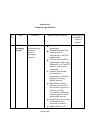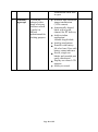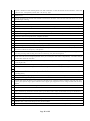
1
E-TENDER NOTICE Smt.G.R.Doshi & Smt.K.M.Mehta Institute of Kidney Diseases & Research Center Dr.H.L.Trivedi Institute of Transplantation Sciences Civil Hospital Campus,Asarwa, Ahmedabad-380016 E-tenders are invited from the reputed manufacturers, direct importers/authorized representative for supply & installation of Medical Equipments/Instruments. All tender documents can be viewed and downloaded free from websitehttp://www.ikdrc-its.org from 05/05/2015 to 20/05/2015 upto 5PM. Dr.H.L.Trivedi Director Page 1 of 35 Smt.G.R.Doshi & Smt.K.M.Mehta Institute of Kidney Diseases & Research Center Dr.H.L.Trivedi Institute of Transplantation Sciences Civil Hospital Campus, Asarwa, Ahmedabad-380016 E-TENDER NOTICE Tender notice No. IKDRC/D-01/RC/2015 E-Tenders are invited by The Director, IKDRC-ITS, Ahmedabad from reputed manufacturers, direct Importers/authorized representative for supply & installation of the following equipments/Instruments List of equipments/Instruments Sr. No. 1. 2. 3. 4. 5. 6. 7. 8. 9. 10. 11. Item Qty. EMD(Rs.) (3% of estimated value) Anesthesia Department Patient warming system Fluid warming cabinet Videolaryngoscope Gynecology Department Upgradation of tray loader system for genetics High End Ultra Sound with up gradation of existing system Endoscopy equipments compatible with existing system Ultra sonic Energy source as per specification Surgical Instruments (list will be available from Gynac OPD Case window, Basement area from 05/05/2015 to 20/05/2015 from 9.00 a.m. to 5.00 p.m.) Radiology Department High End ultrasound and colour Doppler system with fusion technology High End portable ultrasound and colour Doppler system High End ultrasound and colour Doppler system Page 2 of 35 6 22,500/1 30,000/1 10,500/1 84,000/1 3,60,000/1 3,60,000/1 90,000/1 45,000/- 1 2,70,000/2 3,00,000/1 1,50,000/- Key Dates Date and time for downloading of Tender Document from 05/05/2015 to 20/05/2015 upto 5PM. Last date and time for submission of online Tender with deposit 21/05/2015 upto 5 PM. Date and time of opening of online Technical & Commercial Bid 25/05/2015 at 1 PM. Envelope containing the “Technical Supporting Documents” including “Document Fee” & “EMD Fee” to be submitted physically on or before Dt22/05/2015 at below mentioned address: The Director IKDRC-ITS CIVIL hospital campus Asarwa Ahmedabad-380016 Page 3 of 35 Terms & Conditions: 1.Submission of Tender: The tenderer shall submit the tenders in one sealed cover, containing three separate subsealed covers before last date of submission. Part-I:Tender Document Fee Part-II: Technical Bid Part III: EMD 2.Each tender must be accompanied by tender document fee of Rs. 5000 (nonrefundable) by DD of any nationalized/scheduled bank drawn in favour IKDRC. Non-payment of the document fee will make the tenderer liable for disqualification. 3.Earnest Money Deposit: Each tender must be accompanied by EMD in the form of DD in favour of IKDRC payable at Ahmedabad, of any Nationalized Bank / Scheduled Bank. The EMD of the successful bidder shall be returned after the successful completion of contract / order and for unsuccessful bidder(s) it would be returned after award of the contract. Bid(s) received without demand drafts of EMD will be rejected. No interest will be paid on EMD. 4.Rate: Rate should be quoted in Indian Rupees (INR) or in Foreign Currency on DOOR Delivery Basis/CIF Ahmedabad at IKDRC-ITS, Ahmedabad, Inclusive of all the Charges: 5.Validity: The quoted rates must be valid for a period for 90 days from the date of closing of the tender. The overall offer for the tender and bidder(s) quoted price shall remain unchanged during the period of validity. If the bidder quoted the validity shorter than the required period, the same will be treated as unresponsive and it may be rejected. In case the tenderer withdraws, modifies or change his offer during the validity period, bid is liable to be rejected and the earnest money deposit shall be Page 4 of 35 forfeited without assigning any reason thereof. The tenderer should also be ready to extend the validity, if required, without changing any terms, conditions etc. of their original tender. 6. The company should give the certificate that the model quoted is the latest and not obsolete; and spares will be easily available for next 5-7 years. Imported items should be US-FDA or CE (ConformitéEuropéenne) certified. The company must enclose the User list and Performance report for the last 3 years from major hospitals where items are supplied. 7. Warranty / Guarantee: 7.1 Guarantee / Warrantee Period: For the equipment value upto Rs. 5 Lakh The Tenderers must quote for 2 years comprehensive warranty (Including all Spares, Accessories and Labour) from the date of completion of the satisfactory installation. The warranty charges shall not be quoted separately otherwise the offer shall be rejected. Also the bidders are requested to submit their quote (Rates) for subsequent 3 years Comprehensive Maintenance Contract (CMC) (Including All Spares, Accessories and Labour). Failure to comply this condition will entail the rejection of the bids. The price comparison shall be taking into account on basic price and post warranty CMC. 7.2 Guarantee / Warrantee Period: For the equipment value above Rs. 5 Lakh The Tenderers must quote for 5 years comprehensive warranty (Including all Spares, Accessories and Labour) from the date of completion of the satisfactory installation. The warranty charges shall not be quoted separately otherwise the offer shall be rejected. Also the bidders are requested to submit their quote (Rates) for subsequent 5 years Comprehensive Maintenance Contract (CMC) (Including All Spares, Accessories and Labour). Failure to comply this condition will entail the rejection of the bids. The price comparison shall be taking into account on basic price and post warranty CMC. 8. Uptime guarantee: The firm should provide uptime guarantee of 95% . 9. In case of malfunction/breakdown, the company should provide temporary back-up support within 48 hrs of registering the complaint till the time machine is repaired and returned. 10. Downtime penalty Clause a. During the comprehensive warranty period, the guarantee uptime of 95% of 365 days will be ensured. In case the down time exceeds the 5% limit, penalty of extension of guaranty period by two days for each additional day of Page 5 of 35 down time will be enforced. The supplier must undertake to supply all spares for optimal upkeep of the equipment for at least FIVE YEARS after handling over the equipment to the Institute. If accessories / other attachment of the system are procured from the third party, then the vendor must produce cost of accessory / other attachment and the CMC from the third party separately along with the main offer and the third party will have to sign the CMC with the Institute if required. b. The principals or their authorized service providers are required to submit a certificate that they have satisfactory service arrangements and fully trained staff available to support the uptime guarantee. 11. Delivery & Installation: All the items ordered shall be delivered & installed within 90 days from the date of issue of purchase order. All the aspects of safe delivery, installation and commissioning shall be the exclusive responsibility of the supplier. The successful tenderer will provide User manual & Service Manual in English& also arrange for basic required training for supplied items if asked for. If the supplier fails to deliver, install and commission the goods on or before the stipulated date, then a penalty at the rate of 2% per week of the total order value shall be levied subject to maximum of 10% of the total order value. 12. Satisfactory Installation: Satisfactory installation / commissioning and handling over of the equipment means the faultless functioning of the equipment for a minimum period of 30 days after satisfactory installation. 13. The quantity of item given in the tender is tentative, which may be increased or decreased as per the institute’s requirement. 14. Payment Terms will be discussed at the time of placing order. 15. The Institute reserves the right to accept in part or in full or reject any or more tender(s) without assigning any reasons or cancel the tendering process and reject all tender(s) at any time prior to award of contract, without incurring any liability, whatsoever to the affected bidder or bidder(s). 16. IKDRC-ITS reserves the right to ask the tenderers for arranging demonstration of the item for which rates have been quoted before placing purchase order. Page 6 of 35 17. The supplier should provide list of important spare parts and accessories with their part number alongwith installation. Cost of consumables/accessories should be frozen for the period of warranty and CMC. 18. All questions, disputes of differences arising out of and / or in connection with the terms & conditions of the contract, the decision of Director, IKDRC, Ahmedabad will be final and binding to supplier. The legal jurisdiction for any dispute will be Ahmedabad only. I have read and understood all the above terms and conditions of tender and I hereby agree and abide with the same. Date: Signature of Tenderer Place: Name of the Signatory ___________________ Stamp/Seal Page 7 of 35 Annexure-I Technical Specification Sr. No. 1. Item Patient warming system Function Required Specification To prevent hypothermia in paediatric patients, prolonged surgeries Convective forced air technology Should be suitable for intra-operative, postoperative and ICU applications Control unit should be light weight and easily attachable to IV rod/OT table as well as floor mounting Control unit should have at least 4 temperature selections displayed on large digital readout Should have safety features such as Selftest on start-up prior to each use, testing over/under temperature and disconnect visual and audible alarms Should have low energy consumption and noiseless operation High efficiency 0.2 Page 8 of 35 Specification available in offered Model micron air filter to reduces airborne particulate and ensure clean air delivery Airflow (55 CFM) Warm-Up Time of approximately 30 seconds Built-in hour meter to monitor usage for preventive maintenance 2. Fluid warming cabinet To warm IV fluids, blood components, irrigation fluids low heat density electrothermal cable array technology 72 Litre capacity Single chamber with 20 gauge stainless steel exterior casing Easy to use touch pad controls and easy to view LED display Two modes- Injection and irrigation Adjustable temperature range between 37-660 C A dual temperature display showing set point and actual temperature Accurate temperature within +0/1.70 C Audible alarm for over temp situations Insulated glass window doors to maintain temperature and conserve heat energy Internal lighting for observation of inventory with door closed and Page 9 of 35 illumination when door is open 3. Videolary ngoscope To view an enlarged video image of airway structures which is useful in difficult intubationand for teaching purpose Page 10 of 35 Wireless high resolution image transmission (VGA camera) Anatomically shaped blade with a guide channel for ET delivery Adult to infant application/ variable length blade Antifog mechanism Portable with battery life of at least 2 hrs with battery status indicator Sterile, single use blades and reusable part easily disinfected Display on colour LCD monitor Ability to record Item no.4 Up Gradation of Tray Loader system for Genetics: Camera - Basler Camera B/W CCD Digital Camera 1392 x 1040 pixel GenASIs Standard Server component consists of for online data. Dedicated large volume 2TB HDD Storage and software package (File Server component) Includes 5 days onsite labor and retraining. Update of Existing GenASIs Case Data Manager (CDM)Software User License for systems SC110011 /WS110016 / WS110017 Update of Existing GenASIs BandView So/ware User License for systems WS110016 /WS110017 Update of Existing GenASIs FISHView So/ware User License for systems WS110016 /WS110017 Update of Existing MetScan So/ware User License for systems SC110011 Update of Existing GenASIs ReloScan So/ware User License for systems SC110011 Update of Existing GenASIs SpotScan So/ware User License for systems SC110011 Annual Service Support Contract (12 months) Including Free Software Updates Existing System. Serial numbers: SC110011, WS110016, S110017 Microscope frame for transmitted and reflected microscopy with built-in filter holders for four filters (LBD, ND6, ND25 and GF), including hexagonal driver and immersion oil 8cc Dust cover Type 018 Lamp house for 100W halogen with connecting cable 12V100watt Halogen bulb (Philips No.7724) Power cord Trinocular tube Widefield eyepiece 10X F.N. 22 Widefield eyepiece 10X F.N. 22 Ceramic surface mechanical stage with right-hand low drive control (long type) Specimen holder for BX stage, left hand, for two specimens, thick type Quintuple Revolving Nosepiece Swing-out condenser N.A. 0.9-0.16 U Plan Semi Apochromat objective 10X/0.3, WD 10 Plan apochromat objective 1.25X/0.04, WD 5.0 Universal plan flurite objective 100X/1.3, WD 0.2 (spring, oil) Immersion oil 30cc. Low level auto-fluorescence. C-mount camera attachment with 0.63X lens Universal reflected light illuminator with field stop, aperture stop and 8-position mirror unit cassette, including UV protective shield JA5311, shield plate JA5290, Allen wrench AD6162, indication plate JA5820 and shield cap AC0792 (4 pcs) Corresponded to RoHS regulation Page 11 of 35 Neutral density filter for BX3 reflected light illuminator Lamp house for 100W mercury 100W mercury burner 100 Watts Power supply for mercury burner Empty mirror unit Chroma Fluorescence filter for DAPI (420-460nm) Chroma Fluorescence filter for FITC (520-550nm) Chroma Fluorescence filter for TRITC Chroma Fluorescence filter for Aqua Chroma Triple band filter for DAPI/FITC/TRITC Item no. 5 Specification of High End Ultra Sound with Upgradation of existing system Sl. No. Technical Specification 1 System should be the latest "State of the Art" fully digital ultrasound equipment capable of performing OBS-GYN, intra cavitary High resolution scanning specially first trimister scans & Should have Electronic convex 4D volume probe 2 The system should have the following modes : B-Mode (2D), Conventional M-Mode with varying sweep rates, anatomical M-Mode, PW doppler with high PRF (PW), High PRF Doppler Mode , (TD)-Tissue Doppler mode, colour flow doppler mode (CFM), Power Doppler Mode(PD), Directional power Doppler, HD-Flow Doppler Mode (HD-Flow) and B-Flor (BG), B/Colour/FW, volume modes 3D static , Real time 4D Mode, VCI- A, VCI - Curved/ Omniview, STIC & Advance STIC with AMM, eSTIC , 4D Biopsy, Live Bi-plane. 3 Power doppler angio imaging for perfusion studies for visualization of flow in small vessels and should be supported by all transducers. Electronic sector, Electronic Convex, Electronic Linear, Mechanical Volume Sweep, Electronic volume sweep, Matrix technology probe. 4 The System should be having a minimum of 46,000,000 system processing channel technology 5 Tissue Doppler Mode (TD) - Should allow high frame rate acquisition of tissue motion (up to 400 fps) 6 Volume imaging, multislice imaging with variable slice thickness( 0.5 - 10 mm) and multiplanar imaging on all types of 3D and 4D modes. 7 System should have facility for volume 3D/4D with high ultra light 4D convex volume probe and High Frequency 4D volume EV probe (on both gray scale and colour doppler modes) 8 Should be capable of performing live 4D imaging with volume transducers. 4D imaging should be possible in gray scale, colour mode, harmonic mode and with contrast agent imaging. Instant rendering of MPR images should be possible that rival acquired 2D resolution 9 Elastography analysis & Elastography ratio measurement-should be available into the elastography mode. System should have indicator for compression level with side-by side display of 2D image and 2D with graphical representation, elastogram images, preferred in TV probe. 10 System should have facility for dynamic range selections, dynamic resolution selection and different optimization for settings for different tissue compositions, colour coded display with parametric imaging 11 Dynamic range should be 274dB or more with range adjustability by selecting different dynamic contrast curves. Higher dynamic range will be preferred 12 In a 2D scanning - Min depth of field 0-1 cm & Max depth of field 0-36 cm (probe dependent) 13 256 (8 bits) discrete gray levels 14 system should have 23 inch high resolution LED display with DVI interface resolution of FHD 1920 X 1080 Pixel.High brightness with 300 CD/M2, Page 12 of 35 15 2D acquisition frame rate more than 500 frames/sec, colour doppler frame rate more than 300/S 16 The system should have the latest platform of Matrix technology/ Matrix probe availability.& Electronic 4D probe for better Speed and IQ 17 Real time compounding with colour or power doppler imaging 18 Multiple frequency selection for better penetration and resolution for better tissue differentiation and better contrast resolution 19 Post processing tools for annotation, measurement, correction of angle, baseline, sweep speed should be possible on stored images 20 System should have multivariate Tissue Harmonic Imaging including pulse inversion phase cancellation technology and coded harmonics on all transducers 21 It should be able to operate with compound imaging and speckle reduction algorithm 22 System should have on touch tissue contrast resolution adjustment without altering the set presets levels 23 System should have real time compounding imaging technology with minimum 9 transmitted lines of sight. 24 Real time compound imaging should operates in conjuction with Tissue Harmonic imaging, volume modes, panoramic imaging, and duplex doppler and in conjuction with speckle reduction imaging 25 High resolution algorithms for advanced speckle noise reduction, refined tissue pattern displays and fine border definition. 26 Should operate in 2D and 2D/CH/Doppler mixed modes (up to 150 frames per second) and with 3D and contrast agent imaging. 27 This feature shall have operator selectable settings and be capable of displaying in side by side mode with non speckle reduced image 28 Should have trapezoidal imaging and steerable imaging for 2D, colour and doppler with linear probe 29 Panoramic / extended field of view imaging should be available on convex and linear transducers. 30 This mode should build the extended field of view in a real time manner, showing the image as it builds 31 32 33 One button automatic adjustment / optimization for 2D mode, colour mode and doppler mode with auto correction of relevant fields of the mode used Incorporates advanced pulse shaping, coding excitation using new chirp transmit technology and coded harmonics mode for imaging deeper areas and obese patients. Additionally technology if available for imaging obese patients will be preferred The system should have a fast boot up time less than 200 seconds, when switch ON from OFF position and also less than 60 second from STANDBY position. Specify the system booting time less will be preferred 34 System should have high capacity fans with automatic speed for system cooling 35 Year of introduction of the specific model - should be as latest as possible, preferably should have been launches with in 1-2 years 36 Unique user friendly user interface for comfort and fast throughput System controls 37 System should have at least 45 automated and user programmable presents (output power signal processing and calculations) 38 System should have facility to adjust 2D performance instantly for different patient types (thin, average, obese) 39 The system shall display thumbnails on a clipboard with live gray mode while scanning to facilitate exams 40 Pan and zoom facility with high resolution results in both live and frozen images 41 Higher zoom will be preferred with HD zoom functionality upto 22 x zoom 42 Cine loop review facility in individual and mixed modes (frame by frame and in video mode), 2D : up to 10 min (depending on B-Image size and FPS); typical; about 3 min/4000 images (with curved array : 15 cm depth, 43 M mode : upto 20 min motion time (depending on sweep speed and depth) 44 Doppler mode : up to 10 min motion time (depending on sweep speed) 45 Post processing in freeze mode (dynamic Range adjustment, colour display on / off, colour / doppler invert, colour / doppler baseline adjustment, sweept speed, measurement, annotationand pictogram). Post processing of B-Mode images with Speckle Reduction Algorithm Page 13 of 35 46 Real time automatic doppler calculations on touch of a button. Should provide facility to apply automatic doppler analysis retrospectively to frozen spectral data or date retrieved from doppler scrolling. Possibility of manual doppler trace 47 System should have at least 8 calipers with depth information and extensive, customizable measurement and report packages including vascular, abdominal, small parts, urology, pediatrics, ortho, neurology, complete obstetrics, multigestational calculations, gynecology, and fetal heart report packages 48 Calipers should have minimum precision of 0.1mm , small size calipers for measuring < 5mm 49 Calipers of dynamically varying contrast compared to background. Delete last measurement option, curved linear distance measurement 50 51 52 Measurement (Distance and areas) should be possible in real time on frozen and on saved images of the old patients data for better comparison and clinical confidence. System should have facility to save reports along with patient data which can be retrieved later. Measured parameters must be printed directly in from of a report through laser printer or any other standard printers easily available in the market System should have facility of real time biopsy in 4D mode for the better visualization in 2d as well in all the three necessary plan / Axis. The system should be capable of displaying biopsy lines (for all transducers) while performing a 2D scan. 53 3D / 4D MODE 54 system should have Vol scan size max 64 MB for the grey volumes and max 90MB for the color volumes , the reqd memory space depends on the scanning parameters (vol BOX size and quality ( low, mid, height etc ) typical .8-5MB. 55 Volumes - frame/ sec max 359. depending on the scanning parameters . 56 systems should have following acquisition modes ;- 3D static, 3D (2d incl CRI), 3D /CFM (incl CRI), 3D B Flow, 4D real time , 4D BIOSPY, STIC & eSTIC , Vci- Omniview . 57 Speed adjustment on volume imaging 58 different render direction to view the volume image 59 Advanced tool for accurate quantification of irregular regions in 3D and automatically calculates the number and volume of hypo echoic structures to speed follicular assessments 60 Ability to restrict firing of the probe to a particular slice thickness of the region of interest 61 Advanced tool for selection of slice thickness out of complete volume dataset 62 4D fetal echo - 2D + colour + B Flor, STIC + Power doppler mode 63 STIC + CFM doppler mode, STIC + HD Flow Mode, STIC+CRI 64 STIC + CRI+CFM, STIC+CFI+PD 65 STIC+CRI+HD-flow, STIC + B-Flow 66 STIC + multislice mode with cine movement 67 Advanced imaging mode for visualization of hypo echoic areas and get automatic precise volume 68 Simultaneous visualization of 3 planes and 3D to guide the needle to the lesion 69 Transforming nuchal thickness measurement with automation 70 Should have auto 3D/4D rendering as well to get the best of 3D in fraction of second with one touch & should have same technology / feature in the real time 4D as well 71 Advanced Spatio temporal image correlation with STIC anatomical M Mode , & eSTIC 72 Semi automated tool to provide quantifiable NT & IT measurements 73 Automated sonography based technology helps streamline the acquisition of volumetric images of the fetal heart, displaying all recommended views with the push on one button 74 3D automated software dedicated to progression of labour (automatically documents the labour procedure to help users evaluate second stage labour progression (optional) 75 System should have Volume SRI (V-SRI) which helps to provide a high level of speckle reduction utilizing volume/voxels, versus traditional single slice imaging, it helps improve 3d/4d quality in multi-planar studies and also provides an enhanced smoothing effect on rendered images which helps improve diagnostic confidence. 76 System should have free moveable light source around following 3D object , 3D rendering image , VOCAL object Page 14 of 35 77 Systems should have latest rendering modes like hdlive silhouette - to show the internal clinical information of the fetus , HD live flow , transparerancy modes, max - min & xray , light 78 System should have the facility of take the Automatic measurement of the AC, BPD, HC, FL for the better productivity and time save . 79 System should have BI- PLANE mode available with electronic 4D probe with scan angle of B mode angle of 75 deg & Bi Plane angle of 90 deg, 80 81 SYSTEM SHOULD HAVE FOLLOWING PHYSICAL DIMENSIONS 82 The monitor should be 23" high resolution LED monitor with articulating arms with resolution of up to 1280x1024 pixel. 83 • Customizable layout to match workflow needs 84 • Report preview enabling facility for immediate feedback on diagnostic measurements 85 •should have Image presentation in Standard or XL format to allow doctors to see tiny details clearly 86 should have One touch responsiveness. Simplified workflow. 87 • should have 12.1” Touch Panel with multi-touch 88 • should have Electronic TGC and presenting as well for the same 89 •Should have Efficient menu navigation with swipe technology 90 should have Quick and easy 1-button control panel up/down function movement which allows for optimal ergonomics. 91 system should have Probe port illumination which will help in the connectivity and removal of the probe connectors in dark room conditions for the privacy of the patients 92 System should have Fast, secure data management for efficient communication, integrated software Digital video recording including USB recording, Fast USB 3.0 connectivity, Easy DICOM integration. 93 The equipment should be a room based wheeled unit with integrated brake, foot rest, transducer, cable and gel bottle holder and with electronic height adjustment facility for control panel and monitor independently. Transducer and gel bottle holders should be removable 94 There should be a digital brightness and contrast adjustment with preferable three default setting (dark room, semi dark room, bright room) 95 System should have a full size alphanumeric key board with interactive back - lighting. 96 The key board should be floating with rotation of +/- 40 deg from center, and with adjustable height of +200 mm 97 Integrated recording keys for remote control of up to 4 peripherals or DICOM devices one dedicated DVD recording key 98 The system shall have 4 universal probe ports in a convenient ( 3 active at a time) easy to access location with electronic switching facility 99 Must allow digital storage of gray scale as well as colour images (both frozen and cine loops) 100 Facility of reviewing and exporting in different formats 101 System shall support the ability to store digital raw data that allows optimizing imaging parameters such as B Gain, TGC, Colour gain, dynamic range, speckle reduction levels, doppler gain, doppler base line on image recalled from the image archive 102 The system should have on board storage facility for at least 500 GB. 103 The hard drive should be inbuilt. 104 The system shall provide the ability to sort images stored on board based on patient name, exam date, patient id and exam types, patent directly should show network status as print status, archive status, commit status and export to DVD status 105 Possibility to modify / edit patient data during and after exam has been completed 106 Screen and hard copy image distoration of less than 5% 107 Must have integrated CD/DVD writing - burning facility and it could be viewed on any ordinary PC. 108 Should be able to archive data from previously stored CD/DVD 109 DVD /CD drive to store / retrieve images in different formats (TIFF / JPF / AVI / DICOM) / patient reports Page 15 of 35 110 System should be DICOM 3.0 (or higher version) ready (Storing, transfer, print record on CD/DVD, DICOM structured reporting for OS and uro gynecology) including modality work list enabled and also to permit communication between devices of various manufacturer (existing in the department) and a facility of connectivity through Web (minimum network connection speed ot 100 Mbits/s) Details to be provided 111 System should be easily integrated with the departments VIEW POINT 6 patient Data bank. 112 Images must be printed on ink jet printer / laser printer in format of 4-15 spots per page (without using another computer in between). It should send images after each acquire and after end of exam (batch send) 113 114 TRANSDUCERS AND BIOPSY ATTACHMENTS 115 Transducers should be of broadband width with low loss lens and beam former technology for extreme high resolution image. 116 Please specify model number, footprint, bandwidth, imaging frequency, doppler frequency, FOV and weight of each transducer. 117 Lightweight transducers with flexible cables will be preferred 118 119 2D Convex probe - Wide band convex probe with bandwidth of 2-5 Mhz with FOV of 113 Deg 120 4D convex probe - Electronic 4D convex probe with Active matrix array technology with bandwidth of 1-7 Mhz. 121 4D TVTR probe - Wideband high-frequency endocavity volume probe bandwidth of 5-13 mhz for transvaginal examination with reusable biopsy guide 122 2D linear probe - Wide band linear probe with bandwidth of 3-8 Mhz for fetal medician examination 123 B/W Thermal printer of latest model (with CE or FDA mark) for image printouts. Please specify the brand model and specification details 124 Additional important requirement company has to provide service support of the present existing machines installed in the IVF dept for the better patient care and peace of mind for the dept for the successful running 125 price to be quote FOR destination and installation free of cost 126 Warranty - Three years from the date of installation . Page 16 of 35 Item no.6 Page 1-6 Endoscopy Equipments compatible with existing system PART A Camera system – (2 nos). The system should be truly Digital HDTV endoscopic video camera. The system should have the maximum Resolution of 1920 X 1080 pixels, progressive scan and the consistent use of 16: 9 formats for Input & Output to guarantee genuine HDTV. The system should have facility of Optical & Digital Zoom lens to enhance the quality of Image size & cross specialty usage of the camera system, regardless of the telescope used. USB Port for Capturing FULL HD Videos/ HD Stills in External USB drive and direct interface of USB Printer to facilitate direct printouts. System should have facility of controlling additional equipments like light source/ insufflators and recording device from the camera head. System should have facility to offer various visualization modes for surgery and diagnosis by shifting the color spectrum for recognition of the finest tissue Structures and their differentiation. Parallel live display of visualization modes besides white light mode (picture-in-picture). Digital FULL HD camera module should be compatible and fully upgradable to 3D System and to use with Flexible Video Endoscopes. Page 17 of 35 Page 2-6 3 Dimensional camera unit compatible with above camera components 3D with two distal CCD image sensors, direction of view 30°, diameter 10 mm, length 31 cm, for use with 3D CCU - one qty. link module to use with 3D, for use with 3D Telescope, power supply 100 - 120 VAC/200 - 240 VAC, 50/60 Hz including: Mains Cord, length 300 cm Link Cable, length 20 cm – One qty. Clip on glasses- 10 3D glasses – 20 Wire basket - one 3D Monitor – (1 no). 3D Monitor, 32" or more, color systems PAL/NTSC, max. screen resolution 1920 x 1080, image format 16:9, power supply 100 - 240 VAC, 50/60 Hz, Video inputs: 2x DVI, 2x HDSDI, VGA, S-Video, Composite, Video outputs: DVI, 2x HD-SDI, VGA, S-Video, Composite including: External 24 VDC Power Supply Mains Cord 16: 9 widescreen Monitor LED- 26 Inches – (1 no). The monitor should have: HDTV display in original 16: 9 HDTV format. 1080 p/ 50 & 1080 p/60 displays possible. LED crystal display. Max. Resolution of 1920X1080. Screen diagonal – 26”. Desk top with pedestal. Should have the facility of PIP mode. Specifications HD TFT Flat Screen Monitor with stand size 26", Aspect Ratio 16:9 HD format Brightness : 500 cd/m2 Maximum viewing angle : 178° vertical Contrast ratio: 1400 : 1 Reaction Time – 8ms Rated power : 115 watts Power Supply 100-240 VAC Screen Dimensions : 643 x 396 x 87mm Page 18 of 35 Page 3-6 XENON LIGHT SOURCE – (2 nos). Xenon Light Sources 300 watts Lamp type:- Xenon 15V, 300 Watt Color Temperatures 6000K Light Outlets – 1 Light Intensity Adjustment :- Continuously adjustable either manually or automatically by cameras video output signal. Fiber Optic Light Cable. – (2 nos). Thickness 4.8mm Length 300 cm Data Acquisition system and recording- (1 no). Dual channel Documentation System, for recording still images and videos, dual channel up to FULL HD, 2D/3D, including touch screen 10” or more, power supply 100 - 240 VAC, 50/60 Hz consisting of: Appliance Manual Mains Cord License Microsoft License DICOM USB Silicone Keyboard, with touchpad ACC Connecting Cable DVI Connecting Cable 2 HDMI to DVI-CABLE 1. Such system should have 2Tb or more hard disk and should be controllable with camera head from sterile area. Compatible with existing System. Signal management system- (1 no). DEVICE for DVI-D video signal transmission, e.g. an endoscopy camera or room camera, control via a front-button operation. power supply: 100 - 240 VAC, 50/60 Hz Video inputs: 4x DVI-D (DVI jack) Video outputs: 4x DVI-D (DVI jack) consisting of: 400 A Mains Cord, length 300 cm 2x DVI-D Connecting Cable(s), length 200 cm for use as 1 x 4 DVI-D switch or 4 x 4 DVI-D router Compatible with existing System. Editing software to edit HD videos - one Page 19 of 35 Page 4-6 Compact Hysteroscope – (1 no). Compact Hysteroscope, telescope 30°, size 4 mm, with channel for semi rigid 5 Fr. operating instruments, with suction and irrigation valves for single or continuous-flow use, long handle consisting of: Compact Hysteroscope Outer Sheath Suction and Irrigation Valve Monobloc Adaptor Seal, for instrument ports, Contact Hysteroscope –(1 no). Operating and Contact Hysteroscope with forward-oblique telescope 30°, enlarged view, magnification 1x, 50x, diameter 4 mm, length 30 cm, autoclavable, fiber optic light transmission incorporated Accessories Scissors Insert with Outer Sheath, curved, double action jaws, spoon-shaped jaws, length of blades 17 mm, size 5 mm, length 36 cm, sterile, for single use, package of 10 To be used with existing handles – 10 Boxes. Equipment Cart – (2 nos). Equipment Cart, rides on 4 antistatic dual wheels, 2 equipped with locking brakes (front), 2 fixed shelves, 1 drawer unit with lock, integrated cable conduits in both vertical beams, mains switch in vertical beam, 1 set of non-sliding stands, 1 camera holder, power box with socket board with 12 sockets and 12 potential earth connectors Dimensions: Equipment cart: 730 x 1450 x 660 mm (w x h x d), Shelf: 630 x 480 mm (w x d), Caster diameter: 125 mm Consisting of: Equipment Cart Power Box. Page 20 of 35 Page 5-6 PART B Single chip camera system set : (2 nos) 1-Chip Camera-Control Unit (CCU), , with integrated Communication Bus (SCB) and Image Processing Module, Color System PAL/NTSC, Power Supply: 100-240 VAC, 50/60 Hz bestehend aus: Camera Control Unit 400A Mains Cord BNC-Connecting Cable, length 180 cm 547S S-Video (Y/C) Connecting Cable, length 180 cm Special RGB-Connecting Cable, length 180cm Connecting Cables for remote control of Video-Printers Connecting Cable, length 100 cm Keyboard with US-English character set One-Chip Camera Head, color system PAL, soakable, gas-sterilizable, with integrated Parfocal Zoom Lens, f = 25 - 50 mm (2x), 2 freely programmable camera head buttons Cold Light Fountain XENON 175 : (2 nos). Cold Light Fountain XENON 175, with one 175 Watt XENON lamp and one light outlet Power Supply: 100125/220-240 VAC, 50/60 Hz consisting of: XENON 175 400A Mains Cord Fiber Optic Light Cable: (2 nos). Fiber Optic Light Cable, with straight connector, diameter 3.5 mm, length 230 cm Quantity Hysteroscopy set with standard 4 mm scope Forward-Oblique Tele- scope 30°, ø 4 mm, length 30 cm, autoclavable, fiber optic light transmission incorporated, Examination Sheath, ø 5.2 mm, with 1 Luerlock adaptor, for use with Continuous flowExamination sheath Continuous-Flow Examination Sheath, ø 6.2 mm, with 1 LUER-lock adaptor, for use with Examination sheath Operating Sheath, size 5.4 mm, with channel for semirigid 5 Fr. operating instruments, with 1 stopcock and 1 LUER-Lock adaptor, for use with Continuous Flow operating sheath Continuous- Flow Operating Sheath, size 6 mm, with 1 stopcock and 1 LUER-Lock adaptor, for use with operating sheath Biopsy and Grasping Forceps, semirigid, double action jaws, 5 Fr., length 34 cm Scissors, semirigid, blunt, single action jaws, 5 Fr., length 34 cm Bipolar dissection electrode, semirigid, 5 Charr., length 36cm Bipolar High Frequency Cord with 2 x 4 mm banana-plug Needle Electrode, unipolar 5 Fr., length 34 cm 2 2 2 2 2 6 6 6 6 6 Page 21 of 35 Page 6-6 Bipolar High Frequency Cord with 2 x 4 mm banana-plug Unipolar High Frequency Cord, with 4 mm plug, length 300 cm 6 6 Additional Instruments for TCRE SET (UNIPOLAR) Working Element Set consisting of: 1 Working element 1 Cutting Loop, angled 2 1 Coagulating-Electrode, ball end, ø 3 mm 1 Coagulating-Electrode, ball end, ø 5 mm 1 Coagulating Needle Electrode, angled 2 High Frequency Cord 1 Protection Tube, for sterilization and storage of electrodes, curettes and knives Cutting by means of a spring. The thumb support is movable. In rest position the electrode tip is inside the sheath Resectoscope Sheath, including connecting tube for in- and outflow, for continuous irrigation and suction, 26 Fr., oblique beak, rotatable Inner Sheath with ceramic insulation, for use with Working Elements Standard Obturator, for use with Resectoscope Sheaths 2 2 2 Additional Instruments for TCRE SET (BIPOLAR) Telescope 12°, ø 4 mm, length 30 cm, 2 autoclavable, fiber optic light transmission incorporated, Working Element Set, bipolar, cutting by means of a spring, movable thumb support. In rest position the electrode tip is inside the sheath. consisting of: Working Element, bipolar 1 Cutting Loops, bipolar 1 Cutting Electrode, bipolar, pointed Coagulation Electrode half moon®, bipolar, with ball end Bipolar High Frequency Cord Protecting Tube 2 Page 22 of 35 Item no.7 Specification of Ultrasonic Energy Source as per specification Product Specification Ultrasonic cutting and coagulating technology device upgrade with consumable Laparoscopic Coagulating Shears, 5mm Curved, 36cm length with Hand activation facility Item no. 8 SPECIFICATION OF GYNECOLOGICAL SURGERY INSTRUMENTS SET The instruments quoted should be of high quality and standard The Instruments should be imported and of CE certification. Copy of the CE certificate must be enclosed The instruments must be ISO certified and copy to be enclosed Sterilization Container should be quoted alongwith Instrument set. Sterilization Container and Instruments should be of the same parent company. The Sterilization containers should meet international standards and approved for steam sterilization procedures to EN 285: 2008 and validated acctp ISO 17665 Part1:2006 It should have an indicator wherein colour green means the container is "sterile" and when the container is opened, the indicator should automatically change to red colour indicating "unsterile" It should have reusable microbial barriers instead of disposable filters. The microbial barriers should be easy to remove and clean. It should have lateral flow ducts at the top for flow of air. The instruments should remain sterile in the container and the container should be capable of being brought into the Operation Room without any essential packaging. All instruments and Sterilization container must be manufactured by single manufacturer Page 23 of 35 Item no.9 Specification for high end ultrasound and colour doppler system.-quantity required : 1 1. System must be FDA or CE approved product. 2. 5 years warranty including all probes and spares must. 3. The system must be high end and should be latest and state of the art with fully digital technology equipment to incorporate the facility of 2D, M -Mode, CDI, PW Doppler, CW doppler, Power Doppler, directional power angio, Elastography imaging, -Imaging for abdomen, obstetrics & Gynac, Cerebrovascular, peripheral vascular, & superficial parts imaging like breast, scrotum, thyroid, musculoskeletal and adult cardiac. 4. System must be offered with a minimum of 4,00,000 digital processed channels. Technical datasheet should be enclosed in technical bid to support the number of channels on the systems. If not mentioned Please attach a letter from manufacturer along with the technical bid clearly stating the digital processed channels of the offered system. 5. System must have Convex and transvaginal transducer with either single crystal technology or pure wave technology for excellent Image quality on Difficult to image patients. Please mention the crystal technology used in the transducer. Technical data sheet should been closed in technical bid to support the crystal technology. 6. System must be offered with a minimum 21 inch High Resolution Flat Panel Medical grade Display monitor with tilt and swivel facility. Flat monitor should be movable and height adjustable. 7. System should have at -least four Imaging universal active probe ports with electronic switching facility from key board without probe adapter. 8. Operating modes B-mode, M-Mode, B/M Mode, Doppler Mode, Colour flow, Power Doppler, DCA/DPA, B/Colour flow, PW Doppler, CW Doppler, and elastography Imaging. 9. All transducers should have Broad Bandwidth Beam former technology for extreme High Resolution 2D Imaging. Frequency range of Transducers should be 1 to 18 MHz or more. This should be available without the need for frequency switching. 10. B mode & B colour simultaneous should be available side by side real time display of B Mode & Colour flow. Digital zoom facility for region of interest in real time and frozen images. 11. Image storage facility on in build hard disc or MOD/CD/DVD-RW facility should be available. In built hard disk with capacity of 500 GB. System should have extensive image management capability including thumb nail review, Cineloop editing etc. 12. Cine loop as well as cine scroll facility in B mode with storage of 800 or more images should be available. 13. Auto trace & automatic Doppler calculations should be available in Live & frozen images. 14. Should have the state of the art Transmit Real Time Compound Imaging Technology with Multiple transmitted lines of sight, wherein Multiple Coplanar Images from different viewing Angles are obtained and combined into a single compound Image at real-time frame rates for improved visualization. 15. System must be offered with Speckle Reduction Imaging: Image processing technique to remove speckles and clutter artefacts. Page 24 of 35 16. Advanced measurements & calculation package for abdominal, obst./gynac, urology, vascular and cardiac should be available. 17. System should be capable of scanning depth of 30cm. Scanning Depth should be clearly mentioned in the technical quote If not mentioned Please attach a letter from manufacturer along with the technical bid clearly stating the scanning depth of 30cm in the offered system. 18. System must be offered with and 2D frame rate of at least 1000 frames/second. Acquisition frame rate should be clearly mentioned in the technical quote If not mentioned Please attach a letter from manufacturer along with the technical bid clearly stating the frame rate of the offered system. 19. System must be offered with minimum 12 inch high resolution user interface touch panel. 20. System should have THI & should be able to work in combined mode of harmonic imaging and real time compound imaging to get excellent image quality. The system shall offer Tissue Harmonic Imaging in Power Doppler imaging mode for improved sensitivity and specificity in differentiating blood/agent from tissue. 21. Automatic real time & frozen tracing of instantaneous peal velocity & instantaneous mean velocity (or frequency) should be available. Triplex Imaging should be standard on the system. 22. The System should have Panaromic imaging / Sie-scape and extended field of view imaging. 23. The System should be quoted along with Elastography Imaging as standard. Strain based Elastography for the below. 23(a) Strain based Elastography for Breast Imaging accompanied by quantification package software. One touch entry into elastography mode. Elastogram applied as a region of interest box with user control of size and location through entire field of view and measurement of strain rate and total strain rate with graphical display. 23(b) Strain based Elastography for Gynecology Imaging accompanied by quantification package software. One touch entry into elastography mode. Elastogram applied as a region of interest box with user control of size and location through entire field of view and measurement of strain rate and total strain rate with graphical display. 23(c) Strain based Elastography for Urology Imaging accompanied by quantification package software. One touch entry into elastography mode. Elastogram applied as a region of interest box with user control of size and location through entire field of view and measurement of strain rate and total strain rate with graphical display. 24. The System should be quoted with Liver elastography imaging using acoustic push pulses and tracking pulses to assess liver tissue stiffness. The reading must be in both in m/s and kPa. 25. The system should be DICOM ready. System should have capability of HIS and RIS connectivity and should also be connected to the dry chemistry printer available in the department (CR/DR system/ CT/MRI/Mamography). Should provide advanced DICOM connectivity to an enterprise data management system or PACS with advanced DICOM features: DICOM Store, Modality Work list, Performed Procedure Step and Structured Reporting. Please specify the advance DICOM features available on the quoted system. Page 25 of 35 SYSTEM MUST BE THE FOLLOWING TRANSDUCERS 1. 2. 3. 4. 5. 1-5 MHz Broadband Convex Transducer for General Imaging, Abdomen, Renal, OB/GYN imaging with capabilities shear wave or equivalent elastography imaging for assessing liver stiffness. Must have Tissue harmonic imaging. This transducer should have either single crystal technology or pure wave technology for excellent Image quality on Difficult to image patients. Please mention the crystal technology used in the transducer by attaching technical data sheet of transducer. 5-18 MHz Linear Array Transducer for Vascular, breast, Musculoskeletal, small parts imaging with capabilities strain based elastography imaging. Must have Tissue Harmonic Imaging. Please mention the elastography technology used in the transducer by attaching technical datasheet of transducer. 3-10 MHz Broadband Microconvex Transducer for endocavity imaging with capabilities of strain based elastography imaging. Must have Tissue Harmonic Imaging. This transducer should have either single crystal technology or pure wave technology for excellent Image quality on Difficult to image patients. Please mention the crystal technology used in the transducer by attaching technical data sheet of transducer. 3-12MHz Linear Array Transducer for Vascular, intervention, bowel, Musculoskeletal, small parts imaging .Must have Tissue Harmonic Imaging. 5-8 MHZ broad band curved array for pediatric and neonatal applications. System should be supplied with the following peripheral devices: Sony Thermal B/W Printer 2 KVA ONLINE UPS. System should have biopsy kit for convex, linear and endocavity probe. Quantification Tool automated measurements of intima media thickness in carotids and other superficial vessels. Warranty condition: 1.Warranty period uptime of 95% 0f 365 days will be ensured. In case the down time exceeds the 5% limit penalty of extension of warranty period by two days for each additional day of down time will be enforced. The vendor must undertake to supply all spares for optimal upkeep of the equipment for at least 5 years after handling over the unit to the institute. 2.The principals or the authorized service providers are required to submit certificate that they have satisfactory service arrangements and fully trained staff available to support the uptime warranty. Page 26 of 35 Item no.10 Specification for high end portable ultrasound with colour Doppler and echocardiography. Quantity required: 2 1. System should be FDA or CEA approved. 2. 5 years warranty including all probes and all spares. 3. System should be quoted with multiport connection trolley. At least three active probe connectors are must. 4. System must be a state-of-the-art model all digital beam former with super computed signal processing and clinically proven imaging technologies. 5. System must be offered with the following applications. : abdomen, obstetrics &gynac, cerebrovascular, peripheral vascular, adult transcranial, superficial parts like breast, scrotum, thyroid, musculoskeletal , Adult Paediatric, Neonatal Cardiology , TEE Imaging. 6. The System should not weight more than 7 kgs. Systems which are heavier than 7 kgs are liable for Rejection. 7. System must be offered with a minimum of 200000 digital processed channels per image frame. Original technical data sheet should be enclosed in the technical bid to support the number of channels on the systems. If not mentioned Please attach a letter from manufacturer along with the technical bid clearly stating the Channels of the offered system. 8. System must have Convex, transvaginal and cardiology transducer with either single crystal technology or pure wave technology for excellent Image quality on Difficult to image patients. Please mention the crystal technology used in the transducer. Technical data sheet should be enclosed in technical bid to support the crystal technology. 9. System must be offered with a minimum 15 inch High Resolution Flat Panel Display monitor with minimum monitor resolution of 1050 x 1400. Systems offered with smaller screen are liable for rejection. 10. System must be offered with frequency compounding facility. Other equivalent Technology can also be offered. Processing technology in technical bid should be highlighted. 11. System must be offered with 2D, M - mode, Colour M- mode, Anatomical M-mode, Colour Flow, Pulse Wave Doppler, continuous Wave Doppler and Directional Colour Power Doppler. 12. System must be offered with Speckle Reduction Imaging: Image processing technique to remove speckles and clutter artefacts. 13. System must be offered with a very high dynamic range of at least 170 Db to pick up subtle echoes. Original technical data sheet should be enclosed in the technical bid to support the Dynamic range in Db. If not mentioned Please attach a letter from manufacturer along with the technical bid clearly stating the dynamic range of the offered system. 14. Frequency processing facility for the transducers should be 1 - 12 MHz. This must be available without the need for frequency switching. 15. System must be offered with Eight-slide pot control adjustment of TGC curve. 16. Independently selectable gain Control in Lateral plane. (Better technology can also be offered). 17. Triplex Imaging should be standard on the system. 18. System must be offered with a 2D frame rate of at least 750 frames/second. Acquisition frame rate should be clearly mentioned in the technical quote If not mentioned Please attach Page 27 of 35 19. 20. 21. 22. 23. 24. 25. 26. 27. 28. 29. 30. a letter from manufacturer along with the technical bid clearly stating the dynamic range of the offered system, failing which the bid is liable for rejection. System must be offered with Anatomical M-Mode (Angle-corrected M-mode). System must be offered with Pulsed wave Tissue Doppler Imaging (TDI) for velocity mapping of cardiac tissue and vessel wall motion. Must be offered with a single button control for automatic optimization and adjustment of TGC and Receiver Gain to achieve optimal uniformity of image quality and faster scans. This should be demonstrated to the users in Cardiology and vascular exams during technical discussions. System must be offered with Colour TDI - uses colour to display direction and timing of myocardial function. System must be offered with Enhanced Tissue Harmonic Imaging should be standard on the system. This should be based on a real - time digital signal storage and phase cancellation technique to enhance axial and contrast resolution. System should be offered with Fully-integrated stress echo module including User-defined protocols ,Stop and resume a protocol, Re-label, add or remove stages or views during a protocol, Up to eight-stage capture-and-study protocols, LVO with stress, Wall motion scoring and reporting. They should have minimum hard 80 GB disk drive space. System must have inbuilt battery backup 1 hour or more. System should have extensive image management capability including thumb nail review, Cine loop editing etc. System should have facility to transfer images to an integrated CD writer, without any interfacing. Specify if integrated CD writer is available in your technical quote. Print - Should have direct connectivity to Inkjet printer for printing images & report. The unit offered in tender may require technical demonstration. SYSTEM MUST BE THE FOLLOWING TRANSDUCERS. SYSTEM : 1 1. 1-5 MHz Broadband Adult Echo Transducer for Adult Cardiology imaging. Must have Tissue Harmonic Imaging, Must have either single crystal technology or pure wave or matrix technology for excellent Image quality on Difficult to image patients. Must attach original technical datasheet of transducer to specify the above technology used in the transducer. 2. 1-5 MHz Broadband Convex Transducer for General Imaging, Abdomen, Renal, OB/GYN imaging. Must have Tissue Harmonic Imaging. This transducer should have either single crystal technology or pure wave technology for excellent Image quality on Difficult to image patients. Please mention the crystal technology used in the transducer by attaching technical data sheet of transducer. 3. 3-12MHz Linear Array Transducer for Vascular, intervention, bowel, Musculoskeletal, small parts imaging. Must have Tissue Harmonic Imaging SYSTEM :2 1. 1-5 MHz Broadband Convex Transducer for General Imaging, Abdomen, Renal, OB/GYN imaging. Must have Tissue Harmonic Imaging. This transducer should have either single Page 28 of 35 crystal technology or pure wave technology for excellent Image quality on Difficult to image patients. Please mention the crystal technology used in the transducer by attaching technical data sheet of transducer. 2. 3-12MHz Linear Array Transducer for Vascular, intervention, bowel, Musculoskeletal, small parts imaging. Must have Tissue Harmonic Imaging 3. 3-10 MHz Broadband Microconvex Transducer for endocavity imaging. Must have Tissue Harmonic Imaging. This transducer should have either single crystal technology or pure wave technology for excellent Image quality on Difficult to image patients’ .Please mention the crystal technology used in the transducer by attaching technical data sheet of transducer The following upgrades on the same system must be quoted as optional. 4-12 MHz Broadband Paediatric Echo Transducer for Neonatal and Paediatric Cardiology imaging. 2-7MHz Trans-oesophageal Transducer with for Adult applications with Electro cautery Suppression. Must have Tissue Harmonic Imaging. Must have more than 1500 crystals for excellent Image quality provides real-time simultaneous longitudinal and short-axis views on Difficult to image patients .Must attach original technical data sheet of transducer to specify the no of crystal used in the transducer. System should be upgradable to convex, linear intraoperative probe. System should be supplied with the following peripheral devices Sony Thermal B/W Printer. Warranty condition : 1. Warranty period uptime of 95% 0f 365 days will be ensured. In case the down time exceeds the 5% limit penalty of extension of warranty period by two days for each additional day of down time will be enforced. The vendor must undertake to supply all spares for optimal upkeep of the equipment for at least 5 years after handling over the unit to the institute. 2. The principals or the authorized service providers are required to submit certificate that they have satisfactory service arrangements and fully trained staff available to support the uptime warranty. Page 29 of 35 Item no.11 High end ultrasound and colour Doppler system. Quantity required :1 1. System must be FDA or CE approved product. 2. 5 years warranty including all probes and spares must. 3. The system must be high end and should be latest and state of the art with fully digital technology equipment to incorporate the facility of 2D, M -Mode, CDI, PW Doppler, CW doppler, Power Doppler, directional power angio, Contrast Imaging, Elastography imaging, Imaging for abdomen, obstetrics &Gynac, Cerebrovascular, peripheral vascular, & superficial parts imaging like breast, scrotum, thyroid, musculoskeletal and adult cardiac. 4. System must be offered with a minimum of 4,00,000 digital processed channels. Technical datasheet should be enclosed in technical bid to support the number of channels on the systems. If not mentioned Please attach a letter from manufacturer along with the technical bid clearly stating the digital processed channels of the offered system. 5. System must have Convex and transvaginal transducer with either single crystal technology or pure wave technology for excellent Image quality on Difficult to image patients. Please mention the crystal technology used in the transducer. Technical data sheet should been closed in technical bid to support the crystal technology. 6. System must be offered with a minimum 21 inch High Resolution Flat Panel Medical grade Display monitor with tilt and swivel facility. Flat monitor should be movable and height adjustable. 7. System should have at -least four Imaging universal active probe ports with electronic switching facility from key board without probe adapter. 8. Operating modes B-mode, M-Mode, B/M Mode, Doppler Mode, Colour flow, Power Doppler, DCA/DPA, Contrast Imaging, B/Colour flow, PW Doppler, CW Doppler, and elastography Imaging. 9. All transducers should have Broad Bandwidth Beam former technology for extreme High Resolution 2D Imaging. Frequency range of Transducers should be 1 to 18 MHz or more. This should be available without the need for frequency switching. 10. B mode & B colour simultaneous should be available side by side real time display of B -Mode & Colour flow. Digital zoom facility for region of interest in real time and frozen images. 11. Image storage facility on in build hard disc or MOD/CD/DVD-RW facility should be available. Inbuilt hard disk with capacity of 500 GB. System should have extensive image management capability including thumb nail review, Cineloop editing etc. 12. Cine loop as well as cine scroll facility in B mode with storage of 800 or more images should be available. Cineloop frames should also be available for abdominal contrast applications. 13. Auto trace & automatic Doppler calculations should be available in Live & frozen images. 14. Should have the state of the art Transmit Real Time Compound Imaging Technology with Multiple transmitted lines of sight, wherein Multiple Coplanar Images from different viewing angles are obtained and combined into a single compound Image at real-time frame rates for improved visualization. 15. System must be offered with Speckle Reduction Imaging: Image processing technique to remove speckles and clutter artefacts. 16. Advanced measurements & calculation package for abdominal, obst./gynac, urology, vascular and cardiac should be available. 17. System should be capable of scanning depth of 30cm. Scanning Depth should be clearly mentioned in the technical quote If not mentioned Please attach a letter from manufacturer Page 30 of 35 along with the technical bid clearly stating the scanning depth of 30cm in the offered system. 18. System must be offered with a 2D frame rate of at least 1000 frames/second. Acquisition frame rate should be clearly mentioned in the technical quote If not mentioned Please attach a letter from manufacturer along with the technical bid clearly stating the frame rate of the offered system. 19. System must be offered with minimum 12 inch high resolution user interface touch panel. 20. System should have THI & should be able to work in combined mode of harmonic imaging and real time compound imaging to get excellent image quality. The system shall offer Tissue Harmonic Imaging in Power Doppler imaging mode for improved sensitivity and specificity in differentiating blood/agent from tissue. 21. The system should have Contrast Harmonic Imaging and should have optimization settings to detect the Contrast Agents. Please specify other advanced Technologies to perform better Contrast Harmonic Imaging. 22. Automatic real time & frozen tracing of instantaneous peak velocity & instantaneous mean velocity (or frequency) should be available. Triplex Imaging should be standard on the system. 23. The System should have Panaromic imaging / Sie-scape and extended field of view imaging. 24. The System should be quoted along with Elastography Imaging as standard. Strain based Elastography for the below. 24(a) Strain based Elastography for Breast Imaging accompanied by quantification package software. One touch entry into elastography mode. Elastogram applied as a region of interest box with user control of size and location through entire field of view and measurement of strain rate and total strain rate with graphical display. 24(b)Strain based Elastography for Gynaecology Imaging accompanied by quantification package software. One touch entry into elastography mode. Elastogram applied as a region of interest box with user control of size and location through entire field of view and measurement of strain rate and total strain rate with graphical display. 24(c) Strain based Elastography for Urology Imaging accompanied by quantification package software. One touch entry into elastography mode. Elastogram applied as a region of interest box with user control of size and location through entire field of view and measurement of strain rate and total strain rate with graphical display. 25. The System should be quoted with Liver elastography imaging using acoustic push pulses and tracking pulses to assess liver tissue stiffness. The reading must be in both in m/s and kPa. 26. The system should be DICOM ready. System should have capability of HIS and RIS connectivity and should also be connected to the dry chemistry printer available in the department ( CR/DR system/ CT/MRI/Mamography). Should provide advanced DICOM connectivity to an enterprise data management system or PACS with advanced DICOM features: DICOM Store, Modality Work list, Performed Procedure Step and Structured Reporting. Please specify the advance DICOM features available on the quoted system. 27. Live Ultrasound Fusion Imaging with navigation Generate and display fused multimodality images to leverage the combined advantages of resolution, contrast, and real-time feedback from the following modalities. 7a. CT 7b. MR 7c. PET/CT Page 31 of 35 7d. Rotational fluoroscopy 7e.Track the tips of flexible and rigid instruments during minimally invasive needle procedures used clinically in biopsies, ablations and drainages in liver, kidney, and bone 7f. Displays the instrument position, orientation, and trajectory on pre -procedure, intraprocedure, and fused multimodality images to help guide the instrument, even when the target is hard to see or difficult to reach. 7g. Motion compensation (respiratory gating) - Provides advanced software algorithms that track and show patient breathing SYSTEM MUSTBETHE FOLLOWINGTRANSDUCERS 1. 1-5 MHz Broadband Convex Transducer for General Imaging, Abdomen, Renal, OB/GYN imaging with capabilities of CEUS and shear wave or equivalent elastography imaging for assessing liver stiffness. Must have Tissue harmonic imaging. This transducer should have either single crystal technology or pure wave technology for excellent Image quality on Difficult to image patients. Please mention the crystal technology used in the transducer by attaching technical data sheet of transducer. 2. 5-18 MHz Linear Array Transducer for Vascular, breast, Musculoskeletal, small parts imaging with capabilities of CEUS and strain based elastography imaging. Must have Tissue Harmonic Imaging. Please mention the elastography technology used in the transducer by attaching technical datasheet of transducer. 3. 3-10 MHz Broadband Microconvex Transducer for endocavity imaging with capabilities of CEUS and strain based elastography imaging. Must have Tissue Harmonic Imaging. This transducer should have either single crystal technology or pure wave technology for excellent Image quality on Difficult to image patients. Please mention the crystal technology used in the transducer by attaching technical data sheet of transducer. 4. 3-12MHz Linear Array Transducer for Vascular, intervention, bowel, Musculoskeletal, small parts imaging with capabilities of CEUS imaging. Must have Tissue Harmonic Imaging. 5. 7-15 MHz Intra operative linear Array Transducer. Should attach technical data sheet of transducer to specify. Page 32 of 35 System should be supplied with the following peripheral devices: Sony Thermal B/W Printer 2 KVA ONLINE UPS. System should have biopsy kit for convex, linear and endocavity probe. Quantification Tool automated measurements of intima media thickness in carotids and other superficial vessels. Warranty condition 1. Warranty period uptime of 95% 0f 365 days will be ensured. In case the down time exceeds the 5% limit penalty of extension of warranty period by two days for each additional day of down time will be enforced. The vendor must undertake to supply all spares for optimal upkeep of the equipment for at least 5 years after handling over the unit to the institute. 2.The principals or the authorized service providers are required to submit certificate that they have satisfactory service arrangements and fully trained staff available to support the uptime warranty. Page 33 of 35 Annexure-II TECHNICAL BIDDOCUMENTS Name of manufacturer/Importer/ Authorized representative Complete Address & Telephone No./email address Letter from parent firm in case of authorized representative Import lisence No. if direct importer Name address &Ph.No./email of service centre Whether the firm is a registered firm Yes/No (attached copy of certificate) US FDA/CE certificate PAN No. (enclose the attested copy of PAN Card) Service Tax No. (enclose the attested copy of Service Tax Certificate) VAT No. (enclose the attested copy of VAT Certificate) Whether the firm has enclosed the Bank Draft/Pay Order/Banker’s cheque of Earnest Money Deposit. Whether the Firm/Agency has signed each and every page of Tender/NIT Please provide full list of consumables alongwith price Any other information, if necessary Authorized signatory of the bidder with seal. Page 34 of 35 Annexure III Format for Financial Bid (To be submittedon the letterhead of the company / firm separately for each item) S.N. o Name of Item Quantity Rate VAT/Taxes Amount 1.I/We have gone through the terms &conditions as stipulated in the tender enquiry document and confirm to accept and abide the same. . 2.No other charges would be payable by the Institute. Authorized signatory of the bidder withseal. Page 35 of 35