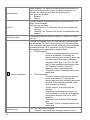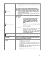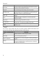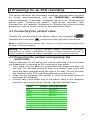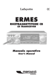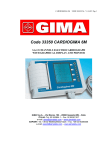
1
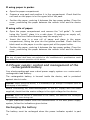
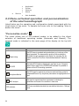
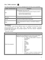
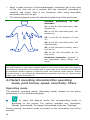
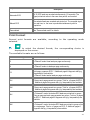
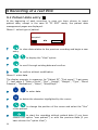
ar2100adv
user manual
english
This User Manual has been prepared with the objective of giving the user all the
information necessary to make the best use of the CARDIOLINE® ar2100adv.
General information
et medical devices SpA, continuously in search of technological improvement and
customer satisfaction, reserves the right to modify this publication without prior
notice at any time.
All rights reserved © et medical devices SpA ITALY.
CARDIOLINE® is a registered trademark of et medical devices SpA
CARDIOLINE® product support services
For any questions about a CARDIOLINE product:
consult the documentation and other printed material included in the package;
consult any guidelines available.
If you find no solution, you can obtain further information by contacting your
CARDIOLINE supplier.
Before calling, check you have the available documentation to hand and the product
nearby. It may also be necessary to supply the following information:
serial number and product reference number, if available;
type of hardware available, including any network hardware fitted;
operating system used, for software products;
exact contents of any error messages displayed;
description of the operation being executed when the problem occurred;
description of any action taken to solve the problem.
um_ar2100adv_cardioline_02_eng1 Rev. 02/sr/GZ 20/03/2007 Ref: 36519098
2
Contents
1 Introduction
5
1.1 How to read the manual
6
1.2 Information and recommendations relating to safe use
7
1.3 The electrocardiograph
Front view
9
9
Side view
9
Parts, symbols and controls
10
2 Installation and initial preparation
13
2.1 Selecting the installation site
13
2.2 Loading the thermal paper
13
2.3 Power supply; control and management of the rechargeable battery
Recharging the battery
14
14
2.4 How to switch on the electrocardiograph
15
2.5 How to switch off the electrocardiograph
Auto power off
15
16
3 Preparation for use: the menu
17
3.1 How to access the menu
17
3.2 Structure of the menu
17
3.3 Menu-activated operation and personalization of the electrocardiograph
20
"Personalise mode"
20
The “ECG archive”
21
“Settings”
21
“Tools”
24
4 Preparing for an ECG recording
25
4.1 Connecting the patient cable
25
4.2 Preparing the patient and applying the electrodes
4.3 Select recording
sensitivity, filters
Operating mode
characteristics
operating
mode,
25
print
format,
speed,
26
26
Print format
27
Speed of recording on paper
28
Sensitivity of recording on paper
28
Recording filters
28
5 Recording of a rest ECG
30
3
5.1 Patient data entry
30
5.2 Recording in manual mode
31
5.3 Recording in automatic mode
31
Automatic calculation of ECG parameters
32
Automatic ECG interpretation
32
Copy of an automatic ECG recording
33
ECG memory: saving a recording
34
ECG memory: archive management
34
Saving to Personal Computer archive
34
5.4 Recording in ECG Autotimer mode
35
5.5 Recording in "PC ECG" mode
36
5.6 “Paper Saving” mode”
36
5.7 Recording in “HRV Analysis" mode
37
5.8 Recording in “Arrhythmia mode”
37
5.9 Defibrillation!
38
6 Management and control of electrocardiograph functionalities
6.1 Disconnected electrodes, potential defibrillation
39
6.2 Batteries low or in need of recharging
39
6.3 Print system control. Out of paper
40
6.4 Status messages and error indication: description and related event
40
6.5 Troubleshooting
41
7 Maintenance
42
7.1 Self-test
42
7.2 Replacing the thermal paper
43
7.3 How to clean the device and the electrodes
43
7.4 Periodic checks
43
8 Technical specifications
Basic accessories supplied
4
39
44
45
1 Introduction
ar2100adv combines optimised performance in multichannel ECG recording
with all the features of reliability, modularity, versatility and upgradeability
that
characterise
the
latest
generation
of
CARDIOLINE®
electrocardiographs.
ar2100adv is an electrocardiograph with dual power supply (mains and
rechargeable internal batteries), which in the basic configuration will:
record an ECG exam in automatic, manual and timed mode;
reproduces the ECG signal on 210 mm paper in various formats thanks
to high resolution thermal printer: 3, 6x1, 6x2, “Full Page 1” (3x4+R for
1 page), “Full Page 3” (3x4+3R for 1 page) and 12 channels;
store the most recent recording in automatic mode and print additional
copies.
Thanks to the flexibility of the software used and to the infrared interface,
the ar2100adv can be adapted at any given moment to suit your individual
requirements. The range of “options” offered is particularly generous and
there are no restrictions or constraints, as the selection can be made either
at the moment of purchase or later on at your clinic or surgery without
having to interrupt day-to-day activity.
In just a few minutes, your ar2100adv can be equipped with:
“memory option”: storage of up to 40 full ECG exams, with no need to
print out immediately on paper (“paper saving” mode);
"ECG parameters option": automatic ECG parameter measurement
program;
"ECG signal interpretive option": a useful and dependable diagnostic
support provided by the program;
"arrhythmia option": a program enabling detection of arrhythmia events
during continuous recording;
“HRV analysis option”: a program enabling detection of variations in
heart rate;
•
“PC archive option": for saving the exam to archive stored in a personal
computer running CARDIOLINE software. The data upload to the PC is
made by use of the wireless “IR” interface; no direct connection to the
PC is required.
•
“PC-ECG option": for real time display of the twelve leads on your
computer screen to allow management of patient medical records and
archiving of exams in digital format using CARDIOLINE software. The
software has an optional module for automatic interpretation of the ECG
signal.
For more information on available options, contact your selected dealer.
-
5
CONGRATULATIONS ON YOUR PURCHASE. Your new computerised
electrocardiograph CARDIOLINE® has been designed and built in
compliance with the applicable regulations in force at the time when et
medical devices SpA, Cavareno (Trento) - ITALY drew up this manual. et
medical devices operates in accordance with the requirements for quality
management systems defined by EN ISO 9001: 2000 and EN ISO 13485:
2003 standards. The system is covered by a Nemko Certification AS (Cert.
N. 800278). Your new electrocardiograph has also been built in compliance
with the Medical Device Directive 93/42/EEC and is therefore marked by the
relevant CE0470 mark.
1.1 How to read the manual
In order to ensure the CARDIOLINE® ar2100adv is operated in a safe and
correct manner, and to appreciate its ease of use and high reliability, the
user instructions must be read carefully.
This documentation describes the functions of your electrocardiograph
including those provided by all the possible "options" available. It is
therefore possible that some of the functions described may not be present
in the model you have purchased. For details of the options, consult the
"firmware configuration" chart that accompanies each individual appliance.
This symbol allows you to identify the functions not provided on all
models, which must be requested specifically at the time of purchase.
This symbol allows you to identify the functional, behavioural and
operational aspects that may be conditioned by the type of configuration
selected during the step of “Preparation for use: the menu”.
When a given key is depicted in the body of a sentence or a paragraph,
press the corresponding key on the device to perform the action.
The structure of this manual allows you to approach the use of the
electrocardiograph according to your level of knowledge. If you have already
had experience with CARDIOLINE® equipment, the initial fast-track part of
each paragraph will allow you to begin working immediately. In the
continuation of the paragraph, on the other hand, the single aspects of
operation are discussed in more depth.
The manual gives detailed information on the use of the model ar2100adv
in traditional ECG procedures, and an introduction to the use of particular
functionalities involving interaction with software and a Personal Computer.
For instructions on the use of the software applications for Personal
Computer, consult the special online guides.
The quick guide to the electrocardiograph (at power-up the display shows
1
Q
the message “ ? Press 1 ”:
to obtain the printout) sums up the
operations linked to the single commands presented in the manual.
6
Further information and clarifications can be requested directly from:
CARDIOLINE® - Supporto Prodotto
Strada Rivoltana Nuova, 53, I - 20060 Vignate (MI) ITALIA
e-mail: [email protected]
tel. +39 02 95 05 181 fax: +39 02 95 66 013
1.2 Information and recommendations relating to
safe use
-
-
-
-
-
-
-
The electrical system used by the device must be in accordance with the
standard in force.
Always use the equipment according to the instructions in this manual.
The device is equipped with a set of standard accessories. For reasons
of safety, reliability and conformity with the Medical Devices Directive
93/42/EEC, use only original accessories or accessories approved by the
manufacturer.
The device is equipped with a special long-life thermal head writing
system, which allows maximum writing precision. To avoid frequent and
costly replacements and repairs, always use the original paper or paper
approved by the manufacturer. The manufacturer will not accept liability
for any damage to the device or any other adverse effect caused by the
use of unsuitable paper.
Do not subject the device to impact or excessive vibrations.
Do not allow liquids to penetrate inside the device. If this should
accidentally occur, have the device tested by an Authorised Assistance
Centre to verify its functional efficiency, before using it again.
Make sure that the value of the supply voltage corresponds to that
indicated on the data plate of the device.
If you are using the device in connection with others, ensure that: all
connections are made by skilled persons; all connections comply with
safety regulations; all other devices connected respond likewise to
regulations. Non-compliance with regulations can cause physical harm to
the patient connected and to the person operating the device. Should it
be difficult to obtain the necessary information for assessing the risk of
the individual connections, apply directly to the manufacturers
concerned or avoid making the connections.
In the event of other equipment being connected directly or indirectly to
the patient, check for the possible risks caused by the sum of the
leakage currents on the body of the patient.
The device is protected against defibrillation discharges in accordance
with IEC standard 601-1-25; to ensure that the signal is restored, use
only original electrodes or electrodes responding to IEC and AAMI
standards.
If an electrosurgical scalpel is in use, the patient cable should be
disconnected from the device.
At all events, when defibrillators or high-frequency surgical devices are
being used at the same time, it is essential to take the greatest care. If
7
-
-
-
-
-
-
-
-
8
there is any doubt when such devices are in use, disconnect the patient
from the electrocardiograph temporarily.
The device recognises the impulses generated by a pacemaker and does
not interfere with its operation, as prescribed by standards in use at the
time of drafting this manual.
Avoid exposing the equipment to extreme temperatures, excessive dust
or dirt, and very salty or damp environments; observe the ambient
conditions described in detail under the "Technical specifications”
heading.
Periodically check the efficiency of all accessories and of the device
itself. Contact the Authorised Assistance Centre whenever the device
seems to be operating irregularly. To prolong the life of your device,
have it checked periodically by an Authorised Assistance Centre.
Warning: The electrocardiograph can be used for intracardial
applications.
Warning: It is therefore necessary before activating the equipment, to
make sure of the connection to ground (normally secured by the power
supply cable). If grounding of the main electrical service is not certain,
do not connect the device and use it powered only by the rechargeable
internal battery.
Warning: do not use the device in the presence of anaesthetics or
volatile gases!
Warning: devices for medical applications must be used only by
persons who by virtue of training or practical experience are able to
ensure maximum safety and effectiveness in operation. Operators must
in any event read this manual carefully and familiarise themselves with
the instrument before using it on a patient.
Warning: the indications obtained using automatic interpreting
programs or other diagnostic aids must be reviewed and countersigned
by a qualified medical person!
Warning: the device is provided with an IR interface for the transfer of
data to other devices. The IR interface must not be masked, even
accidentally, as this will adversely affect its capability and its operation,
interrupting and preventing the correct flow of data.
The manufacturer will acknowledge liability for the safety, reliability and
functional efficiency of the device only if:
o modifications and repairs are performed by the
manufacturer or by an Authorised Assistance Centre;
o the a.c. mains power supply of the building responds to
current regulations;
o the device is operated according to user instructions;
o any accessories in use are those approved by the
manufacturer.
1.3 The electrocardiograph
In order to simplify the installation and the use of your electrocardiograph,
it is recommended that you become familiar with the component parts and
with the logic of its operation.
Front view
5
1
2
4
3
Side view
4
6
7
8
9
9
Parts, symbols and controls
1. Keyboard:
Function
key
Messages & Symbols displayed /
Associated LED
LED on: device connected
to mains power; internal
battery charging
“full” symbol: battery
charged
“part empty” symbol
battery power less than
30%
“empty” symbol: internal
battery flat; the device
must be connected to the
mains power for
recharging
on/ off
-
indicated electrodes not
connected or insufficient
contact; saturation
Auto
-
Automatic recording
Man
-
Manual recording
-
Recording mode selected
in configuration phase
(“Personalised mode”)
-
filter on
25
-
paper speed 25 mm/s
50
-
paper speed 50 mm/s
select start
operating
mode
interrupt
current
operation;
stop
select
operating
mode
Personalised
-
select print
format :
-
Automatic mode:
3x1, 3x2, 6x1, 6x2, 12, Full
Page 1, Full Page 3.
Manual mode:
3, 6, 12.
a.c. mains
and muscle
interference
filter
10
select paper
speed
5
select
recording
sensitivity
-
paper speed 5 mm/s
-
automatic sensitivity: the
device optimises the ratio
between n° channels and
available space
sensitivity 5 mm/mV
sensitivity 10 mm/mV
sensitivity 20 mm/mV
-
5
10
20
copy last
recording
2. Display: to manage operating functions and patient data
Auto
FP1
►
Recording …
♥ 68
Menu
Esc
In normal operating mode:
line 1: information on recording parameters selected;
line 2: operating information and error messages;
line 3: battery charge status, heart rate, menu;
▲▼◄► indicate the presence of additional menu options and
information.
Esc
cancel operation, delete text, return to previous menu.
scroll menus and information.
Menu access and selection. Execute action highlighted on
lower right of display (e.g.Select).
3. QWERTY alphanumeric keyboard for patient data management.
11
SMB
displays symbol map and special characters.
to
to copy symbol into text.
select.
delete text.
confirm.
4. CF type patient cable connector protected against defibrillation as
indicated by the
symbol.
5. Paper compartment door.
: used to re-establish normal operating conditions in
6. Reset button
the event of an error that cannot be managed using the keyboard.
7. “Mains line” connector.
8. Equipotential earth connection / functional.
9. IR infrared interface.
12
2 Installation and initial preparation
This section describes the operations to be performed before using your new
CARDIOLINE® ar2100adv electrocardiograph. Suggestions are given for
"selecting the installation site" and "recommendations for safe use in
conformity with current statutory regulations" are indicated. Also introduced
are the operations involved in preparing the electrocardiograph for use,
such as "loading the thermal paper", "power supply”; “control and
management of the rechargeable internal battery", "switching on and off",
"the menu", "set-up".
2.1 Selecting the installation site
Your
electrocardiograph
complies
with
European
directives
on
electromagnetic compatibility. The absence of emissions damaging to radio
and telecommunications transmissions is therefore assured, as also is
protection from interference emitted by other systems and equipment
Nevertheless, in order to protect your device from other equipments not in
conformity with the aforementioned directives:
avoid the use of mobile phones near the electrocardiograph;
place the electrocardiograph as far as possible from electrical power
lines and sources of static electricity. The ECG signal can be disturbed if
the electrocardiograph is placed near sources of high voltage or
electricity lines;
avoid placing the electrocardiograph close to other diagnostic or
therapeutic equipment (e.g. X-ray machines, ultrasound machines,
electrically operated beds, etc.) that could be a source of excessive
interference and ECG signal distortion;
if it is impossible to position the electrocardiograph at a distance from
other electrical equipment, switch the other equipment off while
recording an ECG.
Also, to avoid the effect of ambient conditions when recording ECG:
-
-
-
record in a room where the temperature is between 20 and 25 degrees
Centigrade. This precaution prevents the patient from feeling cold, which
could increase shivering and contribute to muscle tremor;
record using the battery, disconnecting the device from the mains power
supply. This avoids presence of mains power disturbance of the recorded
ECG signal.
2.2 Loading the thermal paper
CARDIOLINE® ar2100adv is able to reproduce the ECG signal both on
thermal paper in Z-fold packs. Thermal paper in rolls with can also be used.
It’s necessary to insert the page format “Letter”, refers to the following § for
the configuration menu. To correctly load the different types of paper:
13
If using paper in packs:
a.
Open the paper compartment.
b.
Prepare a new pack and position it in the compartment. Check that the
red mark on the paper is on the upper left of the pack.
c.
Position the paper, centring it between the two paper guides. Close the
cover, positioning the paper between the rubber roller and the device
case.
If using rolls of paper:
d.
Open the paper compartment and remove the “roll guide”. To avoid
losing the “guide”, place it in a safe place. If replacing an empty roll,
retrieve the core before throwing away the empty roll.
e.
Insert the core in a new roll of paper and place in the paper
compartment, fitting the pins into the guides provided. Check that the
black mark on the paper is on the upper part of the paper holder.
f.
Position the paper, centring it between the two paper guides. Close the
cover, positioning the paper between the rubber roller and the device
case.
Caution: use only original thermal paper or paper approved by the manufacturer.
The use of paper that does not respond to the manufacturer's specifications could
jeopardise the correct operation of the device.
2.3 Power supply; control and management of the
rechargeable battery
Your electrocardiograph uses a dual power supply system: a.c. mains and a
rechargeable lead battery.
The rechargeable battery is housed inside the device, and is protected
against short circuits.
Caution: before using the device, it is necessary to go through a complete cycle of
recharging of the battery!
Before connecting the electrocardiograph to the a.c. supply with the cable
supplied, check that the mains voltage is the right voltage for the device.
Caution: when the device is connected to the mains, the batteries are recharged
automatically, even during use.
To gain maximum benefit from the characteristics of the dual power supply
system, follow the indications given below.
Recharging the battery
The battery must be recharged when the power indicator symbol is part
14
empty
: the reserve charge is lower than 30%.
Connect the electrocardiograph to the mains: Led
recharging of the battery requires at least 24 hours.
lit. Complete
For longer life, the battery should be allowed to run down and recharged
completely at least every two months.
A complete recharge allows the recording of up to 220 complete ECGs
(automatic recording mode, 6 channel print format, speed 25 mm/s,
complete with analysis, two pages).
If the battery should be completely discharged (symbol
), it is still
possible to make an ECG recording by connecting the device to the a.c.
mains supply.
The average life of the
discharge/recharge cycles.
battery
is
more
than
300
complete
Warning: do not dispose of a spent battery as ordinary refuse or litter. If the battery
appears to need replacing, consult an Authorised Assistance Centre.
Warning: the device must be connected only to mains with earth connection,
realised following the current regulations.
2.4 How to switch on the electrocardiograph
for at least two seconds.
The display lights up. for at least two seconds. The display lights up.
Warning: if the symbols
and
are displayed, internal power is insufficient
and the battery must therefore be recharged by connecting the device to the mains
(see heading “Power supply; ...”). The battery will recharge even if the device is in
use.
2.5 How to switch off the electrocardiograph
.
The display turns off. The settings for the last recording remain stored
in the memory. To see the effect of switching off on the last automatic
recording see "Copy an automatic ECG recording".
Warning: switching off is not enabled 1. during the transmission of an ECG to a PC;
2. during the self-test routine; 3. if "set-up" mode is active. In these cases, first stop
the device
and then switch off.
15
Auto power off
To conserve the battery charge, the electrocardiograph is provided with an
auto power off function that will activate automatically, depending on the
amount of power still available and on the operating mode selected. The
procedure is activated only after the current operation has been completed.
After 10 min. has elapsed without any key being pressed: reserve power
> 30%.
After 1 min. has elapsed without any key being pressed: reserve power
-
between 15 and 30%
.
After 10 sec. have elapsed without any key being pressed: reserve
power < 15%
.
If the auto power off function is activated, last ECG recorded, if any, and the
relative settings, will be held in the memory.
Auto power off is inhibited if:
a recording is in progress in ECG autotimer mode;
a recording is in progress in PC-ECG mode;
a recording is in progress in HRV mode;
a recording is in progress in Arrhythmia mode;
during the self-test routine;
during "set-up";
the device is connected to the mains.
16
3 Preparation for use: the menu
Navigating within the menu of your ar2100adv, there are options for
intuitive personalization of the operating modes using the dedicated display
keys.
To ensure your electrocardiograph can be operated taking advantage of its
simplicity and versatility of use to the maximum, it is advisable to select the
preferred set-up before the first recording is made.
3.1 How to access the menu
From the main page:
Auto
FP1
►
Recording …
♥ 68
Menu
to display the menu.
to scroll through the menu items.
to access the lower level menu or make a selection. Execute
action highlighted on lower right of display (e.g.Select).
Esc
to return to the previous level.
3.2 Structure of the menu
The menu is organised in four sections: “Personalised mode”; “ECG
archive”; “Settings” and “Tools”. The following tree layout of the menu
illustrates the different levels of exploration possible, and the features that
can be selected. Details on the single items are given in subsequent
headings.
Personalised mode
o
Paper Saving
o
PC ECG
o
Arrhythmia Monitor
o
o
HRV analysis
ECG autotimer
o
Arrhythmia Monitor
17
ECG archive
o
View
♦ PC archive
♦ Print
♦ Delete
o
PC archive
o
Print list
o
Clear
Settings
o
Operation Profile
♦ Patient data management
All / None
Patient ID.
First Name
Last Name 1
Last Name 2
Date of Birth
Sex
Height
Weight
Type
Blood Pressure
Medications
Notes
Ward/Dept.
♦ Lead Sequence
Standard
Cabrera
♦ Auto FP
Simultaneous
Sequential
♦ FP Rhythm Leads
Change
•
Lead Selection
♦
♦
18
Configure analysis
ECG parameters
•
Summary
•
ST amplitudes
•
Templates
ECG interpretation
•
Summary
•
Rhythm
•
Interpretation
•
Parameters
•
Templates
None
Configure Copy
Complete
ECG Only
♦
♦
Archive management
Save
•
•
•
Deleting
•
•
Autotimer
♦
o
Automatic
On demand
None
Manual
Automatic
HRV analysis
♦
Analysis Only
Duration: x
Lead
No. Intervals
Duration of intervals: xxx
Print format
•
12 leads
•
3 leads
•
Lead Selection
•
6 leads
•
Lead Selection
mode
RR Advance
RR Delay
Print Advance
Print Delay
Arrhythmia Monitor
Arrhythmia
General
♦ User Name
♦ Device ID
♦ Ward/Dept. Name
♦ Date \ Time
♦ Units
o
Cm / Kg
o
Inch / Pound
♦ Page Format
♦ Mains filter
50 Hz
60 Hz
♦ Display
Brightness
Contrast
Tools
o
About
o
Print Profile
o
Default Configuration
o
Self-test
♦ User
Display
19
♦
Keyboard
Printer
Memory
About
Not Available
Service
3.3 Menu-activated operation and personalization
of the electrocardiograph
Listed below are the operating and configuration details associated with the
single items of the menu. To operate the menu refer to the heading “How to
access the menu”.
"Personalise mode"
The menu allows one of the enabled modes to be added to the direct
selection of traditional operating modes (Automatic and Manual). The
selected mode is indicated in the main page of the display at the top left
.
Options / Actions available
Paper saving
PC-ECG
Arrhythmia Monitor
HRV analysis
ECG autotimer
20
Description
The ECG trace is recorded and saved without any hard copy of the
signal generated. The steps and the quality of the recording are
monitored through messages in the display, the recording can be
printed or saved to PC archive. Feature associated with “memory”
option.
The twelve ECG leads are displayed in real time on your Computer
screen where, thanks to the CARDIOLINE software it is possible to
perform all the ECG recording operations.
The ECG signal is acquired in continuous mode and then printed in
compressed format. Any arrhythmic phenomena are highlighted on the
trace.
The signal is acquired and then reprocessed, indicating the
parameters and trends of the variation in heart rate.
ECGs are recorded automatically at user-defined intervals in “Settings”
menu .
The “ECG archive”
The menu allows the main archive management functions to be operated.
Options / Actions available
View
Description
Displays the ECG list contained in the archive. Scroll the list and select
an exam/patient. The user can now:
1. Archive the selected exam to a Personal Computer (“ArchPC”);
2. Print the selected example (“Print”);
3. Delete the exam selected (“Delete”).
PC archive
Transfers and saves all stored exams to PC.
Print list
Prints the list of exams currently in the memory.
Empty
Deletes all exams held in the memory. A confirmation message is
displayed.
“Settings”
The "Settings" menu allows different users to configure the functions of the
electrocardiograph as best suits their individual working methods.
Configurable properties are grouped together into two menus: “Operation
Profile” and “General”.
“Operation Profile”
Options / Actions available
Patient data management
Description
The method of managing patient data can be selected by the user. The
selected data will be requested at the start of each recording. Select
"All/None" to cancel the request or enable the complete list.
o All / None (to quickly select or de-select all
available data).
o Patient ID
o Name
o Last Name
o Last Name 2
o Date of Birth
o Sex
o Height
o Weight
o Type
o Blood Pressure
o Medications
o Notes
o Ward/Dept. (to modify the reference entered in
the settings phase for the current ECG only)
21
Lead Sequence
Auto FP
FP Rhythm Leads
Analysis configuration
Configure Copy
22
Leads sequence. The sequence of leads printed can be selected. As
well as modifying the order in which the leads are printed out, the
selection also influences the rhythm lead selection menu.
Two options are available:
1. Standard;
2. Cabrera.
The way the signal is represented may also be selected when a "Full
Page" format is selected
Two options are available:
1. “simultaneous” all 12 leads of the 3x4 part have the same time
reference;
2. “sequential”: the 12 leads of the 3x4 part are represented in time
sequence.
The reference leads can be set for rhythm in FP formats (3x4+R and
3x4+3R).
The type of processing of the ECG trace acquired in automatic mode
can be selected. The choice influences the type of document printed.
Two configuration menus are available, linked to the type of automatic
processing available: "ECG Parameters" and "ECG Interpretation".
Processing may also be disabled by selecting "None".
"ECG Parameters".
o "Summary" represents the minimum report and
cannot be de-selected. The following are
reported: date and time of recording, patient data,
note field, main ECG parameters calculated
(heart rate; rhythm type; P, QT, QTc, PR, QRS
and QTr wave amplitudes; frontal vectors; axes).
o "ST Parameters" printout of table of ST
depression values on the twelve leads.
o “Templates”: printout of templates relative to the
twelve leads.
"ECG Interpretation".
o "Summary" represents the minimum report and
cannot be de-selected. The following are
reported: date and time of recording, patient data,
note field, ECG parameters calculated, frontal
vector, indication of normality.
o “Rhythm Analysis”: print rhythm strip and
diagnosis.
o "Interpretation" processing and printing out of
ECG interpretation. In particular: atrial diagnosis,
repolarisation disorders, atrial blocks, QRS-T
evaluation.
o "Parameters" print complete ECG parameters
table.
o “Templates”: printout of templates relative to the
twelve leads.
The "Copy" key can be configured.
1. “Complete”: the last automatic recording is printed out in full.
2.
3.
PC archive
Archive management
HRV analysis
ECG Autotimer
Arrhythmia
"ECG only": only ECG of last automatic recording is printed out.
“Analysis only”: only analysis of last automatic recording is printed
out (if available).
The "PC Archive" function enables the trace recorded and stored
automatically to be transferred to a PC, using CARDIOLINE software.
The functions of the copy key change according to the choice made.
1. "Copy to PC" when the copy key is pressed, the past automatic
recording is transferred to the PC.
2. "Hard copy" pressing the copy key, the last recording is printed
out on paper.
The way in which ECG traces are saved can be set, and the memory
status checked.
"Save"
o "Automatic" trace saved automatically after
acquisition without action by operator The
operation is indicated on the display by the
following messages: "Saving...." and "ECG
saved".
o "On Demand". when recording is complete,
confirm save request displayed. "Save ECG?"
Press OK to continue.
o "None" saving disabled.
"Delete"
o "Manual" files can be deleted using the delete
function on the Look in ECG archive menu.
o "Automatic" archive files are deleted
automatically after successful transmission to a
PC with the ArchPC function.
The following can be set: duration of the test (1 to 5 minutes) and
reference lead for analysis.
The following can be set: number of recordings (intervals); duration of
intervals (i.e. time between recordings); number of leads being printed:
twelve (in format set), three or six (selectable).
The user can set:
the RR advance (in percentage);
the RR delay (in percentage);
the Print Advance: how many seconds of signal to be printed
as reference normal ECG before the first event (min 2 sec.
max 10 sec.);
the Print Delay: how many seconds of normal ECG after the
last event (min 2 sec. max 10 sec.);
Arrhythmia monitor: enable the printout of the abnormal
ECG during the test.
23
”General".
Options / Actions available
User Name
Device ID
Ward/Dept. name
Date \ Time
Units
Page format
a.c. mains filter
Display
Description
The user ID data can also be entered and reproduced on all printed
documents. Space available 30 characters.
An item identifying the device being used can be entered.
The name of the Ward/Dept. performing the recording can be entered.
The name entered can be changed before a recording enabling the
"Ward/Dept." request in the patient data settings.
Accessing the date/time programming masks. Use number keys for
settings.
Set units to be used for management of numerical patient data
(Weight and Height).
Set reference page format (UNI A4 or Letter).
To ensure the device operates correctly select the mains frequency in
the area of use. Two options are available: 50 Hz, 60 Hz.
"Brightness" and "Contract" may be set according to the environmental
conditions of use.
“Tools”
The "Tools" menu allows the user to access system related information and
activate the self-test and setting functions of the device.
Options / Actions available
Description
About
Displays the version of software program installed.
Print Profile
To print out the operation profile set.
Default Configuration
Self-test
24
To restore the ECG to factory configuration, cancelling the settings
selected.
There are two self-test menus available: “User” and “Service”. Do not
run the service self-test without a qualified technician in attendance.
For details, see “Maintenance”.
4 Preparing for an ECG recording
This section describes the preliminary operations required when recording
an at-rest electrocardiogram with the CARDIOLINE® ar2100adv
electrocardiograph. In particular, indications are given for “connecting the
patient cable”, “preparing the patient”, “applying the electrodes”. Also
illustrated are the necessary procedures for choosing the correct recording
parameters, such as “speed, sensitivity and activation of filters”.
4.1 Connecting the patient cable
Connect the terminal plug of the patient cable to the connector
identified with the symbol
, positioned on the right side of the device.
Note: to avoid breaking the patient cable, remove it from the connector gripping it by
the plug, and without tugging.
Warning: the device is protected internally against defibrillation discharges;
restoration of the signal is guaranteed as long as original electrodes are used. To
ensure conditions of safety are always maintained, use only original accessories.
4.2 Preparing the patient and applying the
electrodes
Careful preparation of the patient and correct positioning of the electrodes
are fundamental in obtaining an ECG recording of high quality.
First, make sure the patient is comfortable and relaxed and does not
feel cold. The individual should lie back on a suitably large couch with
arms and hands extended along the sides of the body: this will minimise
the likelihood of the ECG trace being affected by muscle tremor.
Clean the skin thoroughly with alcohol or ether at the areas where the
electrodes will be placed.
Connect each colour-coded plug of the patient cable to the respective
electrode, observing the colour-position matches indicated below:
Colour
Red
Yellow
Green
Black
White White White White White White -
Symbol Electrode position
Red
Yellow
Green
Brown
Black
Violet
R
L
F
N
C1
C2
C3
C4
C5
C6
Right arm
left arm
Left leg
Right leg
V1
V2
V3
V4
V5
V6
25
Apply a small amount of electrocardiograph conductive gel to the area
of the skin that will be in contact with the electrode, spreading it
carefully and evenly (this is not necessary when using disposable
electrodes with built-in gel).
The following figure shows the standard positioning of the electrodes.
Standard
positioning
electrodes
of
the
V1: on the 4th intercostal space, right
parasternal;
V2: on the 4th intercostal space, left
parasternal;
V3: on the 5th rib, between V2 and
V4;
V4: on the 5th intercostal space, left
hemiclavicular;
V5: on the left anterior axillary, same
level as V4;
V6: on the left mid-axillary at the
level of V4;
Peripheral electrodes: generally a
few centimetres above ankles and
wrists.
Warning: make certain that the conductive parts of the electrodes are not in contact
one with another or with other metallic parts. In any event, silver and silver chloride
electrodes are designed and manufactured in such away as to minimise the likelihood
of accidental contact between conductive parts and external metal objects. Ensure
that the device is not affected by disturbances originating from the a.c. mains power
supply (see “Initial preparation”).
4.3 Select recording characteristics operating
mode, print format, speed, sensitivity, filters
Operating mode
The available recording modes (Operating mode) depend on the active
configuration of the electrocardiograph.
to select the desired mode; the corresponding choice is
displayed on the screen. The options available are: Automatic,
Manual, Personalised. To change Personalised mode see "Settings".
During printing, the active mode is printed on the information line of the
printout.
26
Options / Actions available
Description
Automatic ECG
All 12 ECG leads are recorded simultaneously (10 seconds). The
signal printed out refers to the same time period, and is saved.
Manual ECG
The selected leads are recorded and printed out. The recorded signal
is in real time, i.e. the trace is produced simultaneously with its
acquisition.
Personalised
See “Personalised mode” for details.
Print format
Several print formats are available, according to the operating mode
selected.
to select the desired format; the corresponding choice is
displayed on the screen.
The available formats are as follows:
Format Type
Description
3
In "Manual" mode: three leads per page continuously.
6
In "Manual" mode: six leads per page continuously.
12
3x1
3x2
6x1
6x2
In "Automatic" mode: the twelve ECG leads are printed on a single
Total page. pages per ECG: 1. Additional pages if diagnosis aids (e.g.
Interpretation) are available.
In "Manual" mode: twelve leads per page continuously.
In "Automatic" mode: the twelve ECG leads are printed out in groups
of three; each page presents two groups. Total no. of pages per ECG:
2. Additional pages if diagnosis aids (e.g. Interpretation) are available.
In "Automatic" mode: the twelve ECG leads are printed out in groups
of three; each page presents two groups. Total no. of pages per ECG:
4. Additional pages if diagnosis aids (e.g. Interpretation) are available.
In "Automatic" mode: the twelve ECG leads are printed out in groups
of six per page and each page presents two groups. Total no. of pages
per ECG: 1. Additional pages if diagnosis aids (e.g. Interpretation) are
available.
In "Automatic" mode: the twelve ECG leads are printed in groups of six
on a single page. Total no. of pages per ECG: 2. Additional pages if
diagnosis aids (e.g. Interpretation) are available.
27
Full Page 1 (3x4+R)
Full Page 3 (3x4+R)
In "Automatic" mode: the twelve ECG leads are printed on a single
page: 2.5 secs for each lead (3x4) plus 10 secs for the rhythm leads
(up to 3, selected by the user in FP3). The upper part of the page
reports the diagnosis summary (if available). Total no. pages: 1.
Additional pages if extended diagnosis aids (e.g. Interpretation)
available.
The selected format will be applied to all manual, automatic and autotimed
twelve lead recordings.
Speed of recording on paper
to select the speed; the corresponding value appears in the
display. The options available are: 5mm/s, 25 mm/s and 50 mm/s.
During printing, the paper transport speed is indicated on the information
line.
Sensitivity of recording on paper
to select the sensitivity; the corresponding value appears in the
display. The options available are:
, 5 mm/mV, 10 mm/mV and 20
mm/mV.
During printing, the recording sensitivity is indicated on the information line.
Note: selecting
the sensitivity is set automatically by the device in a way that
optimises the recording over the entire width of the paper. In this case, a sensitivity
of 2.5 mm/mV may be used. This option is recommended for multi-channel printing.
To find out how automatic sensitivity is determined, see "Technical Specifications".
Recording filters
If necessary, it is possible to activate filters capable of improving the
legibility of the signal without modifying its morphology. Activation of the
filters has an effect on the printed signal. To guarantee a correct and
accurate analysis, any automatic interpretation of the trace is performed
always and only on the non-filtered ECG signal.
to activate the filters; the corresponding symbol appears in
the display. The filters available have been designed to reduce the
effect of both mains disturbances and muscle tremor.
The special isoelectric anti drift filter (ADF) remains permanently activated.
During printing, the activated filters are indicated on the information line.
28
Warning: the filters of your ECG are very effective in attenuating disturbances and
do not reduce the diagnostic content of the traces. Nonetheless, it is advisable to
eliminate the cause of the interference and not only the visible effect on the trace
(see “Troubleshooting”; “Initial preparation”).
29
5 Recording of a rest ECG
5.1 Patient data entry
At the beginning of each recording, in case you have choose to insert
patient data, except in the case of “PC ECG” mode, the patient data
management pages are displayed.
Phase 1: select type of patient
New patient?
◄►
OK
to clear data relative to the previous recording and begin a new
entry.
to display the “View” option.
to scroll through existing data and confirm:
Esc
to confirm without modification.
Phase 2: enter data
The display prompts, in sequence, for “Patient ID”, “First name”, “Last name
1”, Last name 2 “Date of birth”, “Sex”, "Height", "Weight", "Type", "Blood
Pressure", "Medications", "Notes", "Ward/Dept."
1
3
Q
…
E
to enter data.
to delete the character highlighted by the cursor.
to change the position of the cursor and select the “Sex”
of the patient.
to start the recording without patient data (if you have
chosen the option “new patient”) or with the previous data (if you
have chosen the “option View”).
30
to terminate the data entry step, the recording is
cancelled.
Warning: new data will be saved only when the final item entered is confirmed.
5.2 Recording in manual mode
Having selected “manual” mode (see “Operating mode”):
to start the recording. If the signal has not yet been
initialised, the
symbol will be displayed.
If predict, the patient data entry procedure now starts (see “Patient
data entry”).
to change the leads printed during recording. The names
of the active leads are indicated in the display.
to interrupt the recording (stop).
Note: during a manual recording, it
characteristics: speed, sensitivity, filters.
is
possible
to
change
the
recording
Warning: starting a manual recording cancels the last trace recorded in automatic
mode.
5.3 Recording in automatic mode
An automatic recording allows the user to run available computation and
analysis programs on the traces (" ECG Parameters", " ECG Interpretation"
options), to obtain a copy of the recording, to save the recording ("Memory"
option), and to transfer the ECG to a Computer ("PC archive" option).
Having selected “Automatic” mode (see “Operating mode”):
to start the recording. If the signal has not yet been
initialised, the message “Wait …” will be displayed.
The patient data entry procedure now starts (see “Patient data
entry”).
During the recording, progress messages are displayed: 1.
“Acquisition …”; 2. “Acquisition OK”. The patient can now be
disconnected.
31
to interrupt printing (stop). If the signal has already been
saved (“Acquisition OK”) it will still be possible to print a copy of the
recording.
Automatic calculation of ECG parameters
The program for automatic measurement of the ECG parameters allows a
report of the principal parameters calculated to be obtained at the end of
each automatic recording.
Start: automatic at the end of the recording.
Stop: automatic at the end of printing the report. The message
“Analysis OK” is displayed.
The principal items of information in the report are:
-
computed value of the following parameters: heart rate; rhythm type;
P, QT, QTc, PR, QRS and QTr wave amplitudes, frontal vectors, axes.
-
summary table of ST amplitudes relative to all twelve leads
-
templates of all twelve leads
;
.
Warning: if the parameters cannot be computed, the message “Analysis nd” is
displayed. This situation may be due to excessive noise affecting the ECG trace or to
incorrect positioning of the electrodes.
Automatic ECG interpretation
The automatic ECG interpreter is a function of the analysis program that can
be used to obtain an evaluation at the end of each automatic recording.
Start: automatic at the end of the recording.
Stop: automatic at the end of printing the report. The message
“Analysis OK” is displayed.
Warning: The automatic interpretation program in case of missing patient data,
considers for the analysis a person of 35 years old gender male.
Warning: the computerised analysis must always be validated by the medical
specialist responsible for the ECG examination.
Warning: if the device cannot perform the analysis due to poor signal quality the
following error message appears on the display: "Analysis na" This situation may be
due to excessive noise affecting the ECG trace or to incorrect positioning of the
electrodes.
Note: The ECG interpreter program is structured in four parts:
32
1) processing and filtering of the electrocardiographic signal
2) identification of the waveform and positioning of the markers
3) calculation of the characteristic parameters of the QRST complex
4) processing of the diagnosis and analysis of the rhythm.
The part of the program "processing of the diagnosis and analysis of the rhythm",
provides the evaluation of the trace, and specifically:
a) identification of the parameters that deviate from standard (these parameters are
identified in the final document by an asterisk), for example, duration of P wave,
lengthening of PQ interval, widening of QRS. Furthermore, the data related to the
rhythm is analysed and evaluated and the related indications are given, for example
sinus arrhythmia, sinus rhythm with extra ventricular systole with compensatory
pause, etc. All diagnostic indications described are defined category B, in accordance
with American College of Cardiology conventions;
b) analysis of repolarisation changes, as internal or external, and the degree of
intensity as reflected by variations in the ST-T segment. These diagnostic indications
are defined category;
c) In the EKG program, the category A diagnostic suggestions are obtained using a
multivariate alternative classification, that is a combination of statistical analysis and
a ramified structure decisional technique. By virtue of its characteristics and the
reliability of the results provided, the EKG program is in conformity with the
requirements of IEC standards pertinent to programs for the automatic ECG trace
evaluation.
Copy of an automatic ECG recording
An ECG recorded in automatic mode, and computed ECG parameters if any,
are automatically saved and can therefore be reproduced on paper any
number of times.
to start printing a copy. The message “Copying …” is
displayed.
to interrupt printing (stop).
If the memory does not contain valid data, the message "No data" is
displayed.
Note: the trace is saved without filtering the signal, irrespective of whether the filters
are activated during the recording. The parameters can therefore be modified before
printing: filters, speed and sensitivity.
Warning:
If the “Memory Option” is not installed: each new acquisition cancels the ECG
trace saved previously!
33
If the “Memory Option” is installed: the save procedure is related to the set-up.
After switching off the unit, enter the archive to obtain copy of the ECG recorded.
ECG memory: saving a recording
At the end of the automatic recording the ECG may be saved in the
memory. The archive can contain up to 40 recordings for subsequent
processing or transfer to PC ("Memory option").
Start: automatic, or on demand at the end of a recording (see
“Settings”).
Warning: The message “Memory full” is displayed when the memory space is closed
to be completely filled. After finishing the current operation, free space in the memory
by cancelling exams or transferring the archive to the PC.
ECG memory: archive management
Having selected “ECG Archive”:
to select the menu: 1. View; 2. PC Archive; 3. Print List;
4.Empty.
to confirm the menu.
For details of the options available, see "ECG Archive".
Saving to Personal Computer archive
Warning: to ensure correct data transmission, position the IR adapter of the PC at a
distance of no more than 50cm. Avoid placing objects between the two interfaces.
Your electrocardiograph is capable of transferring stored ECG traces to a
Personal Computer equipped with CARDIOLINE management software. For
details on the use of the application software, consult the specific manual.
If the “Memory Option” is not installed:
Position the electrocardiograph relative to the infrared adapter
(connected previously to the PC) as illustrated in the following figure:
34
50 cm
plus
to start the transfer.
If the “Memory Option” is installed:
The user can either proceed as described above, if the intention is to
transfer the complete ECG archive.
A detailed management of the ECG transmission can be done by using the
“ECG archive” menu.
Position the electrocardiograph as indicated previously.
Select the “ECG archive” menu and proceed as appropriate. For details
of the options available, see “ECG archive”.
5.4 Recording in ECG Autotimer mode
Warning: To obtain a correct recording in “ECG Autotimer” mode, ensure that the
battery and paper load are enough to perform the selected acquisition.
Operating in Autotimer mode, the device can make timed recordings of the
12 standard leads or of a group of three user-selectable leads.
After having selected “ECG Autotimer” mode:
to start the recording the message “Paper, Battery!” will
appear. If the signal has not yet been initialised, the message “Wait
…” will be displayed.
If predict, the patient data entry procedure now starts (see “Patient
data entry”).
The programmed recordings are in real time . The printouts shown
information, alongside the date / time field, relating to the number of
the interval and the time of recording (format: #xx yy min).
to start a recording manually within an interval.
35
to interrupt (stop).
5.5 Recording in "PC ECG" mode
Your ar2100adv becomes a PC-based acquisition system. For details on the
use of the application software, consult the specific manual.
Having selected “PC ECG” mode (see “Operating mode”):
Position the electrocardiograph relative to the infrared adapter
(connected previously to the PC) as illustrated in the following figure:
50 cm
to start transmission.
transmission…” is displayed.
The
message
“PC
ECG
to end transmission.
Warning: to ensure correct data transmission, position the IR adapter of the PC at a
distance of no more than 50cm. Avoid placing objects between the two interfaces.
5.6 “Paper Saving” mode”
If “Paper Saving” mode is selected the device performs and stores an
automatic ECG recording including any ECG calculation and analysis
programs (option "ECG Parameters", "ECG Interpretation") without any
printout.
After selecting “Paper Saving” mode (see “Operating mode”):
to start the recording. If the signal has not yet been
initialised, the message “Wait …” will be displayed.
If predict, the patient data entry procedure now starts (see “Patient
data entry”).
During the recording, progress messages are displayed: 1.
“Acquisition …”; 2. “Wait…..” while the analysis is being performed
36
(the patient can now be disconnected ) 3. “Save…”.
Once the ECG has been stored it can be printed or transferred to a
PC from the “ECG archive” function.
5.7 Recording in “HRV Analysis" mode
Warning: To obtain a correct recording in “HRV Analysis" mode, ensure that the
battery and paper load are enough to perform the selected acquisition.
A recording in HRV mode allow to analyse the measurement of the heart
rate variability in a predicted interval (from 1 to 5 minutes) and to printout
a complete report: full disclosure of the reference ECG lead (selected by the
menu), patient data summary, table of the variability parameters (total R-R
intervals, medium HR, medium R-R interval, maximum R-R interval,
minimum R-R interval, ratio max/min., standard deviation, coefficient of R-R
variability, number of R-R intervals greater than 2.2s and not reported on
the graph) graph of the R-R distribution in the time domain, R-R trend
graph.
Note: only the electrodes referred to the lead set for the test can be connected,
improving patient comfort.
After selecting “HRV Analysis” mode:
To start the recording, the message “Paper, Battery!” will
appear. If the signal has not yet been initialised, the message “Wait
…” will be displayed.
If predict, the patient data entry procedure now starts (see “Patient
data entry”).
The display shows the time from the test start. The test is
automatically terminated at the end of the scheduled time and the
final report is printed out.
to interrupt (stop). No report is printed and the
recorded data are cancelled.
to print copy of the report at the end of the test.
5.8 Recording in “Arrhythmia mode”
Warning: To obtain a correct recording in “Arrhythmia mode” mode, ensure that the
battery and paper load are enough to perform the selected acquisition.
A recording in Arrhythmia mode allow to analyze in real time the ECG signal
37
for a predicted period of time in order to detect potential abnormalities of
the Rhythm in the time domain.
During the test if an abnormal rhythm is detected a printout of the events
can be obtained in continuous until the rhythm become normal again (the
length of the printout depends on the setup). At the end of the test, or after
every 5 minutes, a complete report is printed: full disclosure of the
reference ECG lead (selected by the menu) with marker identification of the
abnormal beats (*), patient data summary, table of the computed
parameters.
Note: connect only the electrodes related to the reference lead in order to increase
the patient comfort.
After selecting “Arrhythmia mode”:
To start the recording, the message “Paper, Battery!” will
appear. If the signal has not yet been initialised, the message “Wait …” will
be displayed.
The patient data entry procedure now starts, if predict (see “Patient data
entry”).
Pressing again the key
after choosing “new patient”, the
recording start without entry the data patient.
The display shows the time from the test start. The test is automatically
terminated at the end of the scheduled time and the final report is printed
out (in any case every 5 minutes if the test is longer than it).
to interrupt (stop)
5.9 Defibrillation!
If defibrillation occurs the
symbol is displayed. Within 10 seconds of the
discharge the signal is automatically restored (if a printout of the signal was
active in manual mode).
Remember always to avoid direct contact between the electrodes of the
defibrillator and those of the electrocardiograph. The original approved
electrodes supplied with the electrocardiograph have been designed so as to
minimise the risk in case of accidental contact.
38
6 Management and control of
electrocardiograph functionalities
6.1 Disconnected electrodes, potential
defibrillation
Saturation events are controlled and monitored by your electrocardiograph.
The response of the electrocardiograph depends on the current phase of
operation.
Stop phase
Symbol
displayed: critical electrode contact. The user can proceed
with the recording; the "critical" electrodes are indicated in the print
report on the information line (for example "
L 1” indicates the critical
nature of the left arm electrode and of electrode C1).
Symbol OL displayed: electrodes disconnected (saturation). It is not
possible to start an automatic recording. A manual recording can be
started; the "disconnected" electrodes are indicated in the display and in
the print report on the information line, and a flat signal will be
reproduced on paper where a lead cannot be acquired due to the
absence of an electrode (e.g. “OL L 1” indicates saturation of the left arm
electrode and of electrode C1).
Manual recording phase
The event is indicated as for the stop phase. When normal conditions have
been restored, the signal is centred.
Automatic recording phase
If the event is detected during acquisition (10s), the ECG is stopped and
automatically returns to the stop phase. If the signal is already buffered, the
print continues without interruption. The event is indicated in the same way
as for all other phases.
Defibrillation
The
symbol is displayed. See "Defibrillation".
6.2 Batteries low or in need of recharging
When the
symbol is displayed the battery must be recharged. the
reserve charge is lower than 30%.
Follow the indications given in the heading “Installation and initial
preparation”.
39
6.3 Print system control. Out of paper
Sensors verify the correct closure of the paper compartment cover and
indicate when the thermal paper is depleted. During a recording, printout is
inhibited automatically and the messages “Out of paper” or “Printer!” are
displayed for 3 seconds approx.
6.4 Status messages and error indication:
description and related event
Listed below are the various error messages displayed and/or printed on
paper when abnormal events occur. Each message is correlated to a specific
condition or phase of operation.
Messages
Message
Description of status / event
Not Available!
Function or action not available for the
selected operating mode
Analysis na
Automatic ECG analysis cannot be
performed due to excessive signal noise
No data!
Impossible to obtain copy of the last
recording
-
Critical electrodes
Waiting for signal to be centred
before printing can start
OL
Disconnected
defibrillation
Out of paper!
Paper finished, insert a new pack / roll
Printer!
Paper compartment cover open or not
properly closed
electrodes
or
Batteries low
Batteries completely discharged
40
potential
6.5 Troubleshooting
The following table summarises certain problems that may occur and the
relative causes.
Problem
Cause
Isoelectric line drift
-
Interference from a.c. mains supply
-
-
Use of electrodes other than originals Use
of electrodes in saturation
Insufficient electrode/skin contact
Electrode surface dirty
Patient moving
Voltage generator too close; presence of
other clinical instruments (e.g. X-rays,
etc.)
Patient in contact with metallic parts or
with other persons
Patient not relaxed
Peripheral electrodes adhering too tightly
End of paper roll
Paper roll incorrectly positioned
Use of non-original paper
Irregular paper transport
-
Analysis impossible
Signal too unstable or noisy
No copy of trace
Recording interrupted before 10 seconds have
elapsed
Abnormal signal
-
Muscle tremors
Defective patient cable
Defective electrodes
41
7 Maintenance
7.1 Self-test
Run the User self-test procedure periodically. This performs a routine check
on the functional efficiency of the display, the keys, the writing system and
the memory. The user can also print out identifying information relative to
the individual device.
In the event of error messages being displayed, contact the CARDIOLINE®
Authorised Assistance Centre, and a technician will investigate and eliminate
the causes of the trouble.
The self-test menu is accessed by selecting “Tools” -> “Self-test” -> “User“.
Before running the self-test procedure, ensure that there is paper loaded.
to select the type of test required.
to start the test.
Tests available:
o
Display: pixel scan. The presence of blank areas signifies faulty
operation of the display.
o
Keyboard: the position of the single keys is simulated in the
display. Pressing a given key, the corresponding area of the
display is energised. A lack of response in any one area
indicates that the relative key is faulty.
o
Printer: the writing system generates two triangular waves, the
character set in the memory, and signals with different speeds
and sensitivities. Irregularities of the printing system are
detectable in non-continuous lines.
o
Memory: a message relating to the status of the memory is
printed.
o
About. The following items of information are printed: model
identification, serial number of the device, details of software,
version and language code.
Warning: do not run the service self-test without a qualified technician in
attendance.
42
7.2 Replacing the thermal paper
When the thermal paper is depleted, the device stops and any attempt to
start recording is inhibited (see "Print system control. "Out of paper”.)
To replace the paper, proceed as indicated in the heading “Installation and
initial preparation”.
7.3 How to clean the device and the electrodes
To clean the device, use a cloth moistened with water or denatured ethyl
alcohol. Do not use other chemical products or household detergents.
For the electrodes: remove the electrodes from the patient cable and wash
under running water. Do not scratch the electrodes and do not wash the
leads box and the patient socket.
Note: the device cannot be sterilised! The electrodes can be sterilised with ethylene
oxide.
7.4 Periodic checks
To ensure correct and long-lasting operation of the device, it is necessary to
have an Authorised Assistance Centre carry out the following checks:
paper drive speed calibration: every year;
cleaning of paper compartment, paper presence sensor and writing
system: every year;
integrity of cables and connectors: every year, by means of an ECG
simulator;
general check of functional efficiency of the device and leakage
currents: every 2 years.Technical information
Technical information
et medical devices SpA undertakes, when requested by qualified persons,
to furnish the circuit diagrams and the list of components used in the
device, and the information necessary to repair the parts of the device
considered serviceable.
43
8 Technical specifications
A.c. mains power supply
Maximum current
absorbed
Mains protection
Internal power source
Internal power supply
protection
Applied part
Defibrillation protection
Input dynamic
Input impedance
Common mode rejection
Frequency response
Time constant
Acquisition
Leads
Signal memory
Recording sensitivity
Monitor
Writing system
Print channels
Print format
Paper transport speed
Thermal paper
Pacemaker recognition
Filters
Serial interface
Keyboard
Interpretation program
Type of use
Operating modes
44
A.C. mains with internal power supply
230 V ± 10% 50/60Hz 115 V ± 10% 50/60 Hz
0.5 mA at 115 V ~ ±10%
0.25 mA at 230 V ~ ±10%
Fuse: T 0.5 A
12 V - 2Ah rechargeable lead battery
Pico fuse SHF SLO-BLO T 5 A Littelfuse
CF type
Internal
± 300 mV @ 0 Hz.± 10 mV in pass band
> 100 MΩ on each electrode
> 100 dB
0.05 - 150 Hz (-3dB)
3.3 s
12 bit
1000 samples/s/channel printing and filters
500 samples/s/channel in calculation and filters
Resolution 5 µV/bit
12 Standard or Cabrera leads
10 seconds for each lead in automatic mode
5 – 10 – 20 mm/mV
Backlit graphic display 120x32 pixels, (61x16 mm), 2,5
inches
Thermal printer, 8 dot/mm
Usable print height 210 mm
12
Automatic mode: 3, 6x1, 6x2, Full Page (3x4+R)x1, 12x1
Manual mode: 3, 6, 12
5 mm/s ± 10%
5 - 25 – 50 mm/s
in rolls: length 17 m, page 210x280, gridded.
Z-fold pack: length 28 m, page 210x140 mm, gridded.
Z-fold pack: length 60 m, page 210x280 mm, gridded.
Recognises pulse in accordance with current IEC
standards
Mains interference: Modified digital notch filter 50 - 60 Hz
Anti-drift: Digital high-pass 0.5 Hz, linear phase
Infrared
Membrane with functional and alphanumeric keyboard
extended
Parameter calculation (optional)
ECG interpretation (optional):
Arrhythmia Program (optional)
HRV: RR variability (optional)
Continuous
Manual: acquisition and printing in real time
Automatic : simultaneous acquisition
Battery capacity
Recharging time
Housing protection
category
Ambient conditions:
- operation
- transport and storage
Dimensions
Weight
Conformity to standards
Timed: acquisition at user-defined intervals
Arrhythmia: arrhythmic event recognition (optional)
PC-ECG: real time acquisition with display at PC
HRV: heart rate variability analysis
Paper Saving: Automatic acquisition and archiving
without printing
Internal battery: 1hour with printing in 6 channel mode
Internal battery: 24 hours 100%
IP 20
Ambient temperature: from +10°C to +40°C
Relative humidity: from 25% to 95% (without
condensation)
Atmospheric pressure: from 700hPa to 1060 hPa
Ambient temperature: from -10°C to +40°C
Relative humidity: from 10% to 95% (without
condensation)
Atmospheric pressure: from 500 to 1060 hPa
325 x 80 x 345 mm (length x height x depth)
4800 grams without paper
EN 60601-1: 1990
EN 60601-1/A1: 1992
EN 60601-1/A2: 1995
EN 60601-1/A13: 1995
General standards for safety of electromedical equipment
EN 60601-1-2: 1993
Standards on electromagnetic compatibility of
electromedical equipment
EN 60601-2-25: 1995
EN 60601-2-25/A1: 1999
Particular safety standards for electrocardiographs
EN 60601-2-51/Ed.1: 2001
Particular standards on essential recording and analysis
performance
safety
of
single
and
multichannel
electrocardiographs.
Basic accessories supplied
-
Patient cable ref. 63050025
6 suction cup electrodes ref. 66030163
4 peripheral electrodes ref. 63030105
Bottle gel ref.66020002
1 pack Z-fold paper 210mm x 150mm x 30 metres ref. 66010027 (not
available)
1 pack Z-fold paper 210mm x 140mm ref. 66010045
ECG ruler
User manual
45
- Page intentionally left blank -
46
- Page intentionally left blank -
47
Marketing & Sales Head Office
Strada Nuova Rivoltana, 53
20060 Vignate (MI)
ITALY
tel. +39 02 95 05 181
fax +39 02 95 66 013
e-mail: [email protected]
www.cardioline.biz
48