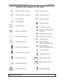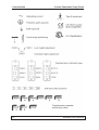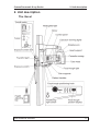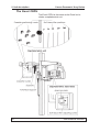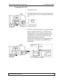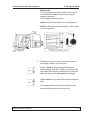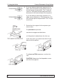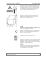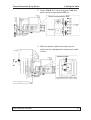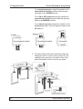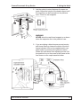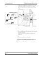
1
Cranex Panoramic X-ray Series
Contents
User’s Manual
for the
Cranex Excel
Cranex Excel Ceph
and
Cranex BaseX
Cassette version
(for film and imaging plates)
Medical Device Directive
93/42/EEC
July 2002
User’s Manual for the
Cranex Excel, Cranex Excel Ceph and Cranex BaseX
Document number 8200900 ver 0702
Original approved English language version
Manufactured by SOREDEX
P.O. BOX 250
FIN-00031 SOREDEX, FINLAND
Tel. +358 10 394 8412
Fax. + 358 9 701 5261
User’s Manual 8200900
i
Cranex Panoramic X-ray Series
Contents
Soredex endeavours to produce product documentation that is accurate and
up to date. However, our policy of continual product development may result
in changes to products that are not reflected in the product documentation.
Therefore, this document should not be regarded as an infallible guide to
current product specifications. Soredex maintains the right to make changes
and alterations without prior notice.
ii
8200900 User’s Manual
Cranex Panoramic X-ray Series
Contents
Contents
1. The Excel, Excel CEPH and BaseX x-ray units ............................. 1
Introduction ......................................................................................................
Using this manual .............................................................................................
Warnings and precautions...............................................................................
Check list before using the unit ......................................................................
General ..........................................................................................................
If you are using film ........................................................................................
If you are using imaging plates .......................................................................
Symbols that appear on the units ...................................................................
1
2
3
5
5
5
6
7
2. Unit description .............................................................................. 9
The Excel .......................................................................................................... 9
The Excel CEPH .............................................................................................. 10
The BaseX ........................................................................................................ 11
The Control panel - Excel and Excel CEPH .................................................. 12
The Control panel - BaseX ............................................................................. 13
Chin Supports ................................................................................................. 14
3. Using the Unit ............................................................................... 15
Switching the unit on and off ........................................................................
Switching the unit on .....................................................................................
Switching the unit off ....................................................................................
Exposure switch lock .....................................................................................
Unlocking the exposure switch ......................................................................
Locking the exposure switch ........................................................................
Operating the unit without x-rays .................................................................
Taking Panoramic exposures ........................................................................
Panoramic exposure programs .....................................................................
Preparing the unit .........................................................................................
Positioning the patient for a panoramic exposure .........................................
Positioning the patient for a Sinus exposure .................................................
Positioning the patient for a TMJ exposure
(Excel and Excel CEPH only) ...................................................................
Taking an exposure ......................................................................................
After exposure ..............................................................................................
Taking Cephalometric exposures (Excel CEPH only) ..................................
Cephalometric exposure programs ...............................................................
Taking a cephalometric exposure .................................................................
User’s Manual 8200900
15
15
15
16
16
16
17
18
18
19
24
31
32
33
36
37
37
38
iii
Cranex Panoramic X-ray Series
Contents
4. Care and Maintenance ................................................................. 44
Cleaning the unit ............................................................................................
Painted surfaces ...........................................................................................
Positioning mirror and light lenses ................................................................
Surfaces that the patient comes into contact with ..........................................
Monitoring the operation of the unit .............................................................
Maintenance ...................................................................................................
Yearly ............................................................................................................
Troubleshooting .............................................................................................
44
44
44
44
45
45
45
46
5. Technical specifications ............................................................ 47
Unit dimensions ............................................................................................ 52
iv
8200900 User’s Manual
Cranex Panoramic X-ray Series
1. Introduction
1. The Excel, Excel CEPH and BaseX x-ray units
Introduction
This manual describes how operate the Cranex
Excel, Cranex Excel CEPH and Cranex BaseX
dental x-ray units.
The Excel is a panoramic dental unit that can take
full and reduced width panoramic exposures, sinus
exposures, partial panoramic exposures and TMJ
exposures.
The Excel CEPH can take the same range of panoramic programs as the Excel, but includes a cephalometric unit.
The BaseX is a simplified version of the Excel. It can
take full and reduced width exposures and sinus
exposures. In addition, height adjustment is manual
and not motor driven as it is with the Excel.
All versions use conventional film or imaging plates
as the image receptor.
Please read these instructions carefully before using
the x-ray unit.
User’s Manual 8200900
5
1. Introduction
Cranex Panoramic X-ray Series
Using this manual
All key illustrations indicate that the key must be
pressed.
A white circle indicates that the indicator light is on.
When the light is on it indicates that the value or
setting is on or has been selected
A black circle indicates that the light is off and the
value or setting is off or has not been selected.
6
8200900 User’s Manual
Cranex Panoramic X-ray Series
1. Introduction
Warnings and precautions
User’s Manual 8200900
•
These dental x-ray units must only be used to
take dental and, where appropriate, cephalometric x-ray exposures. These dental x-ray units
must not be used to take any other x-ray exposures.
•
The x-ray unit or its accessories must not be
modified, altered or remanufactured in any way.
•
The x-ray unit may be dangerous to both patient
and operator unless safe exposure values are
used and correct operating procedures are
observed.
•
As radiation safety and protection requirements
vary from country to country and state to state it
is the responsibility of the operator to ensure that
all local and national radiation safety and protection requirements are met.
•
When taking exposures operators must protect
themselves from radiation.
•
When taking exposures operators must stand at
least two metres (six feet) from the patient.
•
Operators must be able to see and hear the
patient during an exposure.
•
Operators must be able to see the exposure
warning lights and hear the exposure warning
signal during exposures. If the x-ray unit is located in such a position that the operator cannot
see the exposure warning lights, an external
exposure warning light must be used. Contact
your dealer for help.
•
Avoid taking exposures of pregnant women.
7
1. Introduction
8
Cranex Panoramic X-ray Series
•
If you are using film as the image receptor, never
leave the film cassettes open in daylight.
•
If you are using imaging plates as the image
receptor make sure that the imaging plates,
imaging plate cassettes and imaging scanning
device are compatible with each other.
8200900 User’s Manual
Cranex Panoramic X-ray Series
1. Introduction
Check list before using the unit
General
•
Read and familiarize yourself with the Warnings
and Precautions in the introduction.
•
Make sure that you are fully acquainted with the
relevant radiation protection measures.
•
Make sure that all the required radiation protection equipment, such as radiation screens, are of
the approved type and, where necessary, correctly installed.
If you are using film
User’s Manual 8200900
•
Make sure that the film processor you plan to
use is in working order and ready to use.
•
Make sure that the processing chemicals in the
film processor are fresh and are at the correct
processing temperatures and concentrations.
•
Make sure that the safelight in the darkroom is
the correct one for the film you are using.
•
Make sure that the film you are using is fresh.
Do not use old film.
•
Make sure that the film and cassettes/intensifying screens you are using are compatible. Do not
mix films and screens of different colour sensitivity.
•
Make sure that the intensifying screens are clean
and not scratched. If they are scratched do not
use the cassette.
9
1. Introduction
Cranex Panoramic X-ray Series
If you are using imaging plates
10
•
Make sure that the imaging plates that you are
using are compatible with the imaging plate
cassettes.
•
Make sure that the imaging plate scanning device you are using is designed to scan the
imaging plates you are using.
•
Make sure that the imaging plates are clean and
not scratched or damaged. If they are, do not
use them.
8200900 User’s Manual
Cranex Panoramic X-ray Series
1. Introduction
Symbols that appear on the units
User’s Manual 8200900
11
1. Introduction
12
Cranex Panoramic X-ray Series
8200900 User’s Manual
Cranex Panoramic X-ray Series
2. Unit description
2. Unit description
The Excel
User’s Manual 8200900
13
2. Unit description
Cranex Panoramic X-ray Series
The Excel CEPH
The Excel CEPH is the same as the Excel but includes a cephalometric unit
14
User’s Manual 8200900
Cranex Panoramic X-ray Series
2. Unit description
The BaseX
User’s Manual 8200900
15
2. Unit description
Cranex Panoramic X-ray Series
The Control panel - Excel and Excel CEPH
16
User’s Manual 8200900
Cranex Panoramic X-ray Series
2. Unit description
The Control panel - BaseX
User’s Manual 8200900
17
2. Unit description
Cranex Panoramic X-ray Series
Chin Supports
18
User’s Manual 8200900
Cranex Panoramic X-ray Series
3. Using the Units
3. Using the Units
Switching the unit on and off
The on/off switch is located on the side of the unit.
Switching the unit on
Press the on/off switch to the on position (I) to
switch the unit on.
The on/off switch light and display lights for the
preselected spinal compensation, mA and kV values
will come on.
Switching the unit off
Press the on/off switch the off (0) position. All the
lights will go out.
Switch the unit off at the end of every day or when it
is not being used
It is also recommended that the exposure switch
(see next page) is locked when the unit is not being
used.
User’s Manual 8200900
19
3. Using the Units
Cranex Panoramic X-ray Series
Exposure switch lock
The exposure switch lock allows the exposure switch
to be locked. This prevents unauthorized people
from taking exposures even if the unit is switched on.
The exposure switch lock is located on the side of
the unit below the on/off switch.
Unlocking the exposure switch
Insert the key into the exposure switch lock and turn
the key clockwise to the horizontal unlocked position.
Locking the exposure switch
Turn the key anticlockwise to the vertical locked
position and then remove the key.
20
User’s Manual 8200900
Cranex Panoramic X-ray Series
3. Using the Units
Operating the unit without x-rays
In some situations you may have to operate the unit
without x-rays being generated.
To do this, press the test (T) key, the indicator light
will come on. The exposure switch can now be
pressed to demonstrate how the unit operates without x-rays being generated.
User’s Manual 8200900
21
3. Using the Units
Cranex Panoramic X-ray Series
Taking Panoramic exposures
Panoramic exposure programs
The following images can be taken with the Excel,
Excel CEPH and BaseX.
Full-width panorama
Magnification 1.3
Reduced width panorama (-20%) pedo and sinus
Magnification 1.3
The following images can only be taken with the
Excel and Excel Ceph.
Frontal area of the mandible and maxilla
Excel and Excel Ceph only
Magnification 1.3
Right-hand side of the mandible and maxilla
Excel and Excel Ceph only
Magnification 1.3
Left-hand side of the mandible and maxilla
Excel and Excel Ceph only
Magnification 1.3
TMJ, two or four images on the film
Excel and Excel Ceph only
Magnification 1.3
22
User’s Manual 8200900
Cranex Panoramic X-ray Series
3. Using the Units
Preparing the unit
1. Switch the unit on.
2. Press the Return key to drive the rotating unit to
the ready position if it is not already in that position.
3. Push the cassette carriage to the right and then
slide a cassette with film or a cassette with
imaging plate into the cassette carriage. Slide
the cassette in until the right-hand end of the
cassette lines up with the two slots. Arrows
indicate the position of the slots.
Note that film cassettes and imaging plate cassettes are not the same and are NOT interchangeable.
User’s Manual 8200900
23
3. Using the Units
Cranex Panoramic X-ray Series
4. Select the exposure program you require.
Excel and Excel CEPH units only
Press the appropriate program key. The program light will come on. The programs are as
follows:
Normal panoramic or sinus exposure
Reduced width panorama (-20%) pedo or
sinus exposure
Temporomandibular joint (TMJ) exposures,
images on film sides
Temporomandibular joint (TMJ) exposures,
images in film centre
When taking TMJ exposures two pairs of exposures can be taken, mouth open and mouth
closed, on one film. Press one of the TMJ keys
for the first pair of exposures and then press the
other TMJ key and reposition the patient for the
second pair of exposures.
Frontal (anterior) exposure
Partial exposure, right-hand side of patient
Partial exposure, left-hand side of patient
24
User’s Manual 8200900
Cranex Panoramic X-ray Series
3. Using the Units
BaseX only
Turn the program selection knob on the side of
the cassette carriage to select the required
exposure program.
The programs are as follows:
Adult for normal panoramic or sinus exposure
Child for Reduced width panorama (-20%) pedo
or sinus exposure
5. Press the mA key to select the correct mA for
the image receptor you are using.
Select 10 mA for normal-speed film/screen
combinations. Note that 10mA can be used with
high-speed film/screen combinations, but the kV
value will have to be decreased accordingly.
Select 6 mA for high-speed film/screen combinations.
For imaging plates select an mA value for the
required image quality and dose level.
User’s Manual 8200900
25
3. Using the Units
Cranex Panoramic X-ray Series
6. If you are taking a panoramic exposure of an
adult, press the spinal compensation button to
activate spinal compensation. The “I” light will
come on. With spinal compensation the mA is
decreased in the lateral areas of the exposure.
If you are taking a panoramic exposure of a
child, a sinus exposure or TMJ exposures, DO
NOT activate spinal compensation. The “O” light
must be on
7. Select the chin support for the exposure you
wish to take.
For panoramic exposures,
use the chin support and bite block
or if the patient is edentulous, the chin cup
or the chin rest if you cannot use either of the
above with the patient.
For sinus and TMJ exposures, use the chin
rest.
CAUTION: Always use the disposable covers
with the chin supports.
26
User’s Manual 8200900
Cranex Panoramic X-ray Series
3. Using the Units
8. Ask the patient to remove any dentures, jewellery or spectacles and then place a lead apron
over the patient’s back.
9. Adjust the height of the chin rest so that it is level
with the patient’s chin.
Excel and Excel CEPH
With the Excel and Excel CEPH, press the height
adjusting key to position the chin rest.
BaseX
With the BaseX, turn the locking lever downwards to unlock the chin rest. Adjust the position
of the chin rest and then relock it by turning the
locking lever back to the locked position.
User’s Manual 8200900
27
3. Using the Units
Cranex Panoramic X-ray Series
Positioning the patient for a panoramic exposure
1. Ask the patient to step into the unit and grasp
the patient handles.
Slide the cassette carriage to the right as far as
it will go so that you can see the patient.
2. If you are using the chin rest and bite block ask
the patient to place his chin on the chin support
and bite the bite block. Make sure that the patient’s upper and lower teeth are positioned in
the respective notches on the top and bottom of
the bite block.
If you are using the chin cup ask the patient to
position the upper and lower central incisors so
that the biting edges are together.
If you are using the chin rest ask the patient to
position the upper and lower central incisors so
that the biting edges are together and then press
his lower lip against the chin rest.
28
User’s Manual 8200900
Cranex Panoramic X-ray Series
3. Using the Units
3. While the patient is holding the patient handles
ask the patient to step forward a few centimetres so that patient is slightly off balance.
This movement will encourage the patient to
push their head forward in order to maintain
balance.
In this position the patient's cervical vertebrae
will be stretched and straight and the patient will
be in the best position for taking a panoramic
exposure.
If necessary adjust the height of the chin rest to
“stretch” the patient.
4. Open the mirror so that you can see a reflection
of the patient in the mirror.
User’s Manual 8200900
29
3. Using the Units
Cranex Panoramic X-ray Series
5. Press the positioning light button to switch the
patient positioning lights on. The lights will remain
on for 30 seconds. If you need more time, press
the button again.
6. Look at the reflection of the patient in the mirror
and position the midsagittal plane of the patient
so that it is vertical. The patient’s head must be
positioned symmetrically and the patient must be
looking straight ahead.
The patient’s head is not tilted or turned to one
side.
If the unit has a midsagittal light, the plane must
coincide with the light beam.
30
User’s Manual 8200900
Cranex Panoramic X-ray Series
3. Using the Units
7. Close the temple supports by pushing the temple
support lever to the right.
NOTE
When the temple supports are closed the patient’s head is automatically “measured”. This
“measurement” will be used to determine a kV
value when the unit is readied for an exposure.
8. Ask the patient to open his lips so that you can
see his teeth. Estimate the positions of the root
apices of the central upper and lower front incisors.
Adjust the tilt of the patient’s head, by raising or
lowering the chin support, so that both root
apices are on the same vertical axis.
User’s Manual 8200900
31
3. Using the Units
Cranex Panoramic X-ray Series
9. The focal trough light beam indicates the center
of the focal trough, which is 10 mm wide at the
front. The root apices of the central upper and
lower front incisors must now be positioned so
that they are within the focal trough.
To do this, rotate the focal trough positioning
knob backwards or forwards until the root apices
of the central upper and lower front incisors are
within the focal trough.
For patients with NORMAL occlusion the focal
trough light beam will be positioned between the
second and third teeth.
32
User’s Manual 8200900
Cranex Panoramic X-ray Series
3. Using the Units
10. For the dental arch to appear correctly on the
image the Frankfort plane of the patient should
be positioned so that it coincides with or is parallel to the Frankfort plane light. Note however,
that this is only for patients with NORMAL occlusion and is only a very rough guide.
IMPORTANT NOTE
The most important factor in patient positioning is
the position of the upper and lower apices of the
central incisors. They MUST be positioned within
the focal trough even if it means ignoring the
position of the Frankfort plane.
User’s Manual 8200900
33
3. Using the Units
Cranex Panoramic X-ray Series
11. Slide the forehead support towards the patient
until it touches the patent's forehead.
34
User’s Manual 8200900
Cranex Panoramic X-ray Series
3. Using the Units
Positioning the patient for a Sinus exposure
1. For sinus exposures use the chin rest.
Ask the patient to press his upper lip against the
chin rest.
2. The midsagittal plane is positioned the same as
for a panoramic exposure.
3. Position the focal trough light as far backwards
as possible.
4. The Frankfort plane must be horizontal.
User’s Manual 8200900
35
3. Using the Units
Cranex Panoramic X-ray Series
Positioning the patient for TMJ exposures
(Excel and Excel CEPH only)
1. For TMJ exposures use the chin rest.
Ask the patient to press his upper lip against the
chin rest with his mouth closed.
2. The midsagittal plane is positioned the same as
for a panoramic exposure.
3. The focal trough is positioned the same as for a
panoramic exposure.
4. The Frankfort plane must be horizontal.
5. After taking the first pair of TMJ exposures ask
the patient to open his mouth for the second pair
of exposures.
36
User’s Manual 8200900
Cranex Panoramic X-ray Series
3. Using the Units
Taking an exposure
1. Make sure that the temple supports are closed so
that they are holding the patient’s head. Press the
RETURN key to drive the rotating unit to the start
position.
2. Manually slide the cassette carriage to the left
as far as it will go. The READY light will come on
and a kV value based on the patient’s head size
will be automatically selected.
This kV value is for a normal panoramic exposure with spinal compensation ON, using 10mA
and a normal-speed film/screen combination.
IMPORTANT NOTE
The Autoset kV value selected is only a SUGGESTED value based upon the size of the patient’s head. You should always use your own
experience and knowledge when deciding what
kV value to use.
If a high-speed film/screen combination is being
used, decrease the kV value by 6 - 8 kV or use 6
mA instead of 10 mA.
User’s Manual 8200900
37
3. Using the Units
Cranex Panoramic X-ray Series
For different exposure programs and patients
adjust the suggested kV value as follows:
- sinus exposure (spinal compensation OFF),
increase the kV value by 4 - 6 kV.
-TMJ exposure (spinal compensation OFF),
increase the kV value by 2 - 4 kV.
- panoramic exposure heavy boned patient,
increase the suggested value by 2 - 4 kV
- panoramic exposure of an edentulous patient,
decrease the suggested value by 2 - 4 kV.
- panoramic exposure WITHOUT spinal compensation, decrease the suggested value by 2 4 kV.
If you cannot increase or decrease the kV value
by the number of kV required, just select the
highest or lowest kV value possible.
Note that when using film, the age of the
processing chemicals and the film/screen combination being used may also require the kV value
to be adjusted.
3. Ask the patient to press his tongue against his
palate, breathe normally and not move until the
exposure has been completed.
Exposures last approximately 20 seconds.
4. Move at least two metres away from the x-ray
unit and protect yourself from radiation. Make
sure that you are able to see and hear the patient during the exposure.
38
User’s Manual 8200900
Cranex Panoramic X-ray Series
3. Using the Units
5. Press and hold down the exposure switch on the
remote control for the duration of the exposure.
During the exposure cycle you will hear an audible signal and the radiation warning lights will
come on when radiation is generated.
6
When the rotating arm stops moving and the
exposure warning lights go out, remove your
finger from the exposure switch.
NOTE
TMJ Exposures (Excel and Excel CEPH only)
If you are taking TMJ exposures:
Press the RETURN key after you have taken the
first pair of exposures to drive the rotating unit
back to the ready position.
Press the second TMJ exposure key.
Reposition the patient for the second pair of TMJ
exposures (Refer to the section “Positioning the
patient for a TMJ exposure”) and then take these
exposures as described above.
NOTE
When taking TMJ exposures, the audible signal
and the radiation warning lights will ONLY come
on when radiation is generated.
User’s Manual 8200900
39
3. Using the Units
Cranex Panoramic X-ray Series
After exposure
1. Slide the forehead support away from the patient.
2. Open the temple supports.
3. Guide the patient out of the x-ray unit.
4. Remove the cassette from the cassette carriage.
Process the film or if you used an imaging plate,
scan the imaging plate.
40
User’s Manual 8200900
Cranex Panoramic X-ray Series
3. Using the Units
Taking Cephalometric exposures (Excel CEPH only)
Cephalometric exposure programs
Only the Excel CEPH can also be used to take the
following cephalometric images.
Lateral asymmetric vertical
18 x 24 cm / 8” x 10”
or optionally 24 x 30 cm
Anterior-posterior or posterior anterior symmetric
vertical
18 x 24 cm / 8” x 10”
or optionally 24 x 30 cm
Lateral horizontal
18 x 24 cm / 8” x 10”
or optionally 24 x 30 cm
The magnification of all these images is 1.13.
(NOTE: For non US units the magnification can be
set between 1.09 and 1.13 during installation. )
User’s Manual 8200900
41
3. Using the Units
Cranex Panoramic X-ray Series
Taking a cephalometric exposure
1. Select and press the ceph exposure time button
for patient you are taking an exposure of:
- For adults 1.0 - 2.0 seconds
- For children 0.4 - 1.0 seconds
Note
The exposure time will depend on the film/screen
combination being used.
The exposure time indicator light will come on
and the rotating unit and cassette carriage will
move automatically to the ceph position.
CAUTION
DO NOT pull the cassette carriage out past its
stop position. If pulled out too far the unit will
NOT take a ceph exposure. If accidentally pulled
out, push the cassette carriage in as far as it will
go and it will then return automatically to the
correct position.
DO NOT push the rotating arm to the left. If
pushed, it will start rotating. If the rotating arm is
accidentally pushed and it starts to move, pull it
to the right passed the ceph position and it will
move automatically back to the ceph position.
42
User’s Manual 8200900
Cranex Panoramic X-ray Series
3. Using the Units
2. Select 10 mA, a kV value of at least 71 kV and
switch spinal compensation OFF (0).
3. Slide the aperture plate to the aperture you
require for the cephalometric exposure you wish
to take.
User’s Manual 8200900
43
3. Using the Units
Cranex Panoramic X-ray Series
For a lateral exposure using the cassette in the
asymmetrical vertical position slide the aperture plate to the DOWN position.
For a PA or AP exposure using the cassette in
symmetrical vertical position slide the aperture
plate to the MIDDLE position.
For a lateral exposure using the cassette in the
horizontal position slide the aperture plate to the
UP position.
4. Pull the locking catch down and then rotate the
headrest until it is in the correct position for the
cephalometric exposure you plan to take. Also
place disposable covers over the ear posts.
44
User’s Manual 8200900
Cranex Panoramic X-ray Series
3. Using the Units
5. Ask the patient to stand between the open ear
posts. Adjust the height of the head support until
the earposts can be inserted into the patients
ears. Close the ear supports.
WARNING
NEVER drive the ceph head support up or down
when the earposts are in the patient’s ears.
6. If you are taking a lateral exposure and require
soft tissue filtering, determine which of the soft
tissue indicator lines on the cephalometric cassette holder is nearest to the patients nasion.
Rotate the soft tissue filter adjusting screw until
the position line on the screw is the same as the
line chosen on the ceph cassette holder.
User’s Manual 8200900
45
3. Using the Units
Cranex Panoramic X-ray Series
7. Slide the cephalometric cassette into the cassette holder until it lines up with the cassette
positioning mark for the exposure you wish to
take.
8. If you are taking an AP exposure, ask the patient
to close his eyes.
Protect yourself from radiation and take the
exposure.
9. Guide the patient out of the unit.
10. Remove the cassette and process the film or
scan the imaging plate.
46
User’s Manual 8200900
Cranex Panoramic X-ray Series
3. Using the Units
11. To drive the rotating arm back to the panoramic
position press the P key and then the RETURN
key.
User’s Manual 8200900
47
4. Care and Maintenance
Cranex Panoramic X-ray Series
4. Care and Maintenance
Cleaning the unit
WARNING
Switch the unit off before cleaning it.
Painted surfaces
All painted surfaces can be wiped clean with a soft
cloth dampened with a mild detergent.
NEVER use abrasive cleaning agents or polishes on
this equipment.
Positioning mirror and light lenses
The positioning mirror and positioning light lenses are
made of glass. Use a soft cloth dampened with a
mild detergent. NEVER use abrasive cleaning agents
or polishes.
Surfaces that the patient comes into contact with
All surfaces and parts that the patient touches or
comes into contact with must be disinfected after
each patient.
If you need to clean the intensifying screens,
only use cleaners recommended by the screen
manufacturer.
If you need to clean the imaging plates, only use
cleaners recommended by the imaging plate
manufacturer.
48
User’s Manual 8200900
Cranex Panoramic X-ray Series
4. Care and Maintenance
Monitoring the operation of the unit
If any of the unit’s controls or functions fail to operate
or do not operate in the way described in this manual,
switch the unit off, wait 30 seconds and then switch
the unit on again. If the unit still does not operate
correctly contact your service technician for help.
If you hear the exposure warning tone but the exposure warning light does not come on when an exposure is taken, stop using the unit and contact your
service technician for help.
If you do not hear the exposure warning tone when
an exposure is taken, stop using the unit and contact
your service technician for help.
Maintenance
Yearly
Once a year an authorized service technician must
carry out a full inspection of the unit. During the
inspection the following will be carried out:
– a kV/mA test
– a ceph exposure time test (where applicable)
– a beam alignment test
– a ball/pin test
– a check to see that the safety ground is connected
– a check to see that the positioning lights operate
– a check to see that the tube head is not leaking
– a check to see that all covers and mechanical
parts are correctly secured and have not come
loose.
A full description of all the tests and checks is described in the Service Manual.
User’s Manual 8200900
49
4. Care and Maintenance
Cranex Panoramic X-ray Series
Troubleshooting
Problem
The protection light comes on and you are unable to take an exposure.
Cause
There is a problem with the generator.
Solution
Switch the unit off. After a few seconds switch it on again. If the light comes on
again, contact your dealer for assistance.
50
User’s Manual 8200900
Cranex Panoramic X-ray Series
5. Technical Specifications
5. Technical specifications
Classification
Complies with IEC class I type B, IPX0
Complies with IEC 601-1, IEC 601-2-7 and EN 55011 standards
Group 1, class B
Complies with DHHS Radiation Performance Standard, 21CFR Subchapter J.
The unit must be installed in a protected clinical environment.
Description
Dental panoramic and panoramic/cephalometric x-ray units with a high frequency
switching mode x-ray generators. The panoramic version takes panoramic exposures. The panoramic/cephalometric version takes panoramic and cephalometric
exposures and uses a dedicated x-ray source to take cephalometric exposures.
Generator
TUBE
- OPX/105, DE 100/15 or equivalent
FOCAL SPOT
- 0.5 mm IEC 336
TARGET ANGLE
- 5º
TARGET MATERIAL
- Tungsten
OPERATING TUBE POTENTIAL
- Panoramic 63 - 81 kV (±5 kV) at preselected settings
OPERATING TUBE CURRENT
- 6 mA and 10 mA (±1 mA) at preselector,
4.5 mA and 6 mA or 7.5 mA and 10 mA in compensation mode
MAXIMUM TUBE CURRENT
- 11 mA
MAXIMUM OUTPUT POWER
- 945 W nominal
FILTRATION
- minimum filtration 2.5 mm Al
BEAM QUALITY
- HVL over 2.5 mm Al at 81 kV
OUTER SHELL TEMPERATURE
- +50ºC (122ºF) maximum
DUTY CYCLE
- Panoramic: 1:15, 81 kV/10mA
- Cephalometric: 1:20, 81 kV/10mA
User’s Manual 8200900
51
5. Technical Specifications
Cranex Panoramic X-ray Series
Power requirements
INPUT VOLTAGE
- 175-250 VAC (±10%), 50/60 Hz, adjustable, single phase, grounded socket
MAXIMUM LINE CURRENT
- 7 A at 81 kV/10mA
MAXIMUM LINE RESISTANCE
- 1 ohm
MAXIMUM LINE FUSING
- 10 A slow (main fuse 8A slow in device)
LINE SAFETY SWITCH (when required)
- Approved type, min. 10 A 250 VAC
EARTH LEAKAGE CIRCUIT BREAKER (when required)
- Approved type, min. 16 A 250 VAC, breaker activation leakage current in
accordance with local regulations.
Mechanical parameters
PANORAMIC
- SID 520 mm (±10 mm)
- Magnification factor 1.3
CEPHALOMETRIC
- SID 1635 - 1700 mm ±20 mm
- SID, USA only, fixed 1700 mm ±20 mm
- SOD (Source object distance) 1500
- Magnification factor 1.09 - 1.13 (Non US unit, set during installation)
- Magnification factor, USA only, fixed 1.13
WEIGHT
- Panoramic unit 150 kg (330 lb.)
- Pan Ceph unit 195 kg (430 lb.)
DIMENSIONS
- Panoramic unit (HxWxD) 2240 x 1200 x 970 mm (88" x 47" x 38")
- Pan Ceph unit (HxWxD) 2240 x 1200 x 1850 mm (88" x 47" x 74")
VERTICAL HEIGHT OF CHIN REST
- 850-1700 mm (33"-67")
Cassettes
PANORAMIC
- 15 x 30 cm (6" x 12") Optional 24 x 30 cm (10” x 12”) available
CEPHALOMETRIC
- 18 x 24 cm (8" x 10")
- optionally 24 x 30 cm
52
User’s Manual 8200900
Cranex Panoramic X-ray Series
5. Technical Specifications
Recommended image receptors
FILM/SCREEN COMBINATIONS
Kodak T-MAT G (green sensitive) - Kodak Lanex Regular and Medium
IMAGE PLATES
All types of imaging plates intended for extraoral imaging.
Timer
PANORAMIC EXPOSURE TIMES:
50 Hz
Normal
19 s (±15%)
Child
17 s (±15%)
Sides
11 s (±15%)
Middle
10 s (±15%)
TMJ
3.3 + 3.3 s (±15%)
Max 240 mAs
60 Hz
16 s (±15%)
14 s (±15%)
9 s (±15%)
8 s (±15%)
2.8 + 2.8 s (±15%)
Max 202 mAs
CEPHALOMETRIC EXPOSURE TIMES:
- 0.4 - 3.2 s (±15%), ten steps in accordance with R’ 10, series (ISO)
- 4 mAs - 32 mAs nominal, maximum 40.4 mAs
BACK-UP TIME
- 23.5 s (±1.5s)
Leakage technique factors
PANORAMIC
- 81 kV, 2400 mAs/h (81 kV, 10 mA, duty cycle 1:15)
(One normal exposure per 4 minutes cool-down period)
CEPHALOMETRIC
- 81 kV, 1800 mAs/h (81 kV, 10 mA, duty cycle 1:20)
(One 3.2 s exposure per 1 minute cool-down period)
User’s Manual 8200900
53
5. Technical Specifications
Cranex Panoramic X-ray Series
Measurement bases
-
The kV is measured by monitoring the current flowing through a 450 Mohm,
1% feedback resistor connected between the tube anode and ground.
-
The mA is measured by monitoring current in the HT return line, which equals
the tube current.
-
Both parameters are measured using a Soredex kV/mA meter (pt. no.
4800177) or with a digital multimeter (DMM) according to a specified
procedure. The exposure time is measured using a radiation probe positioned in
the primary beam.
Cephalometric collimator
18 x 24 A or 24 x 18 A for lateral projection,
18 x 24 S for P.A. and A.P. projections
24 x 30 aperture optional
8" x 10" A or 10" x 8" A for lateral projections
8" x 10" S for A.P. and P.A. projections
Adjustable soft tissue filter for lateral projection
Operating Room Temperature
- 15ºC (59ºF) to 32ºC (90ºF)
Operating Relative Humidity Maximum
- 95%
Tube housing assembly cooling characteristics
54
User’s Manual 8200900
Cranex Panoramic X-ray Series
Tube rating chart
5. Technical Specifications
Anode thermal characteristics
OPX/105
Tube rating chart
Anode thermal characteristics
DE 100/15 ö
User’s Manual 8200900
55
5. Technical Specifications
Cranex Panoramic X-ray Series
Unit dimensions
56
User’s Manual 8200900