1
Calculations and Patient Worksheets (Reports)
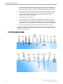
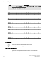
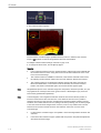
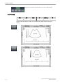
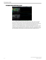
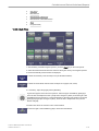
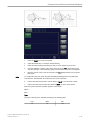
12.6.4.10.3 Echocardiac views
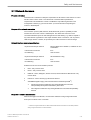
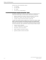
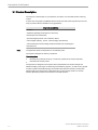
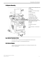
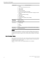
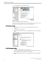
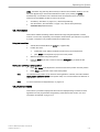
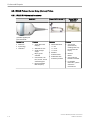
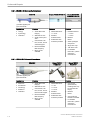
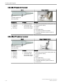
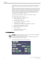
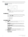
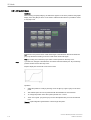
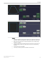
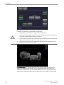
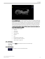
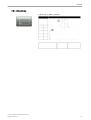
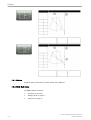
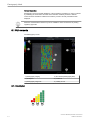
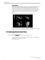
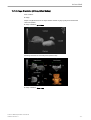
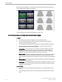
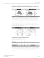
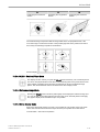
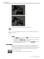
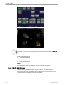
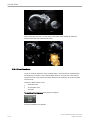
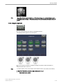
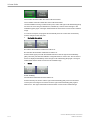
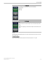
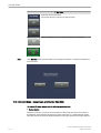
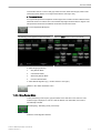
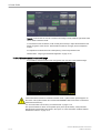
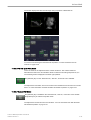
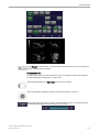
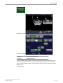
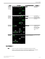
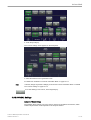
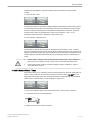
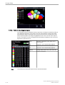
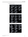
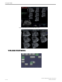
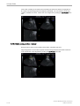
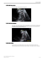
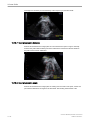
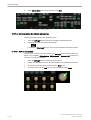
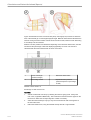
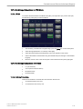
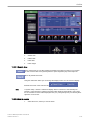
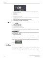
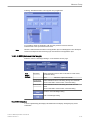
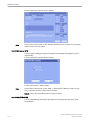
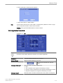
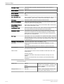
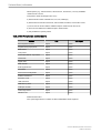
Figure 1 Fetal echocardic views from which the cardiac structures can be measured. (a) Long
Axis view of the left ventricel showing the aortic valve (1) and ascending aorta (2). (b) Aortic
arch view showing the aortic valve (1), ascending aorta (2), descending aorta (3) and inferior
vena cava (4). (c) Short axis view showing the pulmonary valve (1), main (2), right (3) and left
(4) pulmonary arteries. (d) Oblique short axis view, showing the pulmonary trunk and arterial
duct (5). (e) Four chamber view, showing the tricuspid valve (1), right ventricular end-diastolic
dimension (2), right ventricular inlet length (3), right ventricular area (dashed line) (4), mitral
valve (5), left ventricular end-diastolic dimension (6), left ventricular inlet length (7) and left
ventricular area (dotted line) (8). Ao, aorta; Desc Ao, descending aorta; IVC, inferior vena
cava; LA, left atrium; LPA, left pulmonary artery; LV, left ventricle; MPA main pulmonary
artery;RA right atrium; RPA, right pulmonary artery; RV right ventricle.
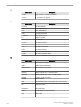
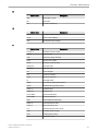
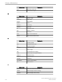
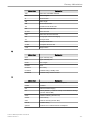
REFERENCE: Schneider C. et. al., “Development of Z-scores for fetal cardiac dimensions
from echocardio-graphy”, Ultrasound Obstet Gynecol. Vol. 26, 2005, pages 599-605.
Fomulas:
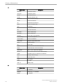
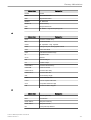
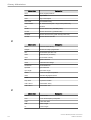
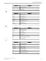
Z-scores = (ln(actual) - ln(predicted cardiac dimensions)) / Root MSE
ln (predicted cardiac dimensions) = m.ln(FL, GA or BPD) + c
FL...femur length; GA...gestational age in completed weeks; BPD...biparietal diameter;
m...multiplier; c...intercept
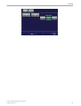
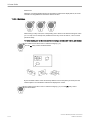
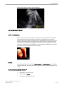
12.6.4.10.4 Fractional Limb
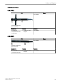
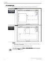
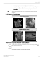
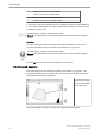
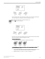
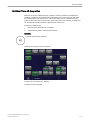
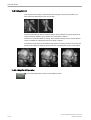
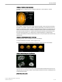
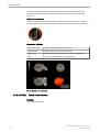
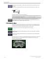
This measurement is for calculating the fetal limbs. Based on this partial volume-calculation
the fetal weight can be estimated.
Voluson™ E8/E8 Expert Basic User Manual
H48691CF Revision 2
12-31