1
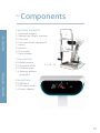
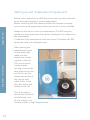
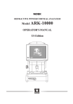
How to get the best use out of me
Let’s get to work!
Eaglet Eye : User Manual
eaglet-eye.com
EMPLOYER
MANUAL
Measurement system for the
Anterior surface of the eye
eaglet-eye.com
Introduction
Thank you for purchasing this Eye Surface Profiler (ESP) system
and for your confidence in Eaglet Eye. The ESP gives you highly
precise measurements of the front surface of the eye, not only of
the cornea but also a large part of the sclera.
We are committed to assuring your complete satisfaction with
the ESP.
Eaglet Eye : User Manual
At Eaglet Eye, we continue our research and development to
further extend the capabilities of all our products and solutions.
This may result in improvements of certain features of the ESP
system that have not yet been updated in the user manual. We
strive to minimize these differences and come with regular
updates of the manual.
For your safety it is essential that you read this manual carefully
and that you familiarize yourself with its content before you start
using this device. In particular, pay close attention to the safety
instructions in this manual.
If you have any questions or desire further information on this
product, please contact us via the website or email us. Our team
will be glad to help you.
The Eaglet Eye team
Eaglet Eye : User Manual
eaglet-eye.com
Description
The Eye Surface Profiler (ESP) is a system using Fourier Transform
Profilometry that measures the anterior surface of the cornea, the
limbus and large parts of the sclera. Up to 20mm diameter of the
visible anterior surface can be measured. The ESP requires
fluorescein instilled in the eye so that the projected light is
scattered back from the eye front surface.
The measurement process consists of 3 single shots in rapid
succession that last a few milliseconds. The ESP calculates a
3-dimensional model of the anterior surface of the eye with on
average more than 250.000 measurement points. From these
measurement points, maps and parameters are calculated
automatically that assist in the evaluation of the shape of the
cornea, limbus and sclera. Separately, the pupil size and iris size
are determined automatically from a top view image.
The ESP measures the complete anterior front surface of the eye
up to 20mm without extrapolation. The corneal limbus is the
border of the cornea and the sclera. The average corneal diameter
is 11.77 +/- 0.37 mm for males and 11.64 +/- 0.47 mm for females.
The new software will detect the limbus automatically and will
give you detailed information about the whole cornea.
The ESP is a Profilometry based system and has therefore no
interference with the shadow of the nose.
Eaglet Eye : User Manual
eaglet-eye.com
Contents
01. Quick Reference Guide
02. Components
Important elements
View window
Connections
03. Safety
Setting up and installation of equipment
Technical details of the ESP
04. Operational instructions
Home screen overview
Database Location
Add a patient
New measurement
Finding an existing patient
Search results
History
Viewing results
Parameters
Sagittal height
Figures
Source
Grid
Bisphere elevation
Bisphere 3D
Tangent angles map
Tangent angles 3D map
Height
03
04
04
04
04
05
07
08
09
09
09
11
11
15
15
15
16
16
20
22
22
23
23
24
24
25
25
eaglet-eye.com
Eaglet Eye : User Manual
Height 3D
Fluorescein simulation map
Tangential curvature
Axial curvature
Refractive power
Corneal elevation
Quad map
Notes
Evaluating functionalities
05. Exporting, Printing and Comparing Results
06. Lens Design Module
07. Troubleshooting
08. Maintenance and service
Care and maintenance
Casing
Optical components
Software installation
Minimum PC requirements
Conditions of warranty
Liability for malfunction and damages
09. Compliance
EMC
CE
Index
Address of manufacturer and service department
26
26
27
28
29
29
30
30
31
32
36
38
39
39
39
39
39
40
40
41
42
42
42
43
45
Eaglet Eye : User Manual
eaglet-eye.com
01
03
Quick Reference
Guide
02
Components
eaglet-eye.com
Important elements
1. Forehead support
2. Optimal eye height indicator
3. Chin rest
4. Chin rest height adjustment
swivel
5. Joystick
6. Fire button
7. View window
1.
2.
7.
3.
4.
6.
5.
Eaglet Eye : User Manual
View window
8. Digital camera
9. Focus help LEDs
10. Fixation cross
11. Measure pattern
projectors
Connections
12. USB Port
13. DC power inlet
14. Power adapter
12. 13. 14.
8.
10.
9.
11.
04
eaglet-eye.com
03
Safety
To assure safe operation, it is imperative that the instrument is
used according to the instructions in this manual. Therefore, you
should become thoroughly familiar with the content of the user
manual before putting the system into operation. The Eye Surface
Profiler (ESP) system must only be operated by trained personnel
capable of using it properly on the basis of their training,
expertise and practical experience.
Eaglet Eye : User Manual
Please save this user manual and ensure that it is accessible to
operating personnel at all times.
This instrument must only be used for purposes as laid down in
this user manual. Before using this instrument for the first time,
you must be instructed on how to use it by our staff or an
authorized dealer.
Only operate the instrument with the original components
supplied by Eaglet Eye and if the instrument is in good working
condition. Should the instrument be defective, do not operate it,
and contact the supplier.
Before maintenance and cleaning the instrument, always pull out
the power plug of the ESP and all equipment attached to it, for
example a printer. Do not connect cables if this proves difficult. If
it is impossible to make a connection, check to see whether the
plug fits into the socket. In case you ascertain a defect, contact
your authorized dealer to have it repaired by our service team.
When disconnecting electrical connections, always pull on the
plugs, not on the cables. Supplementary equipment which is
connected to the analog or digital interfaces of the instrument
05
Eaglet Eye : User Manual
eaglet-eye.com
must comply with relevant specifications. For detailed instruction
on maintenance and cleaning turn to the chapter on Maintenance
and Service in this manual.
If the ESP is coupled with external electrical equipment (for ex. a
printer) this must not result in a decrease of patient safety. If the
coupling of equipment causes the tolerance levels for leakage
current to be exceeded, safety features, which include a
disconnection device, must be provided for.
Do not operate the delivered equipment
• in areas with risk of explosion,
• in the presence of flammable anesthetics or volatile solvents
such as alcohol, benzene or similar substances.
Do not use or store the instrument in damp rooms. Avoid placing
the instrument in the vicinity of dripping, running or spraying
water and ensure that no moisture can penetrate the instrument.
For this reason, do not place any containers filled with fluids near
the instrument. When cleaning the instrument with a damp cloth,
be sure that no moisture enters it.
This instrument is a high-quality technical product containing
sophisticated optics. In order to assure faultless and safe
operation, service inspections must be performed according to the
service plan of Eaglet Eye. See also the chapter on Maintenance
and Service in this manual.
Eaglet Eye emphasizes that the user bears the full responsibility
for the correctness of data measured, calculated or displayed
using the ESP. The manufacturer will not accept claims based on
erroneous data.
06
Setting up and installation of equipment
Eaglet Eye : User Manual
eaglet-eye.com
Before initial operation, the ESP needs to be set up and connected
by an authorized dealer or service department.
Before installing the ESP, please consider the transport, storage
and current room temperature where the device is to be installed.
Make sure the device is at room temperature. The ESP must be
placed at a place prevented from direct (day)light from influencing
the components.
A reflection-free examination must be assured. Therefore, the ESP
should be used in a darkened room.
When placing the
measurement head
on the base plate,
make sure the
wheels are exactly
aligned in the rail.
This can be easily
checked when
holding a card at the
base plate and at the
end of the rail, the
wheels should touch
the card at both
sides. If this is not
the case, adjust the
wheels in the rail.
This is an optical
device and should be
handled with care.
Do not subject it to
vibrations, jolts or high temperatures.
07
Important elements
Seizes (Depth*Width*Height)= 35 cm*35 cm * 48 cm
Weight:
12 kg
Energy and power:
PLUG POWER SUPPLY, 10W, EU, 7.5V
• Power Supply Output Type: Fixed
• Input Voltage AC Min: 90V
• Input Voltage AC Max: 264V
• Output Voltage Nom.: 7.5V
• Output Current Max: 1.14A
• Output Power Max: 8W
• Output Connector: Barrel Plug
• SVHC: No SVHC (16-Jun-2014)
• Applications: General Purpose
• No. of Outputs: 1
• Output Current: 1.14A
• Output Voltage: 7.5V
• Power Rating: 8W
Power consumption:
8W
Eaglet Eye : User Manual
eaglet-eye.com
Model: ESP 20200
08
Eaglet Eye : User Manual
eaglet-eye.com
04
Operational
Instructions
Home screen overview
Database location
When installing the Eye Surface Profiler software, the database for
new measurements will automatically be saved on:
C:\Users\COMPUTERNAME\AppData\Roaming\Eaglet Eye\Eye
Surface Profiler.
This can be changed to a location of preference by clicking
settings - database - move database. See picture below.
The location has to be a mapped drive (for example: C: / F:). This
opens the possibility to open the database on different locations.
09
eaglet-eye.com
In this screen (see picture below) it is also possible to select a
database if the eye care professional saved several databases.
Eaglet Eye : User Manual
The Eye Surface Profiler software home screen consists of
three columns:
Column
Description
Customer column
From this column recent customers can be selected as
well as new patients that can be added
New measurement
column
Enables access to new measurements if a patient is
selected
Results column
Contains a list of recent measurements
10
Add a patient
Before starting a measurement the patient must be entered into
Eaglet-Eye Surface Profiler.
To add a patient, take the steps listed below:
eaglet-eye.com
1. Go to the home screen;
2. Click on the plus sign in the ‘Customer column’;
‘Customer column’ changes to four input fields for the
patient data
3. Enter the required data;
- Given name
- Family name
- Birthdate
- Patient ID
Eaglet Eye : User Manual
4. Click ‘Apply’.
New measurement
A new measurement can be made by pressing the button ‘New
measurement’. This button is disabled if no customer is selected.
Making a new measurement is done by following the steps
listed below:
1. Make sure the patient does not wear contact lenses;
2. Select the patient from the customers (if the patient is not
available enter the patient following the ‘add a patient’
procedure);
The name of the patient appears in the customer column title.
The button ‘New measurement’ is enabled
11
3. Apply fluorescein;
The patients eye reflects light used by the ESP
Make sure that a large part of the sclera is yellow in order to
get the best results
Eaglet Eye : User Manual
eaglet-eye.com
4. Tell patient to place head on chin rest;
5. Adjust chin rest with swivel so that the patient’s eye is roughly
at level of marker;
6. Select new measurement in the ESP-software;
A live image of the patients eye is shown in the ESP-Software
7. Focus device and ask patient to look at the red cross;
The focus lights help you to focus by aligning and
positioning the lights on the eye as is shown on the right.
8. Hit fire button;
The patient sees two flashes and the ESP-software shows the
images. If the images are unsuitable because of the patient
blinking, click ‘discard’
9. Select measurements by clicking a check mark in the box next to
the images and click ‘save’.
12
The ESP software starts processing the images
Eaglet Eye : User Manual
eaglet-eye.com
Instead of clicking the ‘save’ button after a measurement, the
operator can choose the ‘save and add more’ button to expand a
measurement session. The picture below displays a measurement
session of both eyes. If the operator would click the ‘save’ button
(the one in the right), all of these nine measurements will be saved.
13
In order to provide the highest quality of the measured results
there are some tools that will help the operator to optimize the
focus, centration and sharpness of the camera before making a
measurement. In the left bar in the picture above the operator can
choose the ‘eye tracking’ option.
The colour bar on the right gives live indication of the quality of
focus, brightness and sharpness while the operator adjusts the
position of the measurement head. The two dots on the right of
the eye still have to be placed above each other. The green circle
(with green plus) has to be placed between the white focus points.
eaglet-eye.com
Please instruct the patient to look straight ahead at the red cross
in order to get an optimal centration.
Eaglet Eye : User Manual
The measurements are saved in the ESP software.
All three measurements are selected and they can be unselected
by clicking the check mark. Below these three measurements you
can see an indication of the quality of the measurements, 100%
meaning enough light, fluorescein and excellent focusing by
the operator.
14
Finding an existing patient
Eaglet Eye : User Manual
eaglet-eye.com
A patient can be found in the ESP-software by entering the given
name or family name in the ‘Name’ input field.
15
Search results
The patients matching the criteria will appear in the search
results while typing. You can select the patient by clicking the
name from the results.
You can also search by added notes, for example 'keratoconus'.
History
When no data is entered in the ‘Name’ input field, the most recent
selected patients are listed in the left column.
Viewing results
Results can be selected in the Results column. By default the
ESP-Software lists the measurement sessions by date.
Eaglet Eye : User Manual
eaglet-eye.com
When a patient is selected from the left column, measurement
sessions of this patient are listed only. A measurement session can
be expanded.
Parameters
The following figures
will display the
functionalities and
parameters of the
ESP-software.
16
Refractive
Sphere
Eaglet Eye : User Manual
eaglet-eye.com
Astigmatism
SimK
SimK
parameters
Astigmatism
Corneal
Asphericity
Ks
Kf
Qs
Qf
Qavg
The Maloney spherocylinder is used. Spherical
sphere equivalent corresponds to the plus half of
the cylinder (Astigmatism).
The refractive error of the astigmatic eye stems
from a difference in degree
of curvature refraction of the two different
meridians.
(Anterior Curvature) are calculated with
the so called keratometric index, which is known
from Placido topographers, and is equal to 1.3375.
This does not correspond to the actual index of
refraction of the cornea (1.376) since it takes into
account the posterior surface parameters
calculated with keratometric index are called
simulated (SimKf, SimKs, …).Steep (SimKs) and
SimK flat (SimKf) are calculated from the pair of
meridians 90° apart with the greatest difference in
average power, from 0.5 to 2.0 mm distance from
the centre.
This maximizes the Astigmatism parameter, which
is the difference between SimKf and SimKs.
As mentioned above, the astigmatism value is the
difference between SimKs and SimKf.
K values are calculated with actual refractive
index of the cornea (1.376). Ks stands for steep
curve.
AK values are calculated with actual refractive
index of the cornea (1.376). Kf stands for flat curve.
(steep)
(flat)
(average)
e is the symbol for eccentricity
Q is the symbol for asphericity
p is the symbol for shape factor
17
The three are related through the following formula:
Q = p – 1= -e2
Table conic sections
e
p
Q
0<e<1
P>1
Q>0
e=0
P=1
Q=0
0<e<1
1>p>0
0 > Q > -1
Parabola
E=1
P=0
Q = -1
Hyperbola
P>1
P<0
Q < -1
Oblate ellipse
eaglet-eye.com
Sphere
Prolate ellipse
Note that eccentricity (e) cannot distinguish between oblate and
prolate ellipsoid. However e2 is negative for an oblate ellipsoid
and e2 is positive for a prolate ellipsoid.
Eaglet Eye : User Manual
Best-fit Bisphere
Inner radius
Limbus
After subtracting a best fit sphere from the
corneal part, the inner radius gives information
about the radius of the inner sphere (the corneal
sphere).
The limbus is indicated from the differences
between the corneal and the scleral best fit
spheres. There are two spots in the cross section
where the two best fit spheres interfere (see
picture above, the squares surround these two
spots). The first number displays the mean
distance from apex to the center of the
interfering part of the two best fit spheres
18
eaglet-eye.com
Outer radius
Keratoconus
KPI
Eaglet Eye : User Manual
Warning:
Pupil
Limbus
HVID
Pupil Ø
Pupil offset
PAP+, PAP-
PAC+, PAC-
19
(corneal and scleral). The second number
represents the mean length of this interfering
area. This is still an indication.
After subtracting a best fit sphere from the
scleral part, the outer radius gives information
about the radius of the outer sphere (the scleral
sphere).
The keratoconus prediction index is a compilation
index of DSI, OSI, CSI, SAI, SimK1, SimK2, IAI, and
AA (footnote 1). The mathematical formula for the
simulated KPI, using simulated component indices,
is described below:
KPI = 4.62*[0.30 + 0.01 (-41.23 – 0.15*DSI +
1.18*OSI + 1.49*CSI + 4.13*SAI – 0.56*SimKs +
1.08*SimKf – 3.74*IAI + 0.10*AA)] – 60.25
Keratoconus indices are designed to be an adjunct
to a clinical examination, and should
never be used as a sole assessment tool in the
diagnosis of keratoconus (footnote 2).
Indicates the start of the limbus area measured
from the apex, the second value corresponds to the
width of the limbus.
Horizontal Visual Iris Diameter
Pupil diameter
Distance in x and y from the apex to the pupil
centre.
The palpebral aperture relative to the pupil, these
values are the length of
the eye above and below the pupil.
The palpebral aperture relative to the corneal
centre (apex), these values are the length of the
eye above and below the apex.
Footnotes.
1. See Index, Page 43.
2. (Maeda N, Klyce SD, Smolek MK, Thompson HW. Automated keratoconus screening with
corneal topography analysis. Invest Ophthalmol Vis Sci 1994; 35: 2749–57).
Eaglet Eye : User Manual
eaglet-eye.com
Sagittal height
Tsag-Nsag is the new parameter added to the ESP software that
will quantify scleral asymmetry and give eye care professionals
better control over lens fits.
Literature describes the flattening of the sclera on the nasal side
of the eye, compared to the temporal side which shows no such
behaviour, which introduces an asymmetry to the eye surface.
A pronounced asymmetry between the nasal and temporal scleral
curves will impact lens fit.
The image above indicates a horizontal cross section of a typical
eye surface. The sagittal height is calculated over a total chord of
16mm in this example. Tsag and Nsag each get attributed exactly
half of the total chord length and the respective sagittal heights
are determined from the apex outwards.
20
Other sagittal height parameters displayed in the ESP
software are:
Eaglet Eye : User Manual
eaglet-eye.com
3600
21
This is sagittal
height calculated
over as many
meridians as
possible, building
towards the average
sagittal height over
the full 360 degrees
of the eye.
Meridional
This is the sagittal
height for one
single meridian.
MINsag/MAXsag Similar to the 360
degrees sagittal height
is that it includes all available meridians at the
given chord length. Out of the available sagittal
heights it will then select the maximum and its
angle and also the minimum and its angle.
Figures
The following figures will show the different functionalities and
results after the measurement has been made.
Eaglet Eye : User Manual
eaglet-eye.com
The colour scale of the tangent angles maps can be adjusted and
therefore customized to the individual preference of the eye care
practitioner.
Source
This picture of the source shows an excellent centration and focus.
The white dots on the horizontal red line are an indicator for the
centration; the two white dots on the right part of the iris (placed
above each other) indicate the focus of the camera and therefore
the sharpness of the measurement.
22
eaglet-eye.com
Grid
The grid map visualizes the vertical lines, as projected on the eye
and reflected back into the camera. The clear contrast between the
lines indicates that there was enough use of fluorescein during
this measurement, which optimizes the quality. To read the quality
of the measurement, there is a ‘Quality bar’ on the right, which
indicates the centration, focus and brightness of the measurement.
Eaglet Eye : User Manual
Bisphere elevation
23
The bisphere elevation map allows the operator to simulate two
best fit spheres on the eye: a corneal sphere and a scleral sphere.
To get the most detailed version of the measurement you need to
simulate two spheres due to the difference in steepness of the
corneal curvature compared to the scleral curvature.
eaglet-eye.com
Bisphere 3D
The bisphere 3D map enables the operator to look at the surface
of the eye in 3D in micron precision.
Eaglet Eye : User Manual
Tangent angles map
This map shows the surface of the eye in tangent angles and is
able to point out the limbal area. In the parameter list the inner
edge of the limbus and limbus width is indicated.
24
Eaglet Eye : User Manual
eaglet-eye.com
Tangent angles 3D map
The tangent angles map is also offered in 3D version. The colour
scale of the tangent angles maps can be adjusted and therefore
customized to the preferences of the eye care practitioner.
Tangent angles are important to establish the exact landing zone
of the lens. The tangent angles are measured at every meridian
and at every chord length.
Height
In the height map the details of the sagittal heights are displayed
in a 2D map. These are the raw data.
25
eaglet-eye.com
Height 3D
In the height 3D map the details of the sagittal heights are
displayed in a 3D version.
Eaglet Eye : User Manual
Fluorescein simulation map
The Fluorescein simulation map indicates a simulation of
fluorescein patterns of a contact lens on the eye. It takes as input
the front surface of the cornea and the back surface of a contact
lens, and the specified relative positioning of the lens on the
cornea. The simulation allows the eye care practitioner to assess
the fit of the contact lens on the eye. The simulated lens is
spherical in shape.
26
eaglet-eye.com
Tangential curvature
The tangential map is sometimes referred to as the instantaneous,
local or 'true' map. Using colours to represent changes in dioptric
values, it displays the cornea as a topographical illustration.
Eaglet Eye : User Manual
The tangential calculations are based on a different mathematical
approach, therefore this map can more accurately determine and
display the peripheral corneal configuration.
27
There are no assumptions made as to the spherical shape of the
cornea. Compared to axial maps, tangential maps yield smaller
patterns with details that are more centrally located. Tangential
maps also offer a better visualization of the precise location of
corneal defects.
This map is especially useful in following trends in the
postsurgical or pathologic eye. Disadvantage of the tangential
maps is that they tend to be noisy.
Eaglet Eye : User Manual
eaglet-eye.com
Axial curvature
The axial curvature map, also called 'sagittal' or 'power' map, is the
most straight forward of all the topographical maps. It shows
variations in corneal curvature as projections and uses colours to
represent dioptric values.
Warm colours such as orange and red show steeper areas; cool
colours such as blue and green denote the flatter areas. To get a
global view of the corneal curvature as a whole, the axial
curvature map is the best choice.
The downside to the axial curvature map is its tendency to ignore
minor variations in curvature.
28
eaglet-eye.com
Refractive power
The refractive power map utilizes the measured dioptric power
and applies Snell's law to describe the cornea's actual refractive
power. The aspheric countour of the cornea and spherical
abberations can be compensated by a refractive map. The most
important part of the refractive map is the centre. Abberations on
the pupil almost invariably impact the visual performance.
Eaglet Eye : User Manual
Corneal elevation
The ESP is a profilometry based system and provides a qualitative
and quantitative evaluation of corneal curvature of the whole
cornea. An elevation map shows the measured corneal height from
which a best fitted reference sphere has
been subtracted. This map shows the true shape of the eye.
29
eaglet-eye.com
Quad map
The quad map displays the grid map, tangential curvature map,
corneal elevation map and bisphere elevation map at the same
time to compare those four.
Eaglet Eye : User Manual
Notes
The notes map can be used to describe the measurement or to
report on the patient.
30
Evaluating functionalities
eaglet-eye.com
By right-mouse clicking on the picture different evaluating
functionalities appear.
• Show eye allows to view the measured surface on the eye with
the translucency of your choice.
• HVID shows the Horizontal Visual Iris Diameter.
• None deselects all previous clicked options.
• Limbus draws a circle around the calculated limbus.
• Pupil indicates the location of the pupil on the map.
• Conic indicates the flat and steep curves of the eye.
• Grid shows the compass rose angles.
• Values shows a grid of spot values on the map.
• Scleral angles shows the scleral angles of the eye
measurement.
• The asphericity is calculated at 15 degrees intervals.
Eaglet Eye : User Manual
* Any map or cross section can be minimized in order to get a bigger
view of either the cross section or the map.
31
Exporting,
Printing and
Comparing Results
When a patient is selected in the home screen, it is possible to
print, export, remove or compare any results simply by clicking the
check mark on the right.
Eaglet Eye : User Manual
eaglet-eye.com
05
32
The same options are also to be found in the screen after opening
a specific measurement.
Eaglet Eye : User Manual
eaglet-eye.com
Just select one or more measurements in the right bar (see right
picture below) and then choose between the three options below:
print, export or discard. The measurements you have selected will
appear in the small box in the left of the screen.
33
eaglet-eye.com
The printing option enables the eye care practitioner to print both
OD and OS (see printing example below).
Eaglet Eye : User Manual
The export option enables the eye care practitioner to export
measurements in different formats and extensions.
To compare files just select two measurements from the right
bar and click the comparing files option on the left box below the
different maps bar. It is possible to compare two measurements
from any day of one patient. It is not possible to compare
measurements from several patients to each other.
There are three options to compare results.
34
Eaglet Eye : User Manual
eaglet-eye.com
1. Difference
35
2. Side by side (see picture below)
3. Overlap (see picture below.
eaglet-eye.com
06
Lens Design
Module
Warning:
This lens design module is merely a simulation to better
understand the shape of the eye and cannot be used to
manufacture actual lenses.
Eaglet Eye : User Manual
Clicking this button will open the lens design module
In the left box (shape) the eye care practitioner can fill in
curvature values and sagittal height on four different zones or
more. After clicking the ‘calculate button’ these values will be
calculated in a lens design.
36
In the right of the screen you can find the
parameters of the measurement in order to
help the eye care practitioner to choose the
right values for the lens design.
Eaglet Eye : User Manual
eaglet-eye.com
There are three options to display a lens
design for the measurement: fit, lens and
fluorescein simulation.
37
Eaglet Eye : User Manual
eaglet-eye.com
07
Troubleshooting
• When starting ‘New
measurement’ no image
appears
To connect the ESP
hardware a USB 2.0
connection is required.
Reassure if the USB port
to which the hardware is connected complies with the USB
2.0 standard.
• The camera image freezes Check if the camera image is frozen by
moving your hand in front of the ESP camera. Take the steps
listed below:
1. Close ESP software
2. Pull out USB from base of the measurement head
3. Insert USB back into base of the measurement head
4. Start ESP software
• Software is unable to process the image
1. Close the error dialog
2. Close the ESP software
3. Start the ESP software
• To recover the measurements that were lost during the process
error, follow the next steps
1. Open file browser
2. Go to C:\Users\COMPUTERNAME\AppData\Roaming
\Eaglet Eye\Eye Surface Profiler
3. Click measurement.zip
38
Eaglet Eye : User Manual
eaglet-eye.com
08
39
Maintenance
and Service
Care and maintenance
Casing
When cleaning always pull out the plug! Do not use any
aggressive cleansers, which contain chlorine, solvents, abrasive or
caustic substances!
It is best to clean the surface of the casing with a soft cloth and an
anti-static cleansing agent. Otherwise, wipe off outer surfaces with
a damp cloth. In case of residue, remove with a mixture of equal
parts of spirit and distilled water with a small dose of
conventional detergent.
Optical components
There are no parts inside the instrument which should be cleaned
by the user. Opening the casing
without explicit authorization by Eaglet Eye will void the warranty.
Software installation
Eaglet Eye service department will provide you with the necessary
software installation file(s) and thereafter with the required
software update file(s). These will be in the form of .msi files and
will come with complete step-by-step instructions.
eaglet-eye.com
Before installing a new version, please ensure that you de-install
the older version first through the Windows Control Panel. To
remove the older version please follow these steps: Open Programs
and Features by clicking the Start button, then clicking Control
Panel, next clicking Programs, and then clicking Programs and
Features. Now select a program, and then click Uninstall.
Minimum PC requirements
ESP software will only run on Windows 7 or higher. The computer
system should have at least 4 GB of RAM. Also, the computer must
have at least one available USB 2.0 port.
Conditions of warranty
Eaglet Eye : User Manual
This Eaglet Eye product is a high-quality instrument. It was
manufactured with great care using high- quality materials and
modern production technology. It is important that you read the
user manual before operation and observe the safety precautions.
You have a warranty on this instrument in accordance with legal
warranty regulations starting on the day of purchase. This warranty
covers all kinds of malfunction caused by faulty material and
manufacture.
Malfunction caused by improper operation and external influences
is not covered. Should you nevertheless have reason for legitimate
complaint during the period of warranty, the malfunction will be
eliminated at no cost to you.
These warranty claims can be made if a receipt documenting date
of purchase is presented. If unauthorized persons manipulate the
instrument, all warranty claims become void, for improper
alterations and handling can cause considerable damage to the
user and patients.
40
Eaglet Eye : User Manual
eaglet-eye.com
We request that complaints concerning damages which occur
during transport or delivery be made to the shipping firm
immediately and that the damage be documented on the
consignment note so that the claim can be settled properly. Our
terms of trade and delivery are valid as laid down at the time
of purchase.
41
Liability for malfunction and damages
Eaglet Eye only considers itself responsible for the safety, reliability
and proper function of the instrument if it is used in compliance
with this user manual.
There are no parts attached to or inside the instrument that should
be serviced or repaired by the user. If assembly work, expansions,
adjustments, maintenance work, alterations or repairs are carried
out by non-authorized personnel or if the unit is handled
improperly, Eaglet Eye is not liable for any damages that
might occur.
If authorized persons perform the kind of work described above,
documentation of the nature and extent of the repair work must be
demanded of them, if pertinent with a statement regarding
changes made in specifications or application. The document must
include date and particulars of services performed as well as name
and address of firm and signature.
If desired, Eaglet Eye will put wiring diagrams, lists of replacement
parts, additional descriptions and instructions for installation at
the disposal of such authorized persons.
In case of maintenance and repairs, only original Eaglet Eye parts
are to be used.
Eaglet Eye : User Manual
eaglet-eye.com
09
Compliance
EMC
42
Index
eaglet-eye.com
Area Analyzed (AA)
The simulated index is the ratio of the actual data area to the area
circumscribed by a circle 4.5 mm in radius.
Average Central Power (ACP)
ACP is simulated as the average dioptric power of all points within
the central 3 mm.
Central/Surround Index (CSI)
Eaglet Eye : User Manual
The simulated center/surround index reports the difference
between the average area corrected power between the central
area (3-mm diameter) and an annulus surrounding the central area
(3–6 mm).
Differential Sector Index (DSI)
The simulated differential sector index reports the greatest
difference in average area-corrected power between any two 45°
sectors.
Irregular Astigmatism Index (IAI)
Irregular astigmatism index is simulated as the average
summation of area-corrected dioptric variations along every
semi-meridian for the entire analyzed surface and normalized by
the average corneal power and number of all measured points.
IAI
i: semi-meridional position; j: point location relative to center; Pi,j:
corneal power on the point (i,j); A: area which corresponds to
power Pi,j
43
Inferior-Superior index is simulated as the difference between the
inferior and superior average dioptric values approximately 3 mm
peripheral to the corneal vertex as defined by the center of the
map. Opposite Sector Index (OSI) The simulated opposite sector
index reports the greatest difference in average area-corrected
power between opposite 45° sectors Surface Asymmetry Index
(SAI) The surface asymmetry index is simulated as the centrally
weighted average of the summation of differences in corneal
power between corresponding points 180° apart on all meridians.
Standard Deviation Powers (SDP)
SDP is simulated as the standard deviation of all measured powers
present on the map. Surface Regularity Index (SRI) The surface
regularity index is simulated as a difference in power gradient
between successive points on a hemi-meridian that is assigned a
positive value and added to the running sum. This sum is divided
by the number of points that went into the sum and then scaled.
The mathematical formula of the simulated SRI index is:
Eaglet Eye : User Manual
eaglet-eye.com
Inferior-Superior (IS)
44
eaglet-eye.com
Eaglet Eye : User Manual
Address of manufacturer and service
department
You can receive additional information from our service
department and authorized dealers.
Eaglet Eye B.V.
Jules Breukersstraat 21
6041 BP Roermond
The Netherlands
E-Mail: [email protected]
Web contact: www.eaglet-eye.com