1
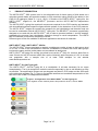
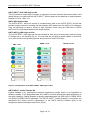
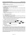
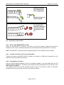
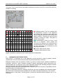
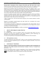
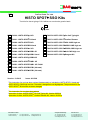
EN Instructions for use HISTO SPOT SSO Kits Test kits for tissue typing of HLA alleles on a molecular genetic basis IVD 0123 REF 726010: HISTO SPOT A 4D REF 726070: HISTO SPOT On-Call Typingkit REF 726011: HISTO SPOT A Xtend REF 726071: HISTO SPOT Coeliac Disease REF 726020: HISTO SPOT B 4D REF 726072: HISTO SPOT ABC CWD high res REF 726021: HISTO SPOT B Xtend REF 726073: HISTO SPOT ABDR CWD high res REF 726030: HISTO SPOT C 4D REF 726074: HISTO SPOT DR/DQ CWD high res REF 726040: HISTO SPOT DRB1 4D REF 726075: HISTO SPOT Null CWD high res REF 726041: HISTO SPOT DRB1 Xtend REF 726098: HISTO SPOT Reagent Kit REF 726045: HISTO SPOT DRB3/4/5 REF 726050: HISTO SPOT DQB1 4D REF 726051: HISTO SPOT DQB1 4D / DQA1 REF 726060: HISTO SPOT DPB1 (96 tests) REF 726061: HISTO SPOT DPB1 (24 tests) Version: 12 /2015 Issue: 2015-08 ® This instruction for use has been revised fundamentally to include the HISTO SPOT Xtend and ® HISTO SPOT CWD high res kits. Therefore, changes are not highlighted. The procedure for the HISTO SPOT® 4D kits has not been changed. This instruction for use does also replace: Instruction for Use: HISTO SPOT® On-Call Typing Kit, Version 03/2014 ® Instruction for Use: HISTO SPOT Coeliac Disease Kit, Version 01/2012 BAG Health Care GmbH Amtsgerichtsstraße 1-5 35423 Lich / Germany Tel.: +49 (0) 6404 / 925 - 0 Fax: +49 (0) 6404 / 925 - 250 www.bag-healthcare.com [email protected] Auftragsannahme/Ordering: Tel.: +49 (0) 6404 / 925 - 450 Fax: +49 (0) 6404 / 925 - 460 [email protected] Customer Service: Tel.: +49 (0) 6404 / 925 - 125 Fax: +49 (0) 6404 / 925 - 421 [email protected] Instructions for use HISTO SPOT SSO kits Version: 12 / 2015 Contents 1. 2. 3. 3.1 3.2 3.3 3.4 3.5 4. 5. 5.1 5.2 5.3 5.4 6. 7. 7.1 7.2 8. 9. 10. 11. 12. 13. PRODUCT DESCRIPTION .................................................................................................... 3 TEST PRINCIPLE.................................................................................................................. 5 MATERIAL ............................................................................................................................. 5 Reagents provided with the HISTO SPOT, HISTO SPOT 4D, HISTO SPOT Xtend, HISTO SPOT Null CWD high res and HISTO SPOT Coeliac Disease kits ......................... 5 Reagents provided with the HISTO SPOT On-Call Typing Kit, the HISTO SPOT ABC CWD high res, the HISTO SPOT ABDR CWD high res and the HISTO SPOT DR/DQ CWD high res kits .................................................................................................................. 5 Reagents provided with the HISTO SPOT Reagent kit ........................................................ 6 Definition of lots and batches ................................................................................................. 6 Reagents and equipment required but not provided .............................................................. 6 STORAGE AND STABILITY .................................................................................................. 6 TEST PROCEDURE .............................................................................................................. 7 Safety conditions and special remarks ................................................................................... 7 DNA isolation ......................................................................................................................... 7 Amplification .......................................................................................................................... 7 5.3.1 HISTO SPOT, HISTO SPOT 4D, HISTO SPOT Xtend, HISTO SPOT Null CWD high res and HISTO SPOT Coeliac Disease kits ............................................ 7 5.3.2 HISTO SPOT On-Call Typing Kit, HISTO SPOT ABC CWD high res, HISTO SPOT ABDR CWD high res and HISTO SPOT DR/DQ CWD high res kit ............. 8 5.3.3 PCR program............................................................................................................. 8 Automated hybridisation assay on the MR.SPOT processor ................................................ 9 5.4.1 Reagent preparation .................................................................................................. 9 5.4.2 Setup of the MR.SPOT processor .......................................................................... 10 5.4.3 Transfer of results to a PC for interpretation ............................................................ 10 5.4.4 Interpretation of results ............................................................................................ 10 WARNINGS AND PRECAUTIONS ...................................................................................... 11 SPECIFIC PERFORMANCE CHARACTERISTICS ............................................................. 12 Evaluation ............................................................................................................................ 12 PCR Amplification reaction .................................................................................................. 12 LIMITATIONS OF THE METHOD ........................................................................................ 12 INTERNAL QUALITY CONTROL ........................................................................................ 13 TROUBLESHOOTING ......................................................................................................... 13 TRADEMARKS USED IN THIS DOCUMENT/PRODUCT .................................................... 13 EXPLANATION OF SYMBOLS USED ON LABELING ....................................................... 14 LITERATURE ..................................................................................................................... 15 Page 2 of 15 Instructions for use HISTO SPOT SSO kits 1. Version: 12 / 2015 PRODUCT DESCRIPTION The HISTO SPOT SSO system is an in vitro diagnostic test for tissue typing of HLA alleles on a molecular genetic basis and provides medium to high resolution typing results of the alleles in the CWD 2.0.0 catalogue (Mack S.J. et al., 2013). It consists of the HISTO SPOT typing kits, the HISTO SPOT reagent kit, the MR.SPOT processor and the HISTO MATCH interpretation software. The HISTO SPOT typing kits contain all components required for the PCR reaction and testwells with immobilized sequence-specific oligonucleotide probes for the detection of the PCR products. The HISTO SPOT reagent kit contains the reagents for the hybridisation and detection and can be used in combination with all HISTO SPOT typing kits. The MR.SPOT processor is specifically designed to be used with the HISTO SPOT kits in order to process between 1 and 96 samples, automating the process from hybridisation, detection through to result interpretation. The HISTO MATCH software is required to interpret the results. Different types of kits are available for different applications and levels of resolution: HISTO SPOT Kits / HISTO SPOT 4D Kits The HISTO SPOT® kits are designed to provide unambiguous results at least at allele group level i.e for the 1st field in the nomenclature. Different combinations of alleles that cross allele groups but have the same positive probe pattern are considered as ambiguous. HISTO SPOT® 4D typing kits usually give allele level results (2nd field) if a common allele filter is used. Criteria for the 4D common allele filter are the following: allele frequency ≥ 0,5% in at least one population with a sample size of at least 1000 samples on the website www.allelefrequencies.net . HISTO SPOT On-Call Typing Kit The HISTO SPOT On-Call Typing Kit is a combination of all tests necessary for an organ transplantation. The kit is designed to make the workflow especially in the on-call situation as easy as possible. The amplification primers are pre-dropped and dried in PCR strips and the SSO tests are combined in a holder (Fig 1). There is a simplified workflow and combined interpretation of this test in the HISTO MATCH interpretation software. Figure 1: Configuration of the HISTO SPOT On-Call Typing Kit The negative control contains primers and probes for HLA-A, -B and –DRB1. Page 3 of 15 Instructions for use HISTO SPOT SSO kits Version: 12 / 2015 HISTO SPOT Null CWD high res Kit This kit provides a simple way to exclude or determine common and well documented class I Null alleles that are not typed with the HISTO SPOT 4D kits (based on the detection of polymorphisms situated in Exons 1 and 4-7). HISTO SPOT Xtend Kits The HISTO SPOT Xtend kits consist of complementary wells to the HISTO SPOT® 4D kits that provide a higher resolution including well documented (WD) alleles from the CWD 2.0.0 catalogue. They can generally be used to achieve a high resolution result using the CWD filter in HISTO MATCH or for resolving ambiguities with single samples. HISTO SPOT CWD high res Kits The HISTO SPOT CWD high res kits are intended for safe and convenient high resolution typing of multiple loci in one teststrip (Fig. 2). The test wells are provided in plastic holders to avoid mixup of wells and the corresponding primers are pre-dried in 8 well PCR strips. ABC: 1 test ABDR: 1 test DR/DQ: 2 tests Figure 2: Configuration of the HISTO SPOT CWD high res kits HISTO SPOT® Coeliac Disease Kit Coeliac disease is an autoimmune reaction triggered by gluten which is an ingredient of different cereals. If not diagnosed early this leads to chronic inflammation and destruction of the small intestine. Coeliac disease is strongly associated with the DQA1*05:01- DQB1*02:01 and DQA1*03-DQB1*03:02 haplotype. The HISTO SPOT® Coeliac Disease kit recognizes the associated HLA alleles and the interpretation in the HISTO MATCH software gives a risk assignment (based on Megiorni and Pizzuti, 2012). Page 4 of 15 Instructions for use HISTO SPOT SSO kits 2. Version: 12 / 2015 TEST PRINCIPLE The test includes four basic steps: - DNA isolation - PCR amplification - hybridisation and detection - data interpretation DNA isolation is performed on the clinical sample, using the DNA isolation method established in the laboratory or using commercial kits. Then the DNA is amplified in a locus specific PCR reaction using the reagents provided with the kit (Mastermix, MgCl2 solution resp. PCR strips with primers, PCR buffer, MgCl2 solution). The specificity of the amplification is governed by a set of biotinylated primers that have been designed to uniquely amplify the chosen HLA locus. After the PCR amplification process, the PCR plate resp. PCR strips containing biotin labelled amplicon is transferred to the MR.SPOT processor. MR.SPOT adds hybridisation buffer to each well and then transfers each amplicon plus hybridisation buffer to a test well containing an array of immobilized sequence-specific oligonucleotide (SSO) probes. These probes are either single oligonucleotide probes or a combination of 2 or more individual probes, immobilised in the same spot (Mosaic Probes) which have been designed to improve the identification of cis located polymorphisms. The biotin labelled amplicon binds to those SSO probes that contain a complementary target sequence and can then be detected by a colourimetric reaction. In order to prevent unspecific binding of the amplicon on the surface of the test wells MR.SPOT has blocked the wells with blocking buffer before transferring the amplicon. After a stringent wash step to remove all unbound amplicon a streptavidin-alkaline phoshatase conjugate is added to the wells and binds to the biotin labelled amplicon captured by the SSO probe. After further wash steps, BCIP/NBT substrate is added which produces a blue-purple colour when converted by the alkaline phosphatase. The resulting coloured dots in the bottom of each test well are photographed by MR.SPOT and the image is transferred into the HISTO MATCH software installed on the PC of the user. The image analysis program of the HISTO MATCH software determines the intensity of each spot in the array and compares it to the intensity of the background. From this data the positive and negative reactions are calculated. The pattern matching program of the HISTO MATCH software determines the HLA type of the sample based on the specific hybridisation pattern. 3. MATERIAL 3.1 Reagents provided with the HISTO SPOT, HISTO SPOT 4D, HISTO SPOT Xtend, HISTO SPOT Null CWD high res and HISTO SPOT Coeliac Disease kits Individually packed test well strips for HLA typing. Each strip contains 8 tests with immobilized, sequence-specific oligonucleotide probes. Mastermix, ready to use, contains biotinylated primers for the chosen locus, dNTPs,Taq polymerase, reaction buffer, 0.05% sodium azide Magnesium Chloride, 6 mM, ready to use Instructions for use, lot specific batch file, hit table and probe information provided on a CD 3.2 Reagents provided with the HISTO SPOT On-Call Typing Kit, the HISTO SPOT ABC CWD high res, the HISTO SPOT ABDR CWD high res and the HISTO SPOT DR/DQ CWD high res kits Individually packed combistrips with test wells for HLA typing combined in a plastic holder. The testwells contain immobilized, sequence-specific oligonucleotide probes. PCR strips with dried primers for the locus specific amplification of the HLA loci to be typed. The primers are matching the HLA loci in the combistrips. PCR caps Page 5 of 15 Instructions for use HISTO SPOT SSO kits Version: 12 / 2015 PCR buffer, ready to use, contains dNTPs, Taq polymerase, reaction buffer, 0,05% sodium azide Magnesium Chloride, 6 mM, ready to use Instructions for use, lot specific batch file, hit table and probe information provided on a CD 3.3 Reagents provided with the HISTO SPOT Reagent kit Blocking Buffer, ready to use, contains 0.001% Proclin® 150. Hybridisation Buffer, ready to use, contains 0.001% dye, 0.1% sodium dodecyl sulphate, 0.001% Proclin® 150. Stringent Wash Buffer, ready to use, contains 0.001% dye, 0.1% sodium dodecyl sulphate, 0.001% Proclin® 150. TBS Wash Buffer (Tris Buffered Saline), ready to use, contains 20 mM Tris, 0.003% dyes, 0.001% Proclin® 150. BCIP® / NBT Substrate, ready to use, (5-bromo-4-chloro-3-indolyl phosphate / nitroblue tetrazolium chloride). Conjugate, Streptavidin Alkaline Phosphatase, concentrate, contains < 0.1% sodium azide (to be diluted 1:1666 in Blocking Buffer). 3.4 Definition of lots and batches Kit: e.g. HISTO SPOT A, defines the locus tested Lot: e.g A084, A085, defines the layout and specificity of the probes that are contained in the kit. A single lot can contain many different batches. Batch: e.g. A085-1, A085-2, A085-3, defines how a probe reacts in comparison to the control probes (cut off values), and defines the manufacture and expiry date of the strips. Single probes may be switched off in some batches within the same lot. This information can be found on the lot-specific CD supplied with the kits (Hit Tables – Excel file). 3.5 Reagents and equipment required but not provided MR.SPOT processor, including HISTO MATCH software, REF 726100 Pipette tips for the MR.SPOT processor, 1000 µl REF 726099 and 200 µl REF 726097 DNA extraction reagents (no salting out method) Skirted PCR plates with lids or adhesive film (HISTO SPOT PCR Frameplates, REF 726220, HISTO SPOT PCR Caps, REF 726090, HISTO SPOT PCR Foils, REF 726089) Thermal cycler Deionized water Variable pipettes (range 0.5 – 1000 µl) and disposable tips 4. STORAGE AND STABILITY The kits will be shipped at room temperature. After delivery all reagents and components of the kits should be stored at 2…8oC. The expiry date is indicated on the label of each reagent and is valid for the originally sealed reagents. The expiry date indicated on the outer box label refers to the reagent with the shortest stability contained in the kit. Individual strips of 8 wells from HISTO SPOT, HISTO SPOT 4D, HISTO SPOT Xtend, HISTO SPOT Null CWD high res and HISTO SPOT Coeliac Disease kits may be opened, the required number of wells for a test run can be snapped off and the unused wells returned to the opened foil bag and stored for future use with the kit. Test wells stored in open foil pouches should be used within 30 days of being opened. The other opened reagents should be used within 3 months. The conjugate dilution must always be prepared afresh for each test run. Page 6 of 15 Instructions for use HISTO SPOT SSO kits 5. TEST PROCEDURE 5.1 Safety conditions and special remarks Version: 12 / 2015 Molecular genetic techniques are particularly sensitive methods and should be performed by well trained personnel, experienced in molecular genetic techniques and histocompatibility testing. The results from these tests must not be used as the sole determinant for making clinical decisions. Transplantation guidelines as well as EFI standards should be followed in order to minimize the risk of false typings, in the particular case of discrepancies in serological and molecular genetic methods. Special safety conditions must be noted in order to avoid contamination and thus false reactions: 5.2 Wear gloves during work (powder-free, if possible). Use new tips with each pipeting step (with integrated filter). Use separate working areas for pre-amplification (DNA isolation and preparation of the reactions) and post-amplification (hybridisation and detection). Preferably use two separate rooms. Amplicon should not be taken back into PCR set up area. Use devices and other materials only at the respective places and do not exchange them. DNA isolation The use of a IVD certified extraction kit for DNA isolation is strongly recommend. Laboratory established standard method for DNA isolation should be validated by the user. Avoid the use of salting out methods, whether possible. Validated DNA extraction methods: Qiagen columns Methods successfully tested in the field: EZ-1 / Geno M6 (Qiagen beads) Promega Maxwell 16 QuatroProbe (BeeRobotics) The presence of heparin potentially inhibits PCR. Therefore, EDTA or Citrate Blood is recommended for typing. The sample DNA should have a concentration of 15-30 ng/µl. The purity indexes should be the following: extinction ratio OD260/OD280: > 1.5 and < 2.0 Higher values indicate the existence of RNA, lower values mean contamination with protein. extinction ratio OD260/OD230: > 1.8 Lower values indicate a possible contamination with carbohydrates, salts or organic solvents. 5.3 Amplification 5.3.1 HISTO SPOT, HISTO SPOT 4D, HISTO SPOT Xtend, HISTO SPOT Null CWD high res and HISTO SPOT Coeliac Disease kits Use skirted PCR plates for the amplification, because they have to be held down at the skirt by a clamp in the MR.SPOT processor afterwards. HISTO SPOT PCR Frameplates have been validated for this application, plates from other suppliers have to be validated by the user. For each sample to be amplified add the following reagents to each PCR tube: 10 µl Mastermix 5 µl MgCl2 5 µl Sample DNA (15-30 ng/µl) Total volume for each amplification reaction is 20 µl. Premix for multiple samples: no. of samples+2 x 10 µl Mastermix no. of samples+2 x 5 µl MgCl2 Page 7 of 15 use 15 µl premix per sample Instructions for use HISTO SPOT SSO kits Version: 12 / 2015 Note: It is important that the DNA concentration is in the range between 15 and 30 ng/µl. Higher concentrations may result in false-positive probe reactions and lower concentrations may cause amplification failures. If a negative control is set up, prepare one PCR reaction with distilled water instead of sample DNA. Seal the amplification tubes with lids or adhesive film and shortly spin down the liquid. Place in the thermal cycler and amplify with the PCR program in chapter 5.3.3. 5.3.2 HISTO SPOT On-Call Typing Kit, HISTO SPOT ABC CWD high res, HISTO SPOT ABDR CWD high res and HISTO SPOT DR/DQ CWD high res kit For each combi test take one PCR strip with pre-dropped amplification primers PCR Primers from the fridge. Make a pre-mix with the following components for each sample: ABC, ABDR, On-Call Typing Kit DR/DQ Kit 80 µl PCR buffer 40 µl MgCl2 40 µl Sample DNA (15-30 ng/µl) 40 µl PCR buffer 20 µl MgCl2 20 µl Sample DNA (15-30 ng/µl) Pipet 20 µl of the pre-mix into each well with the pre-dropped primers and re-suspend the primers with the pre-mix. Note: It is important that the DNA concentration is in the range between 15 and 30 ng/µl. Higher concentrations may result in false-positive probe reactions and lower concentraions may cause amplification failures. For the negative control in well number 8 of the HISTO SPOT On-Call Typing Kit prepare one PCR reaction with distilled water instead of sample DNA: 10 µl PCR buffer 5 µl MgCl2 5 µl H2O Seal the amplification tubes with the supplied caps and shortly spin down the liquid. Place in the thermal cycler and amplify with the PCR program in chapter 5.3.3. 5.3.3 PCR program Programme-Step Time Temperature No. of Cycles First Denaturation 2 Min 96°C 1 Cycle Denaturation 15 Sec 96°C 10 Cycles Annealing + Extension 60 Sec 65°C Denaturation 10 Sec 96°C Annealing 50 Sec 61°C Extension 30 Sec 72°C ∞ 22°C Hold Page 8 of 15 20 Cycles Instructions for use HISTO SPOT SSO kits Version: 12 / 2015 The conditions are the same for all thermal cyclers. However, the overall time required for this step will vary according to the ramping speed of the specific thermal cycler. The following thermal cycler models haven been validated with HISTO SPOT® SSO: Applied Biosystems: PE 9600, PE 9700 (use ramp rate of PE 9600), VeritiTM Biorad: PTC 100 / PTC 200, MyCycler Eppendorf: Mastercycler EP Gradient S If other thermal cyclers are used, the validation has to be done by the user. It is generally recommended to use a ramp rate of 1-2°C/sec. Once the amplification step is complete, the samples may be tested immediately or stored at 2…8oC for up to 5 days. It is not necessary to make a gel to control the amplification. It is also not always helpful because assay results may be good although there was only a very faint band visible on the gel. If a gel should be done anyway, you ahould not take more than 2-3 µl of the amplicon to do this. The amplicon sizes for the different kits are given on the information CDs that can be found in every kit (Hit Table in Excel format: Second sheet “Notes”). 5.4 Automated hybridisation assay on the MR.SPOT processor 5.4.1 Reagent preparation Take HISTO SPOT reagents and HISTO SPOT testwells out of the fridge and allow them to warm to room temperature. Salt crystals may be observed in the hybridisation buffer and in the stringent wash buffer. If crystals are present, warm reagents up to 30°C to dissolve. Warm the whole content of the bottle, not an aliquot. The conjugate has to be diluted 1:1666 in blocking buffer. The conjugate dilution must always be prepared afresh for each test run. The conjugate has to be vortexed and spun down each time before the dilution step! The required volumes of the reagents will vary depending on the number of strips to be tested. MR.SPOT displays the required volumes for the chosen number of strips. Fill the required volumes of the reagents into the corresponding labelled reservoirs. Place the test wells and the PCR plate into the appropriate blocks of the MR.SPOT processor. Note the correct arrangement of the PCR plate. Please make sure that there is no dirt or plastic particles in the reaction plate holder, because this may disturb the heat transfer during hybridization. The test strips from the HISTO SPOT, HISTO SPOT 4D, HISTO SPOT Xtend, HISTO SPOT Null CWD high res and HISTO SPOT Coeliac Disease kits can be separated in single wells according to figure 1, if less than eight tests should be run. If you are using separated wells be careful that they are sitting properly in the reaction plate holder and are not twisted against each other. Please make sure that dummy wells are placed in empty positions up to multiples of four when using single wells or combistrips ! Page 9 of 15 Instructions for use HISTO SPOT SSO kits Version: 12 / 2015 Separate testwells by snapping them off in a right angle. Break off end pieces with the thumb. Do not break the testwells upwards or downwards and do not twist the wells off . Fig. 1: Separating single testwells. 5.4.2 Setup of the MR.SPOT processor Switch on the MR.SPOT processor. The start up screen will appear. Follow the process as indicated on the screen. Details are described in the User Manual for the MR.SPOT processor. Note: The MR.SPOT processor and the reagents should not be exposed to direct sunlight. 5.4.3 Transfer of results to a PC for interpretation Transfer the data to the HISTO MATCH software via network or USB stick as described in the manual for the HISTO MATCH software. 5.4.4 Interpretation of results Open the HISTO MATCH software (if this is not already installed, it can be installed from the CD delivered with the MR.SPOT processor) and interpret the data as described in the manual for the HISTO MATCH software. The images should look like the example shown in figure 2 and figure 3 gives a schematic illustration of the result and the functions of the different probes. Page 10 of 15 Instructions for use HISTO SPOT SSO kits Version: 12 / 2015 The colour of the circles around the probes indicate their function (see IFU for the HISTO MATCH software for details). Figure 2: Image of a result for HLA A the amplification primers in the mastermix and indicate that mastermix was added and that all reagents during the SSO assay were added correctly. Furthermore, they allow the software to locate the image. The pattern is specific for the batch. +: Amplification control for Exon 2 and Exon 3 in duplicate. Those probes are universal for all alleles of the respective locus and show that the PCR was successful. They are also functioning as a reference for the allele specific probes. : Positional probes: They are reacting with : Positive allele specific probe : Negative allele specific probe Figure 3: Schematic illustration of the result and function of the probes 6. WARNINGS AND PRECAUTIONS HISTO SPOT is designed for in vitro diagnostic use and should be used by properly trained, qualified staff. All work should be performed using Good Laboratory Practices. Biological material used for extraction of DNA, e.g. blood or human tissue, should be handled as potentially infectious. When handling biological material appropriate safety precautions are recommended (do not pipet by mouth; wear disposable gloves while handling biological material and performing the test; disinfect hands when finished the test). Biological material should be inactivated before disposal (e.g. in an autoclave). Disposables should be autoclaved or incinerated after use. Spillage of potentially infectious materials should be removed immediately with absorbent paper tissue and the contaminated areas swabbed with a suitable standard disinfectant or 70% alcohol. Material used to clean spills, including gloves, should be inactivated before disposal (e.g. in an autoclave). Page 11 of 15 Instructions for use HISTO SPOT SSO kits Version: 12 / 2015 Blocking Buffer, Hybridisation Buffer, Stringent Wash Buffer and TBS Wash Buffer contain ProClin150 and the Magnesium Chloride Solution contains ProClin300. The reagents contain 0.001% preservative only, nevertheless avoid contact with the skin and mucous membranes. Mastermix and Conjugate contain the preservative sodium azide. The reagents contain < 0.1% sodium azide which is not considered to be a harmful concentration. Nevertheless avoid contact with the skin and mucous membranes. Sodium azide may react with lead and copper plumbing to form explosive metal azides. While disposing of sodium azide containing solutions down laboratory sinks, flush the drains with a large volume of water to prevent azide build-up. All work with reagents should be handled with the appropriate precautions. Wear eye protection, laboratory coats and disposable gloves when handling the reagents. Avoid contact of these materials with the skin, eyes or mucous membranes. If contact does occur, immediately wash with large amounts of water. Burns can occur if left untreated. If spills of reagents occur, dilute with water before wiping dry. Do not expose substrate to metals, oxidising agents. Disposal of all samples, unused reagents and waste should be in accordance with country, federal, state and local regulations. Avoid microbial contamination of reagents when removing aliquots from reagent bottles. The use of sterile disposable pipettes and pipette tips is recommended. Do not use reagents with evidence of turbidity or microbial contamination. Material Safety Data Sheets (MSDS) are available to download at www.bag-healthcare.com . 7. SPECIFIC PERFORMANCE CHARACTERISTICS 7.1 Evaluation For all HISTO SPOT SSO kits evaluation studies with pre-typed DNA samples has been performed. The results were compared to other typing methods (e.g. SSP, sequencing). No discrepancies were observed between the typing methods. For every lot the specificity of each probe was verified with DNA from reference samples. 7.2 PCR Amplification reaction The alleles amplified with each HISTO SPOT SSO kit, the HLA nomenclature release referred to and the exons that are amplified are given in the respective lot specific information. This is found on a CD in each kit. 8. LIMITATIONS OF THE METHOD Because of the high susceptibility of the PCR method to variations in DNA concentration and quality, only DNA samples should be used that have a concentration between 15 and 30 ng/µl and appropriate purity indices (extinction ratio OD260/OD280: between 1.5 and 2.0 / extinction ratio OD260/OD230: > 1.8). Extreme care should be taken to prevent contamination of the kit reagents and other laboratory materials and equipment with amplicons or DNA. Regular wipe tests (e.g. BAG Wipe Test, REF 7091) and negative controls with each assay are strongly recommended. Page 12 of 15 Instructions for use HISTO SPOT SSO kits Version: 12 / 2015 The hybridisation assay is a very temperature-sensitive process. Therefore, the HISTO SPOT SSO kits should only be used in combination with the MR.SPOT processor to ensure correct temperatures and incubation times. All instruments (e.g. pipettes, thermal cyclers, heat blocks; MR.SPOT processor) must be calibrated according to the manufacturers instructions. Accuracy and temperature uniformity of thermal cyclers may be tested with the BAG CYCLER CHECK (REF 7104). 9. INTERNAL QUALITY CONTROL Internal quality control of new lots of the HISTO SPOT SSO kits can be performed using a combination of DNA samples with known HLA type. Internal positive controls are contained in each test well to ensure sucessful amplification and hybridization. Negative controls to detect possible contaminations are recommended. Use a PCR reaction without DNA in the subsequent hybridization assay as a negative control. 10. TROUBLESHOOTING Symptom Instrument Malfunction Error message at data transfer No result No Spots in well Only control spots positive False positive probes Exon dropout No result / inconclusive result due to weak signals 11. Possible problem(s) Numerous Failure in data transfer Failure to grid image Failure to add mastermix to PCR Failure to add DNA to PCR or amplification failure Too much DNA used or conjugate concentration too high (not spun down). DNA concentration too high or DNA degraded Mistake in conjugate dilution or poor amplification Instrument malfunction TRADEMARKS USED IN THIS DOCUMENT/PRODUCT Proclin® is a trademark of Rohm and Haas company. BCIP® is a trademark of Sigma Aldrich Co. Veriti™ is a trademark of Applied Biosystems. Page 13 of 15 Potential Solution(s) Refer to MR.SPOT manual Manually transfer data using USB drive Perform manual gridding Repeat whole assay and check PCR product on gel Repeat whole assay and check PCR product on gel Check DNA concentarion. Spin down conjugate before use Check DNA concentration, run a gel with the DNA Repeat assay. Check hybridisation temperature on instrument Instructions for use HISTO SPOT SSO kits 12. Version: 12 / 2015 EXPLANATION OF SYMBOLS USED ON LABELING IVD For in vitro diagnostic use Storage temperature LOT Batch code Use by REF Catalogue number Consult instructions for use HLA TYPING Intended purpose: HLA typing Mastermix | A Mastermix for amplification of the HLA-A locus Mastermix | B Mastermix for amplification of the HLA-B locus Mastermix | C Mastermix for amplification of the HLA-C locus Mastermix | DRB1 Mastermix for amplification of the HLA-DRB1 locus Mastermix | DQB1 Mastermix for amplification of the HLA-DQB1 locus Mastermix | DQ Mastermix for amplification of the HLA-DQB1 and DQA1 locus Mastermix | DPB1 Mastermix for amplification of the HLA-DPB1 locus Mastermix | DRB3/4/5 Mastermix for amplification of the HLA-DRB3/4/5 loci Mastermix | Coeliac Mastermix for amplification of the specific loci Mastermix | Null Mastermix for amplification of the HLA Null alleles Testwells | A Testwells with bound probes to type the HLA-A locus Testwells | A Xtend Testwells with bound probes to type the HLA-A locus Testwells | B Testwells with bound probes to type the HLA-B locus Testwells | B Xtend Testwells with bound probes to type the HLA-B locus Testwells | C Testwells with bound probes to type the HLA-C locus Testwells | DRB1 Testwells with bound probes to type the HLA-DRB1 locus Testwells | DRB1 Xtend Testwells with bound probes to type the HLA-DRB1 locus Testwells | DQB1 Testwells with bound probes to type the HLA-DQB1 Testwells | DQ Testwells with bound probes to type the HLA-DQB1 and DQA1 locus Testwells | DPB1 Testwells with bound probes to type the HLA-DPB1 locus Testwells | DRB3/4/5 Testwells with bound probes to type the HLA-DRB3/4/5 loci Page 14 of 15 Instructions for use HISTO SPOT SSO kits Version: 12 / 2015 Testwells | Coeliac Testwells with bound probes to type the specific alleles Testwells | Null Testwells with bound probes to detect HLA Null alleles Combistrip PCR Primers Testwells with bound probes for typing of several HLA loci (depending on the typingkit) PCR strips with dried primers for amplification of several HLA loci (depending on the typing kit) PCR Caps PCR Caps PCR Buffer PCR Buffer MgCl2 Magnesium chloride solution BLOCKBUF Blocking Buffer HYBBUF Hybridisation Buffer STRGWASH Stringent wash Buffer TBSWASH TBS Wash Buffer (Tris buffered saline) SUBS BCIP® / NBT Substrate CONJ Conjugate, Streptavidin Alkaline Phosphatase ® HISTO SPOT INFORMATION CD 13. CD, includes Instructions for use, lot specific batch file, hit table and probe information LITERATURE Mack S.J. et al., Common and well-documented HLA alleles: 2012 update to the CWD catalogue, Tissue Antigens 81/4, 183-257 (2013) Megiorni and Pizzuti, HLA-DQA1 and HLA-DQB1 in Celiac disease predisposition: practical implications of the HLA molecular typingJournal of Biomedical Science 19:88 (2012) Instructions for use in other languages see http://www.bag-healthcare.com http://service.bag-healthcare.com or phone +49 (0)6404-925-125 Page 15 of 15