1
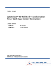
Product Manual
OxiSelect™ 96-Well Comet Assay Kit
Catalog Number
STA-355
96 assays
STA-355-5
5 x 96 assays
FOR RESEARCH USE ONLY
Not for use in diagnostic procedures
Introduction
DNA damage, due to environmental factors and normal metabolic processes inside the cell, occurs at a
rate of 1,000 to 1,000,000 molecular lesions per cell per day. While this counts for only a small part of
the human genome's approximately 6 billion bases (3 billion base pairs), unrepaired lesions to critical
genes can impede a cell's ability to carry out its function and appreciably increase the likelihood of
cancer.
The comet assay, or single cell gel electrophoresis assay (SCGE), is a common technique for
measurement of DNA damage in individual cells. Under an electrophoretic field, damaged cellular
DNA (containing fragments and strand breaks) is separated from intact DNA, yielding a classic “comet
tail” shape under the microscope. Extent of DNA damage is usually visually estimated by comet tail
measurement; however, image analysis software is also available for measuring various parameters.
The OxiSelect™ 96-Well Comet Assay is a fast and sensitive kit for the measurement of cellular DNA
damage. Each kit provides sufficient reagents to perform up to 96 assays.
Assay Principle
Cell Biolabs’ OxiSelect™ 96-Well Comet Assay is a single cell gel electrophoresis assay (SCGE) for
simple evaluation of cellular DNA damage. First, individual cells are mixed with molten agarose
before application to the OxiSelect™ 96-Well Comet Slide. These embedded cells are then treated with
a lysis buffer and alkaline solution, which relaxes and denatures the DNA. Finally, the samples are
electrophoresed in a horizontal chamber to separate intact DNA from damaged fragments. Following
electrophoresis, the samples are dried, stained with a DNA dye, and visualized by epifluorescence
microscopy. Under these conditions, the damaged DNA (containing cleavage and strand breaks) will
migrate further than intact DNA and produce a “comet tail” shape (see Figure 1). Each kit provides
sufficient reagents to perform up to 96 assays.
2
Figure 1: Assay Principle
3
Related Products
1. STA-320: OxiSelect™ Oxidative DNA Damage ELISA Kit (8-OHdG Quantitation)
2. STA-321: OxiSelect™ DNA Double-Strand Break (DSB) Staining Kit
3. STA-324: OxiSelect™ Oxidative DNA Damage Quantitation Kit (AP sites)
4. STA-325: OxiSelect™ Oxidative RNA Damage ELISA Kit (8-OHG Quantitation)
5. STA-350: OxiSelect™ Comet Assay Kit (3-Well Slides), 15 Assays
6. STA-351: OxiSelect™ Comet Assay Kit (3-Well Slides), 75 Assays
7. STA-352: OxiSelect™ Comet Assay Slides (3-Well), 5 Slides
8. STA-353: OxiSelect™ Comet Assay Slides (3-Well), 25 Slides
9. STA-354: OxiSelect™ Comet Assay Control Cells
10. STA-356: OxiSelect™ 96-Well Comet Assay Slide
Kit Components
1. OxiSelect™ 96-Well Comet Slide (Part No. STA-356): One slide.
2. OxiSelect™ Comet Agarose (Part No. 235002): One sterile 15 mL bottle.
3. Vista Green DNA Dye, 10000X (Part No. 235003): One 5 µL vial.
4. EDTA Solution, 500 mM (Part No. 235004): One 50 mL bottle.
5. 10X Lysis Solution (Part No. 235005): One 20 mL bottle.
Materials Not Supplied
1. NaCl powder
2. NaOH pellets
3. 10 N NaOH for pH adjustment
4. DMSO (optional)
5. 70% Ethanol
6. TE Buffer (10 mM Tris, pH 7.5, 1 mM EDTA)
7. PBS (without Mg2+ and Ca2+) and DI H2O
8. EDTA (disodium salt)
9. 37ºC and boiling water baths
10. Horizontal electrophoresis chamber
11. Adjustable single channel micropipettes with disposable tips
12. Adjustable multichannel micropipette with disposable tips
13. Epifluorescence microscope with FITC filter
4
Storage
Upon receipt, store the Vista Green DNA Dye at -20 ºC. Store all other kit components at room
temperature until their expiration dates.
Preparation of Reagents
•
OxiSelect™ Comet Agarose: Heat the Comet Agarose bottle at 90-95ºC in a water bath for 20
minutes, or until agarose liquefies. Transfer the bottle to a 37ºC water bath for 20 minutes and
maintain until needed.
•
Vista Green DNA Dye: Prepare a 1X Vista Green DNA Staining Solution by diluting the provided
stock 1:10000 in TE Buffer (10 mM Tris, pH 7.5, 1 mM EDTA). The solution can be stored at 4ºC
for up to 3 weeks, protected from light.
•
Lysis Buffer: To prepare 100 mL of 1X Lysis Buffer
NaCl
14.6 g
EDTA Solution (provided)
20.0 mL
10X Lysis Solution (provided)
10.0 mL
DMSO
10.0 mL (optional for heme containing samples)
DI H2O
Adjust volume to 90 mL
Mix thoroughly to dissolve NaCl. Slowly adjust the Lysis Buffer to pH 10.0 with 10 N
NaOH, then QS to 100 mL with DI H2O. Chill Lysis Buffer to 4ºC before use.
Note: Buffer will appear cloudy at room temperature, but will clear at 4ºC. pH will also
remain ~10.0.
•
Alkaline Solution: To prepare 100 mL of Alkaline Solution
NaOH
1.2 g
EDTA Solution (provided)
0.2 mL
DI H2O
Adjust volume to 100 mL
Mix thoroughly to dissolve NaOH. Chill Alkaline Solution to 4ºC before use.
•
Electrophoresis Running Solution: Choose the appropriate electrophoresis solution based on the
desired running conditions and assay sensitivity. TBE is preferred for analysis of apoptosis and
enables use of the tail length, rather than the tail moment, for data analysis. TBE electrophoresis
will detect single-stranded and double-stranded DNA breaks, and may detect a few AP sites.
Alkaline electrophoresis is more sensitive and will detect smaller amounts of DNA damage.
Alkaline electrophoresis will detect single-stranded and double-stranded DNA breaks, the majority
of AP sites, and alkali labile DNA adducts.
To prepare 1 L of Electrophoresis Solution
1. TBE Electrophoresis Solution
Tris Base
10.8 g
5
Boric Acid
5.5 g
EDTA (disodium salt)
0.93 g
DI H2O
Adjust volume to 1 L
Mix thoroughly to dissolve solids. Chill TBE Running Solution to 4ºC before use.
OR 2. Alkaline Electrophoresis Solution (300 mM NaOH, pH >13, 1 mM EDTA)
NaOH
12.0 g
EDTA Solution (provided)
DI H2O
2.0 mL
Adjust volume to 1 L
Mix thoroughly to dissolve NaOH. Chill Alkaline Running Solution to 4ºC before use.
Special Precautions
To avoid ultraviolet light damage to cell samples, perform the assay under low/dim light conditions.
Preparation of Samples and Slides
1. Prepare Lysis Buffer, Alkaline Solution, and Electrophoresis Running Solution (see Preparation of
Reagents) prior to performing the assay. Chill all solutions to 4ºC thoroughly.
2. Heat OxiSelect™ Comet Agarose to 90-95ºC in a water bath for 20 minutes, or until agarose
liquefies. Cool the agarose by transferring the bottle to a 37ºC water bath for 20 minutes.
3. Heat OxiSelect™ 96-Well Comet Slide at 37ºC for 15 minutes to promote agarose attachment.
4. Prepare cell samples, including controls, as follows:
•
Suspension Cells: Centrifuge cells at 700 x g for 2 minutes and discard supernatant. Wash cell
pellet once with ice-cold PBS (without Mg2+ and Ca2+), centrifuge, and discard the supernatant.
Finally, resuspend the cells at 1 x 10⁵ cells/mL in ice-cold PBS (without Mg2+ and Ca2+).
•
Adherent Cells: Gently remove cells from flask/dish by scraping with a rubber policeman.
Transfer cell suspension to a conical tube and centrifuge at 700 x g for 2 minutes, discarding the
supernatant. Wash cell pellet once with ice-cold PBS (without Mg2+ and Ca2+), centrifuge, and
discard the supernatant. Finally, resuspend the cells at 1 x 10⁵ cells/mL in ice-cold PBS
(without Mg2+ and Ca2+).
•
Tissue Preparation: Using dissection scissors, mince a small piece of tissue in 1-2 mL of ice
cold PBS containing 20 mM EDTA (without Mg2+ and Ca2+). Allow the tissue/cell suspension
to stand for 5 minutes before transferring the supernatant to a centrifuge tube; avoid transferring
debris. Centrifuge, discarding the supernatant, and then resuspend the cells at 1 x 10⁵ cells/mL
in ice-cold PBS (without Mg2+ and Ca2+).
5. In a pre-warmed 96-well plate, combine cell samples with Comet Agarose (step 2) at 1:10 ratio
(v/v). Titrate to mix, and immediately pipette 20 µL/well onto the OxiSelect™ 96-Well Comet
Slide using a multichannel micropipette. Ensure complete well coverage by spreading the
suspension over the well with the pipette tip.
6
Note: For multiple samples, maintain suspensions at 37ºC to avoid gelation. Titrate samples again
just prior to slide addition.
6. Maintaining the slide horizontally, transfer the slide to 4ºC in the dark for 15 minutes.
7. Carefully, transfer the slide to a small basin/container containing pre-chilled Lysis Buffer (~50-100
mL/slide). Immerse the slide in the buffer for 30-60 minutes at 4ºC in the dark.
8. Carefully, aspirate the Lysis Buffer from the container and replace with pre-chilled Alkaline
Solution (~50-100 mL/slide). Immerse the slide in the solution for 30 minutes at 4ºC in the dark.
Assay Protocol
I. TBE Electrophoresis
1. Aspirate the Alkaline Solution from the container and replace with pre-chilled TBE
Electrophoresis Solution. Immerse the slide for 5 minutes, and then repeat once more.
2. Maintaining the slide horizontally, carefully transfer the slide to a horizontal electrophoresis
chamber. Fill the chamber with cold TBE Electrophoresis Solution until the buffer level covers
the slide.
3. Apply voltage to the chamber for 10-15 minutes at 1 volt/cm (e.g. if the chamber electrodes are
35 cm apart, you would then apply 35 volts to the slide)
4. Maintaining the slide horizontally, carefully transfer the slide from the electrophoresis chamber
to a clean, small basin/container containing pre-chilled DI H2O (~50-100 mL/slide). Immerse
the slide for 2 minutes, aspirate, and then repeat twice more.
5. Aspirate the final water rinse and replace with cold 70% Ethanol for 5 minutes.
6. Maintaining the slide horizontally, remove the slide from the 70% Ethanol and allow to air dry.
7. Once the agarose and slide is completely dry, add 50 µL/well of diluted Vista Green DNA Dye
(see Preparation of Reagents). Incubate at room temperature for 15 minutes.
8. View slides by epifluorescence microscopy using a FITC filter.
II. Alkaline Electrophoresis
1. Maintaining the slide horizontally, carefully transfer the slide from the Alkaline Solution to a
horizontal electrophoresis chamber. Fill the chamber with cold Alkaline Electrophoresis
Solution until the buffer level covers the slide.
2. Apply voltage to the chamber for 15-30 minutes at 1 volt/cm (e.g. if the chamber electrodes are
35 cm apart, you would then apply 35 volts to the slide). Additionally, adjust the volume of
Alkaline Electrophoresis Solution to produce a current setting of 300 mA.
3. Maintaining the slide horizontally, carefully transfer the slide from the electrophoresis chamber
to a clean, small basin/container containing pre-chilled DI H2O (~50-100 mL/slide). Immerse
the slide for 2 minutes, aspirate, and then repeat twice more.
4. Aspirate the final water rinse and replace with cold 70% Ethanol for 5 minutes.
5. Maintaining the slide horizontally, remove the slide from the 70% Ethanol and allow to air dry.
7
6. Once the agarose and slide is completely dry, add 50 µL/well of diluted Vista Green DNA Dye
(see Preparation of Reagents). Incubate at room temperature for 15 minutes.
7. View slides by epifluorescence microscopy using a FITC filter.
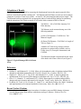
Example of Results
The following figures demonstrate typical OxiSelect™ 96-Well Comet Assay Kit results. One should
use the data below for reference only. This data should not be used to interpret actual results.
Figure 2. Etoposide Treatment of Jurkat Cells. Jurkat cells were untreated (left) or treated (right)
with 20 µM Etoposide for 4 hours before performing Comet Assay (alkaline electrophoresis conditions,
33 V/300 mA for 15 minutes).
8
Calculation of Results
The DNA damage is quantified by measuring the displacement between the genetic material of the
nucleus ('comet head') and the resulting 'tail'. Tail Moment and Tail DNA% are the two most common
parameters to analyze Comet assay results. At least 50 -100 cells should be analyzed per sample. The
Tail Moment has been suggested to be an appropriate index of induced DNA damage in considering
both the migration of the genetic material as well as the relative amount of DNA in the tail.
Tail DNA% = 100 x Tail DNA Intensity/Cell
DNA Intensity
Tail Moment can be measured using one of the
following methods:
(a) Olive Tail Moment = Tail DNA% x Tail
Moment Length*
(b) Extent Tail Moment = Tail DNA% x Length of
Tail (see Figure 3)
A number of Comet Assay analysis software
programs are commercially available, such as
Comet Assay IV (Perceptive Instruments) and
CASPlab.
*Tail Moment Length is measured from the center
of the head to the center of the tail (see Figure 3)
Figure 3: Typical Damaged DNA in Comet
Assay.
References
1. Ostling, O., and Johanson, K. J. (1984). Micro gel electrophoretic study of radiation induced DNA
damages in individual mammalian cells. Biochem. Biophys. Res. Commun. 123, 291–298.
2. Singh, N. P., McCoy, M. T., Tice, R. R., and Schneider, E. L. (1988). A simple technique for
quantification of low levels of DNA damage in individual cells. Exp. Cell. Res. 175, 184–191.
3. Olive, P. L., Banath, J. P., and Durand, R. E. (1990a). Heterogeneity in radiation induced DNA
damage and repair in tumor and normal cells using the "Comet" assay. Radiat. Res. 122, 86–94.
4. De Boeck, M., Touil, N., De Visscher, G., Vande, P. A., and Kirsch-Volders, M. (2000). Validation
and implementation of an internal standard in Comet assay. Mutat. Res. 469, 181–197.
Recent Product Citations
1. Jones, D. A. et al. (2014). Changes in markers of oxidative stress and DNA damage in human
visceral adipose tissue from subjects with obesity and type 2 diabetes. Diabetes Res Clin
Pract. 106:627-633.
9
2. Aydın, E. et al. (2014). The effect of carvacrol on healthy neurons and N2a cancer cells: some
biochemical, anticancerogenicity and genotoxicity studies. Cytotechnology. 66:149-157.
3. Tyagi, A. et al. (2011). Resveratrol selectively induces DNA Damage, independent of Smad4
expression, in its efficacy against human head and neck squamous cell carcinoma. Clin. Cancer
Res. 17:5402-5411.
Warranty
These products are warranted to perform as described in their labeling and in Cell Biolabs literature when used in
accordance with their instructions. THERE ARE NO WARRANTIES THAT EXTEND BEYOND THIS EXPRESSED
WARRANTY AND CELL BIOLABS DISCLAIMS ANY IMPLIED WARRANTY OF MERCHANTABILITY OR
WARRANTY OF FITNESS FOR PARTICULAR PURPOSE. CELL BIOLABS’s sole obligation and purchaser’s
exclusive remedy for breach of this warranty shall be, at the option of CELL BIOLABS, to repair or replace the products. In
no event shall CELL BIOLABS be liable for any proximate, incidental or consequential damages in connection with the
products.
Contact Information
Cell Biolabs, Inc.
7758 Arjons Drive
San Diego, CA 92126
Worldwide: +1 858-271-6500
USA Toll-Free: 1-888-CBL-0505
E-mail: [email protected]
www.cellbiolabs.com
2008-2015: Cell Biolabs, Inc. - All rights reserved. No part of these works may be reproduced in any form without
permissions in writing.
10