1
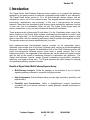
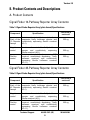
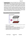
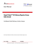
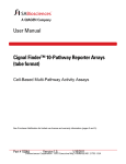
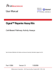
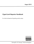
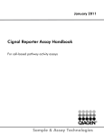
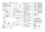
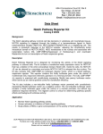
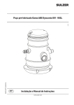
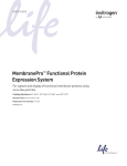
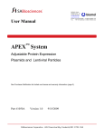
User Manual Cignal Finder™ Multi-Pathway Reporter Arrays (Plate Format) Cell-Based Multi-Pathway Activity Assays See Purchaser Notification for limited use license and warranty information. Part # 1036A Version 1.5 1/10/2011 Cignal Finder™ Multi-Pathway Reporter Arrays (plate format) Cell-Based Multi-Pathway Activity Assays User Manual (For Catalog Numbers CCA-1XXL or CCA-901L) Ordering and Technical Service Contact Information: Tel: Fax: On-line Order: E-MAIL: 1-888-503-3187 (US) 301-682-9200 (outside US) 1-888-465-9859 (US) 301-682-7300 (outside US) www.SABiosciences.com [email protected] (to place an order) [email protected] (for technical support) You may place orders by fax, e-mail or from our website. Each order should include the following information: Your contact information (name, phone, email address) Product name, catalog number and quantity Purchase order number or credit card information (Visa or MasterCard) Shipping address Billing address For more information, visit us at www.SABiosciences.com SABiosciences, a QIAGEN company 6951 Executive Way, Suite 100; Frederick, MD 21703; USA Technical Support: [email protected] 2 www.SABiosciences.com CONTENTS I. Introduction 4 II. Product Contents and Descriptions 6 III. Additional Materials Required 9 IV. Protocol 10 A. Before you begin 10 B. Protocol 12 Appendix: Cignal Finder Multi-Pathway Array Product Descriptions Technical Support: 888.503.3187 (US) 3 301.682.9200 14 Version 1.5 I. Introduction The Cignal Finder Multi-Pathway Reporter Arrays enable you to pinpoint the pathways regulated by the gene products or chemical compounds being studied in your laboratory. The Cignal Finder Arrays consist of 10 or 45 dual-luciferase reporter assays, and are designed for use in one of four research areas. The targeted research areas are cancer, immunology, development, and toxicology. In this era of post-genomics life science research, many labs are investigating how diverse signal transduction pathways function on their own, and in combination, within the cell. The Cignal Finder Arrays equip life science researchers to carry out such studies with speed and confidence. These arrays are cell culture-ready 96-well plates. For the 10-pathway arrays, each of the twelve columns of the 96-well plate contains a pathway-focused reporter or control dried down in all eight wells. For the 45-pathway array, each pathway reporter assay is dried down in two wells, with the remaining wells being used for positive and negative controls. The reporter assays are reverse transfected into your cells. Each pathway-focused dual-luciferase reporter encodes for the mammalian codonoptimized, non-secreted form of the firefly luciferase gene, carrying a protein-destabilizing sequence. Cells rapidly degrade the destabilized form of the firefly luciferase protein and hence the background luciferase activity (noise level) is greatly reduced. Due to low background activity, the magnitude of the response that can be measured (signal-to-noise ratio) as well as the speed of measuring changes in transcription are enhanced. The Cignal dual-luciferase reporter assays provide outstanding reproducibility, sensitivity, specificity, and signal-to-noise ratio. The Cignal reporters are useful assays for carrying out quantitative pathway regulation studies. Benefits of Cignal Finder Multi-Pathway Reporter Arrays Multi-Pathway Analysis: Profile the changes in the activities of ten or forty-five signaling pathways relevant to a specific biological process High Performance: Dual-luciferase assay provides high sensitivity, specificity, and reproducibility Flexibility and Convenience: Utilize a straightforward reverse transfection procedure with your favorite cell lines to rapidly generate valuable mechanism of action data Technical Support: 888.503.3187 (US) 4 301.682.9200 Version 1.5 Figure 1: Overview of Cignal Finder™ 10-Pathway Reporter Array Protocol. Technical Support: 888.503.3187 (US) 5 301.682.9200 Version 1.5 II. Product Contents and Descriptions A. Product Contents Cignal Finder 10-Pathway Reporter Array Contents: Table 1: Cignal Finder Reporter Array (plate format) Specifications Component Total DNA in each well Specification A mixture of an inducible transcription factor Each of the responsive firefly luciferase reporter and 10 Reporter constitutively expressing Renilla construct Assays (40:1). Negative control Positive control A mixture of non-inducible firefly luciferase reporter and constitutively expressing Renilla construct (40:1). A mixture of a constitutively expressing GFP construct, constitutively expressing firefly luciferase construct, and constitutively expressing Renilla luciferase construct (40:1:1). 200 ng 200 ng 200 ng Cignal Finder 45-Pathway Reporter Array Contents: Table 2: Cignal Finder Reporter Array (plate format) Specifications Component Total DNA in each well Specification A mixture of an inducible transcription factor Each of the responsive firefly luciferase reporter and 45 Reporter constitutively expressing Renilla construct Assays (40:1). Negative control Positive control A mixture of non-inducible firefly luciferase reporter and constitutively expressing Renilla construct (40:1). A mixture of a constitutively expressing GFP construct, constitutively expressing firefly luciferase construct, and constitutively expressing Renilla luciferase construct (40:1:1). Technical Support: 888.503.3187 (US) 6 200 ng 200 ng 200 ng 301.682.9200 Version 1.5 NOTE: All constructs are transfection-grade and are ready for transient transfection. These constructs are specifically designed to inhibit transformation and are NOT MEANT for introduction and amplification in bacteria. Each kit also includes a white self-adhesive sealing tape for each plate included in the kit. This tape should be affixed to the bottom of each plate immediately prior to reading the plate in a plate-reading luminometer, in order to maximize the signal-to-noise ratio of each reading. B. Description of Individual Cignal Reporter Assays: Each Cignal Reporter Assay Kit includes the following components: 1. Reporter: Each reporter is a mixture of an inducible transcription factor responsive construct and constitutively expressing Renilla luciferase construct (40:1). The inducible transcription factor-responsive construct encodes the firefly luciferase reporter gene under the control of a basal promoter element (TATA box) joined to tandem repeats of a specific Transcriptional Response Element (TRE; Figure 2A). This construct monitors both increases and decreases in the activity of a key transcription factor, which is a downstream target of a specific signaling pathway. The constitutively expressing Renilla construct encodes the Renilla luciferase reporter gene under the control of a CMV immediate early enhancer/promoter (Figure 2B) and acts as an internal control for normalizing transfection efficiencies and monitoring cell viability. It is also useful to confirm transfection and to verify active luciferase in the transfected culture. 2. Negative control: The negative control is a mixture of non-inducible reporter construct and constitutively expressing Renilla luciferase construct (40:1). The noninducible reporter construct encodes firefly luciferase under the control of a basal promoter element (TATA box), without any additional transcriptional response elements (Figure 2C). The negative control is critical to identifying specific effects and determining background reporter activity. 3. Positive control: The positive control is a constitutively expressing GFP construct (Figure 2D), pre-mixed with a constitutively expressing firefly luciferase construct (Figure 2E), and a constitutively expressing Renilla luciferase construct (Figure 2B) (40:1:1). The positive control is necessary for visual confirmation of transfection. It is also useful for transfection optimization studies. The expression of the GFP from the positive control construct can be monitored by fluorescence microscopy using an excitation filter of 470 ± 20 nm (470 / 40 nm) and an emission filter of 515 nm (long pass). Technical Support: 888.503.3187 (US) 7 301.682.9200 Version 1.5 A. B. Tandem repeats of TREs TATA box Firefly Luc CMV immediate early enhancer/promoter Renilla Luc TATA box C. Firefly Luc D. CMV immediate early enhancer/promoter MGFP E. CMV immediate early enhancer/promoter Firefly Luc Figure 2: Schematic representation of constructs involved in the Cignal Reporter Assay. (A) The inducible transcription factor-responsive construct expressing firefly luciferase, (B) The constitutively expressing Renilla luciferase construct, (C) The noninducible firefly luciferase reporter construct, (D) The constitutively expressing GFP construct, and (E) The constitutively expressing firefly luciferase construct. IMPORTANT NOTE: There are a few reports in the literature of the CMV regulatory element being activated by certain stimuli (see below). SABIosciences recommends that you confirm that the stimulus used in each Cignal reporter assay does not induce the CMV regulatory element, in order to confirm that the CMV-Renilla construct is the appropriate normalization construct for the experiment. This can be done empirically by testing the impact of a stimulus on the Cignal positive control reporters, which are each under the control of the CMV enhancer/promoter cassette. If stimulus is one of the very few reported activators of the CMV regulatory element, SABiosciences advises contacting technical support. W. Bruening, B. Giasson, W. Mushynski, and H. D. Durham. 1998. Nucleic Acids Research 26(2):486-489. Activation of stress-activated MAP protein kinases up-regulates expression of transgenes driven by the cytomegalovirus immediate/early promoter Madhu S. Malo, Moushumi Mozumder, Alexander Chen, Golam Mostafa, Xiao Bo Zhang, Richard A. Hodin. 2006. Analytical Biochemistry 350:307309. pFRL7: An ideal vector for eukaryotic promoter analysis Technical Support: 888.503.3187 (US) 8 301.682.9200 Version 1.5 III. Additional Materials Required: Mammalian cell line cultured in the appropriate growth medium Cell culture medium and standard cell culture supplies Multi-channel pipettor and pipettor reservoirs Transfection reagent [Recommended reagent: SureFECT Transfection Reagent (SABiosciences, Cat. No. SA-01), however, other transfection reagents work equally well] Polystyrene test tubes (BD FALCON, Cat # 352099) Opti-MEM® I Reduced Serum Medium (Invitrogen, Cat. No. 31985-062) Fetal bovine serum (FBS) Non-essential amino acids (NEAA) (Invitrogen, Cat. No. 11140-050) Penicillin/Streptomycin Hemacytometer Dual-Luciferase® Assay System o Dual-Luciferase® Reporter Assay System (Promega, Cat. No. E1910) This system requires cell lysis, and is well-suited for the rapid quantitation of both luciferase reporters when using luminometers with reagent auto-injectors. o Dual-Glo® Luciferase Assay System (Promega, Cat. No. E2920) This system is used to assay for both luciferase reporters on intact cells in growth medium. This system can be used with any luminometer, including those without reagent auto-injectors. 96-well white opaque flat bottom microtiter plate Luminometer Technical Support: 888.503.3187 (US) 9 301.682.9200 Version 1.5 IV. Protocol: A. Before you begin: 1. Cell line selection: The Cignal Reporter Assay may be used with various mammalian cell lines. Cell lines show a great deal of variation in the levels of signaling proteins. The transcriptional activator activities in the cell line used will determine the sensitivity of the assay. A cell line should be selected based on the functionality of the signal transduction pathway under investigation, as well as for the “transfectability” of the cell line (see below). 2. Transfection reagent selection: SABiosciences recommends the use of SureFECT (SABiosciences, Cat. No. SA-01) as a transfection reagent. The Cignal Reporter Assay, however, also performs equally well with other transfection reagents. When using alternative transfection reagents, please refer to the manufacturer’s instructions on the use of those reagents. 3. Optimization of transfection conditions: The sensitivity of the Cignal Reporter Assay depends on the transfection efficiency. The transfection efficiency, in turn, primarily depends upon cell line used. Therefore, it is very important to optimize the transfection conditions for each cell type under study. Variables to consider, when optimizing the transfection conditions include cell density, cell viability, amount of DNA, ratio of DNA to transfection reagent, transfection complex formation time, and transfection incubation time (see the detailed protocols for our recommendations). The positive control construct included with each Cignal Reporter Assay can be used for determining the optimal transfection conditions. 4. Optimization of assay condition: The response rate in the Cignal Reporter Assay depends on the assay conditions (conditions of the experimental treatment). To obtain maximum response given by any stimulus, perform dosing and time-course studies. The optimal amount of stimulus and the time of treatment must be obtained empirically for each experiment (see different protocols for our recommendations). 5. Important recommendations for best results: A. Perform all transfections in triplicate to minimize variability among treatment groups. B. Include positive and negative controls in each experiment to obtain reliable results. C. Use low-passage cells that are actively growing and are greater than 90% viable, for maximal transfection efficiencies. D. Do not add antibiotics to media during transfection, as this may cause cell death. E. Take care to always seed the same number of cells in each well, in order to maximize the reproducibility of your experiment. F. Serum induces various signaling pathways, leading to cross-talk and high background. Therefore, use reduced amounts of serum (0.5%) in the assay medium during the experimental treatment to minimize these serum effects. Technical Support: 888.503.3187 (US) 10 301.682.9200 Version 1.5 6. Transfection Protocols: In order to use the Cignal Finder 10 or 45-Pathway Arrays in the plate format, a reverse transfection method must be employed. This approach involves seeding the cell line of interest onto the transfection complexes in a one day procedure. This is in contrast to traditional transfection methods, in which cells are seeded on the first day of the experiment and transfection complexes are added to the cells the following day. The SureFECT transfection reagent has been specifically developed as a reverse transfection reagent. Optimized reverse transfection protocols using the SureFECT transfection reagent are described throughout the Cignal Finder Reporter Arrays User Manual. Utilizing reverse transfection procedures results in both a time savings as well as improved reproducibility, when compared to traditional forward transfection methods. Conditions for using transfection reagents from other vendors in reverse transfection protocols may also be developed. This will require initial process optimization studies. Below is a general protocol overview for reverse transfection of the Cignal Finder 10- or 45-Pathway Reporter Arrays. Reverse Transfection Protocol Overview (1 DAY PROCEDURE) Add 50 μL of Opti-MEM to each well of Cignal Finder array plate to resuspend reporter constructs Dilute SureFECT into Opti-MEM Add 50 μL of diluted SureFECT to 50 μL of resuspended reporter constructs, mix well and incubate at room temperature for 20 minutes Trypsinize (if necessary), count, and suspend cells to appropriate density Immediately seed 50 μL of suspended cells to each well Replace growth medium after 16-24 hours of transfection Technical Support: 888.503.3187 (US) 11 301.682.9200 Version 1.5 B. PROTOCOL The following protocol is designed to reverse transfect an adherent cell line, HEK293, using SureFECT Transfection Reagent (Cat. No. SA-01). If you are using a transfection reagent other than SureFECT follow their manufacturer’s protocol for optimizing transfection. This is just a general guideline; the optimal conditions/amounts should be optimized according to the cell type and the study requirements. Read the protocol completely before starting the experiment. IMPORTANT: (1) Do not add antibiotics to media during transfection as this causes cell death. (2) Avoid the use of DMEM medium. 1. Add 50 µl of Opti-MEM® to each well of the Cignal Finder Array plate (avoid using DMEM). Resuspend the reporter assay constructs by gently tapping the side of the plate, while slightly rocking the plate back and forth, then left to right, five times each and incubate it for 5 minutes at room temperature. 2. SABiosciences uses 0.3 µl of SureFECT in 50 µl of Opti-MEM® per well for each individual transfection. In order to prepare sufficient SureFECT for an entire 96-well plate, SABiosciences recommends diluting 32.4 µl of SureFECT into 5400 µl of Opti-MEM® (sufficient for 108 transfections). Mix gently by inverting tube slowly and set the tube at room temperature for 5 minutes. 3. After the 5 minute incubation, add 50 µl of diluted SureFECT into each well containing 50 µl of the diluted nucleic acids (1:1 ratio). 4. Mix by gently tapping the sides of the plate for at least 30 seconds and incubate for 20 minutes at room temperature to allow complex formation to occur. 5. Meanwhile, wash cells in a culture dish once with Dulbecco’s PBS without calcium and magnesium, and treat with 1-3 ml trypsin-EDTA for 2-5 minutes at 37ºC in a humidified atmosphere containing 5% CO2. Suspend the cells in 7-9 ml of Opti-MEM® containing 5% of fetal bovine serum, then centrifuge the cells down, remove the supernatant, and resuspend the cells to 6 × 10⁵ cells/ml in Opti-MEM® containing 10% of fetal bovine serum and 1% NEAA***. To ensure reproducible transfection results, it is important to accurately measure the cell density with a hemacytometer or an automated cytometry device. 6. After the 20 minute incubation for complex formation is completed, mix the cell suspension by several inversions of the tube containing the cells or by gentle pipeting of the cell suspension. 7. Add 50 µl of prepared cell suspension (3 × 10⁴ cells in Opti-MEM® containing 10% of fetal bovine serum) to each well containing constructs-SureFECT complexes. This gives a final volume in each well of 150 µl. Mix gently by rocking the plate back and forth, then left to right. Do not move the plate in a circular motion, as this may cause the cells to preferentially sediment around the edges of each well. 8. Incubate cells at 37°C in a 5% CO2 incubator for 16-24 hours. Technical Support: 888.503.3187 (US) 12 301.682.9200 Version 1.5 9. After 16-24 hours of transfection, change the medium to complete growth medium (DMEM with 10% FBS, 0.1mM NEAA, 1mM Sodium pyruvate, 100 U/ml penicillin and 100 µg/ml streptomycin). 10. Carry out the luciferase assay using either the Dual-Luciferase Reporter Assay System or Dual-Glo Luciferase Assay System from Promega. Follow the manufacturer’s protocol for developing the assay. Please see specific recommendations in the Important Notes section below, for some general recommendations on when to carry out the luciferase assays for different types of studies. Each Cignal Finder Array plate comes along with a white self-adhesive sticker, which should be attached to the bottom of the plate before reading the luciferase activity. Using the sticker to cover the optical bottom of the 96-well plate helps to maximize the signal-to-noise ratio of each reading. Important Notes: Listed below are general recommendations for different experimental designs. 1. To determine the effect of siRNA/shRNA on different cell signaling pathways, we recommend doing transient co-transfection of siRNA/shRNA and reporter constructs. For this one can add 2 pmol of siRNA or 200 ng of shRNA plasmid to the resuspended reporter construct in step 1 of the protocol. The luciferase assay can be developed 48-72 hours after the co-transfection. Please remember to include negative control siRNA/shRNA to assist in the interpretation of your results. 2. To determine the effect of cDNA overexpression on different cell signaling pathways, we recommend doing the transient co-transfection of experimental vector and reporter constructs. For this one can add 100-200 ng of experimental vector to the resuspended reporter construct in step 1 of the protocol. The luciferase assay can be developed 36-48 hours after the co-transfection. Please remember to include negative control vector(empty vector) to assist in the interpretation of your results. 3. To determine the effect of recombinant protein or small peptide on different cell signaling pathways, we recommend changing the cell medium to assay medium (OptiMEM® containing 0.5% of fetal bovine serum, 1% NEAA, 100 U/ml Penicillin and 100 µg/ml Streptomycin) instead of growth medium in step 9 and treating the transfected cells with 3 or 4 different concentrations of recombinant protein or small peptide 6 to 24 hours prior to assay development. 4. To determine the effect of small chemicals on different cell signaling pathways, we recommend changing the cell medium to assay medium (Opti-MEM® containing 0.5% of fetal bovine serum, 1% NEAA, 100 U/ml Penicillin and 100 µg/ml Streptomycin) instead of growth medium in step 9 and treating the transfected cells with 3 or 4 different concentrations of small chemicals 6 to 24 hours prior to assay development. For any other troubleshooting or technical questions about the Cignal Reporter Assay, please call one of our Technical Support representatives at 1-888-503-3187 or 301-682-9200 or email at [email protected]. Technical Support: 888.503.3187 (US) 13 301.682.9200 Version 1.5 Appendix: Cignal Finder 10-Pathway Reporter Arrays Cignal Finder Cancer 10-Pathway Reporter Array (Tube Format: CCA-001L; Plate Format: CCA-101L) Transcriptional Regulatory Element (TRE) Pathway Wnt Notch p53/DNA Damage TGFβ Cell Cycle/pRb-E2F NFB Myc/Max Hypoxia TCF/LEF response element RBP-J binding element p53 response element SMAD response element E2F binding element NFB binding element E-box binding element HIF response element MAPK/ERK MAPK/JNK Serum response element (SRE) AP-1 binding element Transcription Factor TCF/LEF RBP-J p53 SMAD2/SMAD3/SMAD4 E2F/DP1 NFB Myc/Max Hypoxia-inducible factor-1 (HIF-1) Elk-1/SRF AP-1 Cignal Finder Immune Signaling 10-Pathway Reporter Array (Tube Format: CCA-008L; Plate Format: CCA-108L) Transcriptional Regulatory Element (TRE) Pathway NFB Type I Interferon Interferon Gamma IL-6 Interferon Regulation TGFβ cAMP/PKA ++ PKC/Ca C/EBP Glucocorticoid Receptor NFB binding element Interferon stimulated response element (ISRE) Interferon gamma activation sequence (GAS) STAT3 binding element IRF-1 binding element SMAD response element cAMP regulatory element (CRE) NFAT response element C/EBP binding element Glucocorticoid response element (GRE) Transcription Factor NFB STAT1/STAT2 STAT1/STAT1 STAT3 IRF-1 SMAD2/SMAD3/SMAD4 CREB NFAT C/EBP Glucocorticoid Receptor (GR) Cignal Finder Development 10-Pathway Reporter Array (Tube Format: CCA-003L; Plate Format: CCA-103L) Transcriptional Regulatory Element (TRE) Pathway Notch Wnt Myc/Max NFB TGFβ Cell Cycle/pRb-E2F C/EBP cAMP/PKA MAPK/ERK MAPK/JNK RBP-J binding element TCF/LEF response element E-box binding element NFB binding element SMAD response element E2F binding element C/EBP binding element cAMP regulatory element (CRE) Serum response element (SRE) AP-1 binding element Technical Support: 888.503.3187 (US) 14 Transcription Factor RBP-J TCF/LEF Myc/Max NFB SMAD2/SMAD3/SMAD4 E2F/DP1 C/EBP CREB Elk-1/SRF AP-1 301.682.9200 Version 1.5 Cignal Finder Stem Cell & Differentiation 10-Pathway Reporter Array (Tube Format: CCA-006L; Plate Format: CCA-106L) Transcriptional Regulatory Element (TRE) Pathway Oct4 Nanog KLF4 Sox2 Myc/Max Hedgehog Notch Wnt Pax6 MEF2 Oct4 binding element Nanog binding element KLF4 binding element Sox2 binding element E-box binding element Gli binding element RBP-J binding element TCF/LEF response element Pax6 binding element MEF2 binding element Transcription Factor Oct4 Nanog KLF4 Sox2 Myc/Max Gli RBP-Jk TCF/LEF Pax6 MEF2 Cignal Finder Nuclear Receptors 10-Pathway Reporter Array (Tube Format: CCA-005L; Plate Format: CCA-105L) Pathway Estrogen Androgen PPAR Retinoic Acid Vitamin D Glucocorticoid Progesterone Retinoid X Liver X Hepatocyte Nuclear Factor 4 Transcriptional Regulatory Element (TRE) Estrogen Response Element Androgen Response Element PPARbinding element Retinoic Acid Response Element Vitamin D Response Element Glucocorticoid Response Element Progesterone Response Element RXR binding element LXR binding element HNF4 binding element Transcription Factor Estrogen Receptor (ER) Androgen Receptor (AR) PPAR RAR Vitamin D Receptor (VDR) Glucocorticoid Receptor (GR) Progesterone Receptor (PR) RXR LXRa HNF4 Cignal Finder Stress & Toxicity 10-Pathway Reporter Array (Tube Format: CCA-007L; Plate Format: CCA-107L) Pathway Antioxidant Response DNA Damage NFB Hypoxia ER Stress Heavy Metal Stress Heat Shock Glucocorticoid MAPK/JNK Xenobiotic Transcriptional Regulatory Element (TRE) Antioxidant Response Element (ARE) p53 response element NFB binding element HIF response element ER Stress Response Element (ERSE) MTF1 binding element Heat Shock Response Element (HSE) Glucocorticoid response element (GRE) AP-1 binding element Xenobiotic Response Element Technical Support: 888.503.3187 (US) 15 Transcription Factor Nrf2/Nrf1 p53 NFB HIF-1a CBF/NF-Y/YY1 MTF1 HSF-1 Glucocorticoid Receptor (GR) AP-1 AhR 301.682.9200 Version 1.5 Technical Support: 888.503.3187 (US) 17 301.682.9200 Version 1.5 Technical Support: 888.503.3187 (US) 18 301.682.9200 Version 1.5 LIMITED PRODUCT WARRANTY This warranty limits our liability to replace this product in the event the product fails to perform due to any manufacturing defect. SABiosciences Corporation makes no other warranties of any kind, expressed or implied, including without limitation, warranties of merchantability or fitness for a particular purpose. SABiosciences Corporation shall not be liable for any direct, indirect, consequential or incidental damages arising out of the use, the results of use or the inability to use this product. Luciferase Limited Use Label License READ THIS FIRST BEFORE OPENING PRODUCT Firefly and/or Renilla Luciferase and Monster Green Limited Use Label License For research use only. The terms of the limited license conveyed with the purchase of this product are as follows: Researchers may use this product in their own research and they may transfer derivatives to others for such research use provided that at the time of transfer a copy of this label license is given to the recipients and the recipients agree to be bound by the conditions of this label license. Researchers shall have no right to modify or otherwise create variations of the nucleotide sequence of the luciferase gene or Monster Green® gene except that Researchers may: (1) clone heterologous DNA sequences at either or both ends of said luciferase or Monster Green® gene so as to create fused gene sequences provided that the coding sequence of the resulting luciferase or Monster Green gene has no more than four deoxynucleotides missing at the affected terminus when compared to the intact luciferase or Monster Green® gene sequence, and (2) insert and remove nucleic acid sequences in furtherance of splicing research predicated on the inactivation or reconstitution of the luminescent activity of the encoded luciferase. In addition, Researchers must do one of the following: (1) use luminescent assay reagents purchased from Promega Corporation for all determinations of luminescence activity resulting from the research use of this product and its derivatives; or, (2) contact Promega Corporation to obtain a license for the use of the product and its derivatives. No other use or transfer of this product or its derivatives is authorized without the express written consent of Promega Corporation including, without limitation, Commercial Use. Commercial Use means any and all uses of this product and derivatives by a party for monetary or other consideration and may include, but is not limited to use in: (1) product manufacture; and (2) to provide a service, information or data; and/or resale of the product or its derivatives, whether or not such product or derivatives are resold for use in research. With respect to such Commercial Use, or any diagnostic, therapeutic or prophylactic uses, please contact Promega Corporation for supply and licensing information. If the purchaser is not willing to accept the conditions of this limited use statement, SABiosciences is willing to accept the return of the unopened product and provide the purchaser with a full refund. However, in the event the product is opened, then the purchaser agrees to be bound by the conditions of this limited use statement. The above license relates to Promega Corporation patents and/or patent applications on improvements to the luciferase and Monster Green® gene. NOTICE TO PURCHASER I This product is intended for research purposes only and is not intended for drug or diagnostic purposes or for human use. The purchase of Cignal Reporter Assay kits includes a limited, nonexclusive license to use the kit components for research use only. This license does not grant rights to use the kit components for reproduction of any constructs, to modify kit components for resale or to manufacture commercial products without written approval of SABiosciences Corporation. No other license, expressed, implied or by estoppel, is granted. NOTICE TO PURCHASER II The Dual-Luciferase™ Reporter Assay and Monster Green™ Fluorescent Protein are trademarks of Promega Corporation. Technical Support: 888.503.3187 (US) 19 301.682.9200 Version 1.5 Cignal Finder™ Multi-Pathway Reporter Arrays (Plate Format) Part #1036A Version 1.5 Technical Support: 1/10/2011 888.503.3187 (US) 20 301.682.9200