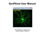
1
Product Manual Radius™ 384-Well Cell Migration Assay Catalog Number CBA-127 384 assays CBA-127-5 5 x 384 assays FOR RESEARCH USE ONLY Not for use in diagnostic procedures Introduction Cell migration is a highly integrated, multistep process that orchestrates embryonic morphogenesis, tissue repair and regeneration. It plays a pivotal role in the disease progression of cancer, mental retardation, atherosclerosis, and arthritis. The initial response of a cell to a migration-promoting agent is to polarize and extend protrusions in the direction of the attractant; these protrusions can consist of large, broad lamellipodia or spike-like filopodia. In either case, these protrusions are driven by actin polymerization and can be stabilized by extracellular matrix (ECM) adhesion or cell-cell interactions (via transmembrane receptors). The Radius™ Cell Migration Assay Kit utilizes a proprietary 384-well plate to monitor the migratory properties of cells. Each plate well contains a 0.68 mm non-toxic, biocompatible hydrogel spot (Radius™ Gel) where cells cannot attach. When adherent cells are seeded in the Radius™ Cell Migration well, they attach outside of the Radius™ Gel coated area. Once firm cell attachment is achieved, the hydrogel is quickly removed to expose a cell-free region to study cell migration/closure. This format provides a robust in vitro system to measure 2-D cell migration, screen potential inhibitors and study cytoskeleton reorganization events. Additional features of the Radius™ Cell Migration Assay: • • • • • • • • • • Exclusive coating method which produces consistent gel spot size - 0.68 mm diameter ± 0.014 mm (2%) Qualitative, quantitative, real-time or endpoint analysis Radius™ Cell Migration Plate does not need to be used all at once – unused wells can be used in future experiments, up to a total of 3 migration experiment cycles Compatible with all cell stains, dyes, and labels Complete migration zone closure achievable in 15-30 hours (actual time is cell line dependent) Analyze by phase contrast or fluorescence microscopy Compatible with HCS/HCI instrumentation Adaptable to liquid handling equipment Optimized for 10X magnification (entire Radius™ Gel migration area is viewed in a 10X magnification field) Radius™ Gel removal is controlled and extremely fast (migration starts almost simultaneously between wells) The Radius™ Cell Migration Assay Kit is designed for High Content Analysis applications or imaging software and is adaptable to liquid handling equipment. Each kit provides sufficient quantities to perform 384 migration tests. 2 Assay Principle 3 Related Products 1. CBA-100: CytoSelect™ 24-Well Cell Migration Assay (Boyden Chamber Assay, Colorimetric) 2. CBA-101: CytoSelect™ 24-Well Cell Migration Assay (Boyden Chamber Assay, Fluorometric) 3. CBA-106: CytoSelect™ 96-Well Cell Migration Assay (Boyden Chamber Assay, Fluorometric) 4. CBA-110: CytoSelect™ 24-Well Cell Invasion Assay (Boyden Chamber Assay, Colorimetric) 5. CBA-120: CytoSelect™ 24-Well Wound Healing Assay 6. CBA-125: Radius™ 24-Well Cell Migration Assay 7. CBA-126: Radius™ 96-Well Cell Migration Assay 8. CBA-130: CytoSelect™ 96-Well Cell Transformation Assay (Soft Agar Colony Formation) Radius™ 384-well Cell Migration Plate Dimensions Well Volume Well Depth Well Diameter - Top Well Diameter - Bottom Plate Length Plate Width Plate Height A1 Row Offset A1 Column Offset Well Center to Well Center Spacing Flange or Skirt Height Well Bottom Elevation Well Bottom Thickness Well Bottom Area 112 µL 11.43 mm 3.63 mm 2.67 mm 127.8 mm 85.5 mm 14.2 mm 8.99 mm 12.12 mm 4.5 mm 6.096 mm 2.79 mm 0.635 mm 0.056 cm2 Kit Components 1. Radius™ 384-well Cell Migration Plate (Part No. 112701): One 384-well black-walled, clearbottom tissue culture treated plate with each well containing one Radius™ non-toxic, biocompatible hydrogel spot (384 gel spots total per plate) 2. Radius™ Gel Pretreatment Solution (Part No. 112502): One Sterile Bottle – 13.0 mL 3. Radius™ Wash Solution (Part No. 112702): One Sterile Bottle – 25.0 mL 4. Radius™ Gel Removal Solution, 75X (Part No. 112703): One Sterile Tube – 500 µL 5. DAPI Fluorescence Stain, 1000X (Part No. 112002): One Amber Tube – 30 µL 6. Fixation Solution (Part No. 122402): One Bottle – 20.0 mL 4 Materials Not Supplied 1. Adherent migratory cell lines and culture medium 2. Multichannel pipette or liquid handling system 3. Cell culture incubator (37ºC, 5% CO2 atmosphere) 4. Inverted light microscope with a digital camera 5. Optional: Microscope stage or cage incubator 6. Optional: Inverted fluorescence microscope with DAPI filter (350nm/470nm) 7. Optional: Imaging Software for measuring cell migration 8. Optional: HCS/HCI Instrument Storage Upon receipt, aliquot and store the Radius™ Gel Removal Solution and DAPI Fluorescence Stain at -20ºC (avoid multiple freeze/thaw cycles), and transfer the Fixation Solution to 4ºC. All other kit components should be stored at room temperature until the kit’s expiration date. Preparation of Reagents • • 3X Radius™ Gel Removal Solution: Just prior to use, prepare a 3X Radius™ Gel Removal Solution by diluting the provided 75X stock 1:25 in complete culture medium. 1X DAPI Fluorescence Stain: Just prior to use, prepare a 1X DAPI Fluorescence Stain by diluting the provided 1000X stock 1:1000 in PBS. Assay Protocol (Must be under sterile conditions) I. Pretreatment of Radius™ Migration Plate 1. Under sterile conditions, remove the Radius™ 384-well Cell Migration Plate from its packaging. Note: If wells of the plate have already been used for a previous experiment, the unused wells must be completely moisture-free before proceeding. Please allow the covered plate to dry at room temperature for 1 hour. Each plate can handle up to 3 migration experiment cycles. 2. Determine which wells will be assayed (it is recommended that all samples be tested in triplicate). Slowly add 25 µL of Radius™ Gel Pretreatment Solution to each well by carefully pipetting down the wall of the well. Note: The Radius™ Gel Pretreatment Solution should not be added to any wells that will not be used immediately. 3. Cover the plate and incubate at room temperature for 20 minutes. 5 4. Carefully aspirate the Radius™ Gel Pretreatment Solution from the wells. Do not allow wells to dry. Note: Avoid potential damage to the Radius™ Gel Spot (located near the center) by aspirating from the edge of the well. 5. Slowly add 50 µL of Radius™ Wash Solution to each well. Proceed to the Cell Seeding Section below. Note: Wells can remain in the Radius™ Wash Solution for up to 1 hour. II. Cell Seeding 1. Harvest and resuspend cells in culture medium at 0.5 - 2 x 10⁵ cells/ml. Note: Cell seeding density is highly cell line dependant, factoring in cell size, spreading and division. Ideally, the desired monolayer confluency at the start of migration (after Radius™ Gel Removal step) should be 80-90%. Optimization is recommended and can be done in a standard 384-well cell culture plate prior to migration assays. 2. Carefully aspirate the Radius™ Wash Solution from the wells (step 5 above). Do not allow wells to dry. Note: Avoid potential damage to the Radius™ Gel Spot (located near the center) by aspirating from the edge of the well. 3. Slowly add 50 µL of the cell suspension to each well by carefully pipetting down the wall of the well. 4. Transfer the plate to a cell culture incubator for 4-24 hours to allow firm attachment/spreading. Take care to avoid shaking or bumping the plate. III. Radius™ Gel Removal 1. Carefully remove the Radius™ Migration Plate from cell culture incubator. 2. Prepare sufficient 3X Radius™ Gel Removal Solution for all wells by diluting the stock 1:25 in culture medium (See Preparation of Reagents Section). 3. Slowly add 25 µL of 3X Radius™ Gel Removal Solution to each well. 4. Transfer the plate to a cell culture incubator for 30 minutes to allow complete gel removal. 5. Carefully aspirate the 1X Radius™ Gel Removal Solution/media from each well and replace with 50 µL mL of fresh media. Do not allow wells to dry. Agents that inhibit or stimulate cell migration may also be added directly to the wells. 6. At this point, pre-migration images may be captured with an inverted microscope, imaging software, or HCI/HCS instrument. 7. Transfer the plate back to the cell culture incubator/microscope stage incubator during the migration process. 8. Monitor the migration closure by endpoint or real-time analysis. For time course experiments, live cell compatible dyes or labels are required (e.g. Calcein AM, GFP, RFP). For endpoint experiments, fixed cell detection should be used (Cell Stain, DAPI, TRITC-phalloidin). 6 IV. (Optional) DAPI Fluorescence Labeling 1. Aspirate the media from the wells and add 50 µL mL of Fixation Solution to each. 2. Allow the cells to fix for 10 minutes at room temperature. Aspirate and discard the solution. 3. Carefully wash each well 3 times with 75 µL of PBS. 4. Prepare sufficient 1X DAPI Fluorescence Stain for all wells by diluting the stock 1:1000 in PBS (See Preparation of Reagents Section). 5. Add 50 µL of 1X DAPI Stain to each well to be stained. 6. Incubate 15 minutes at room temperature. 7. Carefully wash each well 3 times with 75 µL of PBS. 8. Add 75 µL PBS to each well to keep cells hydrated. 9. Examine wells under an inverted fluorescence microscope with DAPI filter (350nm/470nm) Analysis of Results There are a number of software programs available for the analysis of cell migration images. One of these is CellProfiler™ Cell Image Analysis Software offered free-of-charge by the Broad Institute*. You may find more information on this program online at www.cellprofiler.org. In order to analyze data from our Radius™ Cell Migration Assays, the CellProfiler™ software must be customized. For your convenience, we have developed add-ons that will customize the program for you. Please visit our website at www.cellbiolabs.com, type “Cellprofiler” in the Search box, and follow the instructions to download the appropriate add-on. *CellProfiler™ is a trademark of the Broad Institute. There is no relationship between Cell Biolabs, Inc. and the Broad Institute. Cell Biolabs offers these add-ons as a courtesy to our customers who wish to analyze data obtained using our Radius™ Cell Migration Assays. 7 Example of Results The following figures demonstrate typical results with the Radius™ 384-well Cell Migration Assay Kit. One should use the data below for reference only. This data should not be used to interpret actual results. Figure 1. Various Detection Methods with Radius™ Cell Migration Assay. HeLa cells were seeded overnight. After Radius™ Gel removal, cells were stained with DAPI, Cell Stain Solution (not included in kit), and Calcein AM (not included in kit) according to the assay protocol. 8 100 80 75 % Inhibition % Closure 100 50 25 0 0.00 60 40 20 0 0.01 0.10 1.00 0.00 Cytochalasin D (uM) 0.01 0.10 1.00 Cytochalasin D (uM) Figure 2. Inhibition of NIH3T3 Cell Migration by Cytochalasin D. NIH3T3 cells were seeded at 5,000 cells/well overnight. After Radius™ Gel removal, the cells were allowed to migrate for 24 hrs in the presence of various Cytochalasin D concentrations. 9 References 1. Ridley AJ, Schwartz MA, Burridge K, Firtel RA, Ginsberg MH, Borisy G, Parsons JT, Horwitz AR. (2003) Science 302, 1704-9. 2. Horwitz R, Webb D. (2003) Curr Biol. 13, R756-9. 3. Lauffenburger DA, Horwitz AF. (1996) Cell 84, 359-369. Warranty These products are warranted to perform as described in their labeling and in Cell Biolabs literature when used in accordance with their instructions. THERE ARE NO WARRANTIES THAT EXTEND BEYOND THIS EXPRESSED WARRANTY AND CELL BIOLABS DISCLAIMS ANY IMPLIED WARRANTY OF MERCHANTABILITY OR WARRANTY OF FITNESS FOR PARTICULAR PURPOSE. CELL BIOLABS’ sole obligation and purchaser’s exclusive remedy for breach of this warranty shall be, at the option of CELL BIOLABS, to repair or replace the products. In no event shall CELL BIOLABS be liable for any proximate, incidental or consequential damages in connection with the products. Contact Information Cell Biolabs, Inc. 7758 Arjons Drive San Diego, CA 92126 Worldwide: +1 858-271-6500 USA Toll-Free: 1-888-CBL-0505 E-mail: [email protected] www.cellbiolabs.com 2011: Cell Biolabs, Inc. - All rights reserved. No part of these works may be reproduced in any form without permissions in writing. 10