Download Ultrasound Compatible Lumbar Puncture/ Epidural Simulator
Transcript
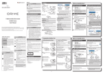
M43E
Ultrasound Compatible Lumbar Puncture/
Epidural Simulator
Instruction
Manual
Contents
● Please read
General information
P.1
● Preparation
P.2-6
Before training
● Training
● After training
P.7
P.8-10
Ultrasound compatible puncture block is anatomically correct and offers realistic image of ultrasound.
It includes lumbar vertebrae, spinous process, superior articular process, transverse process, epidural
space and spinal dura mater.
● Features
1. Ultrasonic landmarks of lumbar spine can be visualized.
2. Replacement parts are durable for multiple procedures.
3. This simulator can be positioned in the upright or lateral position.
4. Translucent block makes the needle trace visible.
5. The lumbar region model provides a platform for wide training opportunities, by adding
interchangeable training blocks for landmark and fluoroscopic procedures.
* Epidural space is designed to be filled with water for better sonographic imaging. Skip this process if you
would like to perform loss of resistance technique.
Please read
General information
Set includes
Before your first use, ensure that you have all components listed below.
⑦
①
⑧
②
③
1
1
③ Ultrasound skin cover
1
④ Irrigation bottle
⑤ Syringe
1
⑥ Stand
1
⑦ Irrigator bag
1
⑧ Support base
1
(Lateral position)
⑨ Support base
(Sitting position)
Storage case (no image)
Instruction manual
⑥
④
① Lower torso manikin
② Ultrasound puncture pad
1
1
1
⑨
⑤
Caution: Please use the ultrasound skin cover (11348-230) for training. Skin cover for landmark procedure
(11348-150) is not ultrasound compatible.
*
The lower torso manikin ①
is the same model as that of the M43B: Lumbar Puncture Simulator II.
DOs and DON’T s
DO s
DON’Ts
Handle with care
Never wipe the phantom or models with
thinner or organic solvent.
The materials for phantom and models are special
composition of soft resin. Please handle with care
at all times.
Do not mark on the the components except
for the ultrasound skin cover with pen or leave
printed materials contacted on their surface.
Ink marks on the models will be irremovable.
Cleaning and care
Please clean the phantom completely every time
after you finish the training. Remaining ultrasound
gel on the phantom may deteriorate the product.
*Skin marking on the ultrasound skin cover can be removed.
Keep the training set at room temperature,
away from heat, moisture and direct sunlight.
Please note: The color of the phantom may change overtime. However, please be assured that this
is not deterioration of the material and the ultrasonic features of the phantom will
stay unaffected.
1
Preparation
Before training
1 Remove the puncture pad
*Product is packaged as shown on the left.
1. Disengage the magnets and remove the skin
cover from the manikin.
2. Remove the clear plastic sheet slowly from one
side of the puncture pad.
* Be sure to hold the magnets.
* Work with both hands. Disengage the remaining to magnets one by one. Do not pull by the skin.
3. Hold the puncture pad by the plastic edges and
remove from the lower torso manikin.
2
Preparation
Before training
2 Prepare the stand
1. Assemble the stand.
2. Hang the irrigator bag on the stand.
3 Preparation of the US puncture pad (Lumbar puncture)
Ultrasound puncture pad
Plug for the syringe
Plug for the syringe (for subarachnoid space)
(for epidural space)
Connector to the
irrigator bag
(for subarachnoid
space)
Connector to the
irrigation bottle
1. Connect the tip of the tube from the
irrigator bag to the central connector
at the head-end of puncture pad.
*Insert the tube completely to prevent
the tube from detaching during training
sessions.
Put the tube tip
up to here.
pad
3
Preparation
Before training
2 Preparation of the US puncture pad (Lumbar puncture)
2. Fill the irrigator bag with water until the
surface reaches to the 200mL line.
3. Connect the syringe to the plug for subarachnoid space
on the opposite side of the irrigator bag tube. Insert the
syringe, turn clockwise and lock in place.
200mL
Syringe
Insert and turn clockwise
pad
4. Open the clamp.
Open the clamp
Detach
5. Pull the syringe slowly to draw water into the pad. To remove any air bubbles remaining in the tube,
tilt the pad and continue to pull the syringe slowly. Also check from the back side of the pad to see
if any bubbles remain in the tube.
6. Close the clamp.
7. Turn the syringe counter-clockwise to unlock
and remove from the puncture pad.
Close the clamp
4
Preparation
Before training
3 Preparation of the US puncture pad (Epidural)
* Epidural space is designed to be filled with water for better sonographic imaging. Skip this process if you
would like to perform loss of resistance technique.
1. Open the lid and fill the bottle with water till 1/3
of its hight. Then reset the lid. Be sure to twist the
bottle part for opening and closing the lid.
*Twisting the lid of the bottle may damage the tubing.
2. Connect the tip of the tube from the irrigation
bottle to the connector at the head-end of the
puncture pad.
3. Connect the syringe to the plug for epidural
space on the puncture pad. Insert the syringe,
turn clockwise and lock in place.
Detach
Syringe
pad
4. Pull the syringe slowly to draw water into the pad.
To remove any air bubbles remaining in the pad
or the tube, tilt the pad and continue to pull the
syringe slowly.
5
Insert and turn clockwise
5. Turn the syringe counter-clockwise to unlock and
remove from the puncture pad.
Preparation
Before training
4 Set the puncture pad in the manikin
1. Hold the puncture pad by the plastic edges and insert
into the lower torso manikin.
* Be careful not to fold the tubing.
5 Attach the skin cover to the lower torso manikin
1. Noting the marks at the back of the skin cover
(L,R,↑), attach it to the lower torso manikin.
2. Attach two corners of the skin cover to the manikin
with the magnets. As you lay the skin down, press out
any remaining air between the skin and the puncture
pad and attach the other two corners.
* The orientation marks come on the inner side of the skin cover.
*Overtime, the surface of the
pad may become less adhesive.
When this occurs, apply
ultrasound gel on the pad
surface to help the pad attach
to the skin.
*Be sure to wash and remove all
remaining gel after training/
before storage. Dried gel may
damage the products.
6
Training
2 Place the lower torso manikin on a support base
ventral side
The higher side
comes to the head
end of the manikin.
head end
dorsal side
head end
higher
lower
The sitting position support base is
designed to come the front end of the
lower torso manikin.
ventral side
ventral side
The higher side
comes to the ventral
side of the manikin.
dorsal side
higher
lower
3 Training session
Training Skills
○ Ultrasound-guided
lumbar puncture
epidural anaesthesia
○ CSF collection and CSF pressure measurement
○ Ultrasound-guided
The ultrasound skin cover may be marked with a surgical skin
marker, and markings can be removed.
Do not mark on the the components except for the ultrasound
skin cover. Ink marks will be irremovable.
● Lumbar
puncture
Please be sure to leave the clamp to the
irrigator bag opened during training.
* Refill the irrigator bag with more water
once the water has run out.
● Epidural
anaesthesia
Epidural space is designed to be filled with water for better
sonographic imaging. Skip this process if you would like to
perform loss of resistance technique.
Successful performance can be confirmed by observing the
injected air flows into the irigation bottle.
Caution: Please use the ultrasound skin cover (11348-230) for training. Skin cover for landmark procedure (11348-150) is not
ultrasound compatible.
7
After Training
1 After Training
1. Wipe off any remaining ultrasound gel from
the skin or manikin using wet tissues.
2. Remove the ultrasound skin cover.
a. Disengage the magnets at the two adjacent
corners. Be sure to hold the magnets and not
to pull by the skin.
b. Peel the skin cover slowly off of the manikin.
3. Hold the puncture pad by the plastic edges and
remove from the lower torso manikin.
8
* Work with both hands. Disengage the remaining to
magnets one by one. Do not pull by the skin.
After Training
1 After Training
●
Remove water from subarachnoid space
5. Open the clamp.
4. Connect a syringe filled with air to the plug for
subarachnoid space of the puncture pad.
6. Slowly push air from the syringe into the water-filled
pad until all water has been removed from the pad.
●
7. Close the clamp.
Remove water from epidural space
8. Discard any remaining water in the irrigation
bottle. Twist the bottom part of the bottle to
remove the top.
9. Connect a syringe to the plug for epidural space of the puncture pad. Pull the syringe slowly until
all water has been removed from the pad.
9
After Training
1 After Training
●
Cleaning and Storage
10. Remove the tube attached to the puncture pad.
11. Please be sure to wipe all remaining ultrasound
gel on the ultrasound puncture pad with wet
tissues.
12. Cover the surface of the puncture pad with
a clear plastic sheet and press out any remaining
air between the sheet and pad.
* Please gently wipe off any remaining gel on the pad and wash with water if necessary. Please do not rub on the
pad harshly, as it may damage the surface and affect the ultrasound images.
* Remove all remaining gel before storing. Any remaining gel substance will create a layer on the pad surface and
affect the ultrasound images.
13. Discard any remaining water in the irrigator
bag.
14. Store the puncture pad with the protective
sheet in the lower torso manikin.
10
Caution
Do not mark on the phantom with pen or leave printed materials contacted on its surface.
Ink marks on the phantom will be irremovable.
The contents of this manual are subject to change without prior notice.
No part of this instruction manual may be reproduced or transmitted in any form without permission
from the manufacturer. Please contact manufacturer for extra copies of this manual which may contain
important updates and revisions.
Please contact manufacturer with any discrepancies, typos, or mistakes in this manual or product
feedback. Your cooperation is greatly appreciated.
URL:http://www.kyotokagaku.com
e-mail:[email protected]
Worldwide Inquiries & Ordering
Kyotokagaku Head Office and Factories:
15 Kitanekoya-cho, Fushimi-ku, Kyoto, 612-8388, JAPAN
Tel: +81-75-605-2510 Fax: +81-75-605-2519
Kyotokagaku USA Office: USA and Canada sales and services
3109 Lomita Boulevard, Torrance, CA 90505-5108, USA
Tel: 1-310-325-8860 Fax: 1-310-325-8867
2015.01


![[PDF:0.6MB]](http://vs1.manualzilla.com/store/data/005664304_1-5124c75bfbffa9dc3657cce05bb0013f-150x150.png)

