1
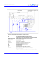
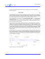
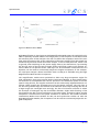
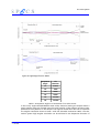
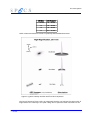
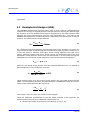
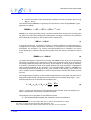
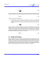
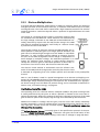
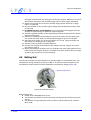
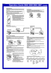
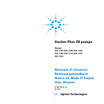
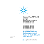
Spectrometer
Figure 7: Medium Area Mode
High Magnification is (see Figure 8: High Magnification Mode page 15) particularly suitable for spatially resolved studies. The image plane of the sample is in coincidence with
the entrance plane of the analyzer. The user can define the acceptance area of the analyzer with the entrance slit. As the trajectories of electrons emitted from the sample are
influenced by electrical fields around the sample, T1 has a fixed potential, which is set
to ground, after switching on the power supply. Due to lens aberrations, rays entering
the lens far away of the lens axis at larger angles could find a path to the analyzer entrance. With an Iris aperture these “bad rays” can be eliminated. Furthermore the Iris
Aperture can be used to continuously adjust the angular acceptance of the analyzer.
For small spot analysis a lateral resolution down to 100 μm is available using the High
Magnification Mode and the Iris aperture.
The magnification modes were optimized to allow very large acceptance angles for
high transmission from point sources (Point Transmission Modes). In these modes angular resolution is accomplished with the Iris aperture in the diffraction plane of the lens
system. Using thie Iris, the angular resolution can be continuously adjusted between ±1°
and ±9° while keeping the acceptance area on the sample constant. The rays close to
the lens axis (paraxial rays) are focused at the Gaussian focus. Rays entering the lens at
a larger angle are converged more strongly. The disc of minimum confusion is where
the envelope of emergent rays has its smallest diameter. Slight under-focusing of the
lens displaces the disk of least confusion to the image plane. Thereby a higher angular
acceptance is achieved but the spatial resolution is worsened. This makes the Point
Transmission Modes most suitable for AES, ISS and synchrotron studies. As with the
High Magnification Modes, the Iris Aperture can be used to continuously adjust the angular acceptance.
14
PHOIBOS