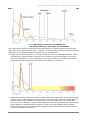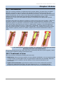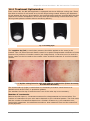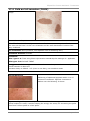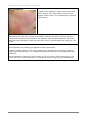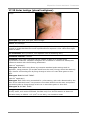
1
Ellipse I2PL and Laser products Clinical workbook Ellipse IPL Clinical workbook: Clinical workbook Ellipse A/S Agern Allé 11 DK-2970 Hørsholm Denmark www.ellipse.com For further information please e-mail [email protected] Telephone +45 45 76 88 08 Fax +45 45 76 88 89 Release date: 26-09-2013 An imprint of Ellipse A/S. © 2013 Ellipse A/S. All rights reserved. ENG This clinical workbook is additional information for background knowledge and additional training. This is not the Instructions for Use (IFU). The official intended use and treatment guide is stated in the printed manuals sent with your particular system. DK Denne kliniske brugervejledning indeholder yderligere oplysninger og baggrundsviden og anvendes for yderligere uddannelse. Dette er ikke bruger manualen. Den officielle påtænkte anvendelse og behandlings guide (Brugermanual) er angivet i de trykte manualer sendt med systemet. ES Este manual clínico es información adicional a la formación recibida y los conocimientos adquiridos anteriormente.El uso adecuado y una guía de tratamientos están detallados en los manuales enviados con este sistema. FR Ce manuel clinique constitue une information additionnelle pour la formation et la connaissance de fond. L’utilisation officielle prévue et le guide de traitement sont définis dans les manuels imprimés accompagnant ce système. IT Questa manuale clinico contiene informazioni aggiuntive per la conoscenza di base e la formazione. La destinazione d'uso e la guida ai trattamento sono indicate nei manuali stampati inviati con questo sistema. PL W niniejszym podręczniku klinicznym znajdują się dodatkowe informacje uzupełniające wiedzę podstawową i szkolenia. Oficjalne przeznaczenie oraz wskazówki do przeprowadzania zabiegów znajdują się w drukowanych wersjach instrukcji użytkownika wysyłanych razem z systemem. NL Dit klinisch werkboek bevat extra informatie met betrekking tot achtergrond kennis- en training. De officiële gebruikershandleiding wordt bij het systeem geleverd. Page 2 1MAN8219–A03– ENG Ellipse IPL Clinical workbook: Table of contents Chapter 1 Introduction......................................................................7 1.1 How to use this workbook ............................................................................... 7 1.2 Sources of Information.................................................................................... 7 1.3 Ellipse A/S ..................................................................................................... 7 1.4 Quality policy ................................................................................................. 8 1.5 Legal notice ................................................................................................... 8 Chapter 2 Anatomy ...........................................................................9 2.1 Skin anatomy ................................................................................................ 9 2.2 Layers of the skin ........................................................................................... 9 2.3 Skin type..................................................................................................... 11 2.4 Sun Tan ...................................................................................................... 12 2.5 Hair anatomy ............................................................................................... 12 2.6 Hair growth ................................................................................................. 13 Chapter 3 Light-tissue Interaction ..................................................17 3.1 Physics – light as electromagnetic radiation (EMR) ........................................... 17 3.2 Laser and intense pulsed light (IPL) ................................................................ 19 3.3 Light – tissue interaction ............................................................................... 19 3.4 5-ALA and protoporphyrins ............................................................................ 25 Chapter 4 Successful treatments with Ellipse I2PL products ...........27 4.1 Introduction................................................................................................. 27 4.2 Pre-treatment care ....................................................................................... 27 4.3 Pre-treatment information ............................................................................. 27 4.4 General check list immediately before treatment .............................................. 29 4.5 General check list for treatments .................................................................... 29 4.6 General check list after treatment .................................................................. 31 4.7 Choice of applicator ...................................................................................... 31 4.8 Contraindications ......................................................................................... 32 Chapter 5 Treatment factors determined by system used ...............35 5.1 Introduction – the Ellipse Plus Range .............................................................. 35 5.2 General ....................................................................................................... 35 5.3 Normal mode – all Ellipse systems .................................................................. 38 5.4 Expert mode ................................................................................................ 38 Chapter 6 Hair removal ...................................................................43 6.1 Introduction................................................................................................. 43 6.2 Causes of unwanted hair ............................................................................... 43 6.3 Hair removal methods ................................................................................... 43 1MAN8219–C08–ENG Page i Ellipse IPL Clinical workbook: 6.4 Hair removal using Ellipse I2PL ....................................................................... 44 Chapter 7 Vascular lesions ..............................................................55 7.1 Introduction................................................................................................. 55 7.2 Causes of vascular lesions ............................................................................. 55 7.3 Treatment of vascular lesions ........................................................................ 55 7.4 Removal of vascular lesions using Ellipse I2PL .................................................. 56 7.5 Successful vascular treatment using Ellipse I2PL products .................................. 57 Chapter 8 Photo rejuvenation .........................................................63 8.1 Introduction................................................................................................. 63 8.2 Treatment of sun damaged skin ..................................................................... 63 8.3 Treatment of sun-damaged skin using Ellipse I2PL ............................................ 63 8.4 Successful treatment of sun-damaged skin with Ellipse I2PL ............................... 65 Chapter 9 Pigmented lesions ...........................................................73 9.1 Introduction................................................................................................. 73 9.2 Successful treatment of pigmented lesions with Ellipse I2PL ............................... 74 Chapter 10 Acne ..............................................................................79 10.1 Introduction .............................................................................................. 79 10.2 Treatment of acne ..................................................................................... 79 10.3 Leeds acne grading scale ............................................................................ 80 10.4 Acne treatment using Ellipse I2PL ................................................................ 81 Chapter 11 Rosacea ........................................................................85 11.1 Introduction .............................................................................................. 85 11.2 Treatment of rosacea ................................................................................. 86 11.3 Rosacea treatment using Ellipse I2PL ............................................................ 86 Chapter 12 Poikiloderma of Civatte .................................................89 12.1 Introduction .............................................................................................. 89 Chapter 13 Photodynamic Therapy (PDT) .......................................91 13.1 Introduction .............................................................................................. 91 13.2 Ellipse as an approved light source. ............................................................. 91 13.3 PDT treatment – additional contraindications. ............................................... 92 13.4 PDT treatment of actinic keratosis using Ellipse I2PL....................................... 93 13.5 PDT-enhanced treatment of acne vulgaris using Ellipse I2PL ............................ 93 13.6 PDT-enhanced rejuvenation using Ellipse I2PL ............................................... 94 Chapter 14 Nd:YAG Vascular Treatments ........................................95 14.1 Introduction .............................................................................................. 95 14.2 Treatment of telangiectasias, venulectasias and reticular vessels ..................... 95 14.3 Treatment of Venous Lakes and resistant Port Wine Stain ............................... 96 14.4 Treatment Optimization .............................................................................. 97 Page ii 1MAN8219–C08– ENG Ellipse IPL Clinical workbook: Chapter 15 Onychomycosis with Nd:YAG ........................................99 15.1 Introduction .............................................................................................. 99 Chapter 16 Desired and adverse effects ........................................103 16.1 Introduction ............................................................................................ 103 16.2 Therapeutic window ................................................................................. 103 16.3 Hair removal (HR+, HR-L+ and HR-D+ applicators) ....................................... 104 16.4 Vascular treatment (VL+ or PR+ applicator) ................................................. 104 16.5 Pigment treatment (PL+ applicator) ............................................................ 105 16.6 Photo rejuvenation (PR+ or VL+ applicator) ................................................. 106 16.7 Acne treatment ....................................................................................... 107 Chapter 17 Treatments .................................................................109 17.1 Recommended Use of I2PL handpieces for SPT+ ........................................... 109 17.2 Recommended Use of I2PL handpieces for I2PL, PPT and MultiFlex.................. 110 17.3 Becker’s nevi .......................................................................................... 113 17.4 Café au lait macules (CALM) ..................................................................... 115 17.5 Cherry angioma....................................................................................... 117 17.6 Dark circles under the eyes ....................................................................... 119 17.7 Diffuse redness ....................................................................................... 121 17.8 Ephelides ................................................................................................ 123 17.9 Epidermal melasma ................................................................................. 125 17.10 Facial telangiectasias............................................................................. 127 17.11 Hemangioma of infancy ......................................................................... 129 17.12 Hemosiderin ......................................................................................... 131 17.13 Leg telangiectasias................................................................................ 133 17.14 Phlebectasia ......................................................................................... 135 17.15 Poikiloderma of civatte .......................................................................... 137 17.16 Port wine stain ..................................................................................... 139 17.17 Pyogenic granuloma .............................................................................. 141 17.18 Rosacea ............................................................................................... 143 17.19 Seborrheic keratosis.............................................................................. 145 17.20 Solar lentigo (plural lentigines) ............................................................... 147 17.21 Spider angioma .................................................................................... 149 17.22 Sun-damaged (Pigmented) skin .............................................................. 151 17.23 Stretchmarks (striae) and scars (cicatrices) ............................................. 153 17.24 Venous lakes ........................................................................................ 155 Chapter 18 Appendices .................................................................157 18.1 Photo documentation ............................................................................... 157 18.2 Woods lamp ............................................................................................ 158 1MAN8219–C08–ENG Page iii Ellipse IPL Clinical workbook: 18.3 Additional treatment notes for patients with skin types 3-5 ........................... 158 18.4 List of drugs that may cause photosensitivity in patients .............................. 159 Chapter 19 Glossary of terms ........................................................165 Table of figures Fig 1. Cross-section through skin ............................................................................ 10 Fig 2. Fitzpatrick skin type scale.............................................................................. 11 Fig 3. Section of human hair ................................................................................... 12 Fig 4. Cross section of hair in skin ........................................................................... 13 Fig 5. Hair follicles placed three together .................................................................. 13 Fig 6. The hair growth cycle.................................................................................... 13 Fig 7. Richard-Meharg hair growth table ................................................................... 15 Fig 8. Example of a Mechanical Wave....................................................................... 17 Fig 9. Spectrum of electromagnetic radiation ............................................................ 17 Fig 10. Laser types and wavelengths generated ....................................................... 18 Fig 11. Contrast between laser and IPL ................................................................... 19 Fig 12. Lights interaction with tissue ....................................................................... 19 Fig 13. Penetration depth depends on spot size ........................................................ 20 Fig 14. Absorption as a function of wavelength ........................................................ 21 Fig 15. Cooling rates............................................................................................. 22 Fig 16. Absorption spectra of chromophores showing commonly used lasers for treatments................................................................................................ 23 Fig 17. Typical single mode filtering ........................................................................ 23 Fig 18. Example of dual mode filtering .................................................................... 24 Fig 19. Spectra of haemoglobin (Hb) and oxyhemoglobin (HbO2) absorption ............... 24 Fig 20. Spectra of Protoporphyrin IX (PpIX) absorption ............................................. 25 Fig 21. Wavelengths of Ellipse I2PL applicators ......................................................... 31 Fig 22. Comparison of surface area in small and large targets .................................... 36 Fig 23. Estimated relaxation as a function of target diameter ..................................... 36 Fig 24. Intensity for treatments of different sized targets, with the same energy setting 37 Fig 25. Standard pulse timing for treating pigmented lesions (PL applicator)................ 39 Fig 26. Long pulse with low intensity (15 J/cm2) ...................................................... 40 Fig 27. Too short a pulse time with high intensity (15 J/cm2) ..................................... 41 Fig 28. Choice of hair removal applicator for different Fitzpatrick skin types ................ 45 Fig 29. Default pulse times .................................................................................... 46 Fig 30. Treated area with perifollicular oedema and erythema. Note hairs on surface that are exploding out of the hair follicle due to too short pulse time. ..................... 46 Fig 31. Perifollicular oedema and erythema ............................................................. 48 Fig 32. General erythema ...................................................................................... 48 Fig 33. Erythema in a skin type 4 patient ................................................................ 48 Fig 34. Applicator with pressure ............................................................................. 49 Fig 35. Hair treatment intervals ............................................................................. 50 Fig 36. Therapeutic window (hair removal) .............................................................. 51 Fig 37. Consent form (hair removal) ....................................................................... 54 Fig 38. Classification of treatable vascular lesions..................................................... 55 Fig 39. Comparison of Vascular Treatment Applicators .............................................. 56 Page iv 1MAN8219–C08– ENG Ellipse IPL Clinical workbook: Fig 40. Vascular Skin Reaction Speed a) Before Shot ; b) <1second after ; c) 2 seconds after ............................................................................................ 59 Fig 41. Port Wine Stain showing longer lasting blueing of vessel ................................ 60 Fig 42. Applicator lightly touching the skin .............................................................. 60 Fig 43. Therapeutic window (vascular) .................................................................... 61 Fig 44. Applicators for sun-damaged skin ................................................................ 64 Fig 45. Adverse reaction to PR+ applicator in suntanned skin ..................................... 65 Fig 46. Instant Colour change and erythema in diffuse redness (left). Progressive change in colour of pigment a) Pre-treatment b) after 1 minute c) after 12 hours ......... 67 Fig 47. Reduction in energy compared to “normal” facial areas .................................. 68 Fig 48. Applicator lightly touching the skin .............................................................. 69 Fig 49. Consent form (skin treatments) ................................................................... 71 Fig 50. Applicators for pigmented lesions ................................................................ 74 Fig 51. Progression of skin reaction with VL+ or PR+ applicator a) pretreatment b) after 1 minute 2) after 12 hours ............................................... 75 Fig 52. Progression of skin reaction with PL applicator .............................................. 76 Fig 53. Applicator in contact with skin ..................................................................... 76 Fig 54. A normal pore becomes blocked by a blackhead, leading to increased bacteria production and inflammation. ...................................................................... 79 Fig 55. Grades of facial acne .................................................................................. 80 Fig 56. Grades of acne on the back......................................................................... 80 Fig 57. Grades of acne on the chest ........................................................................ 80 Fig 58. Applicator in contact with skin without pressure ............................................ 83 Fig 59. Applicator in contact with skin without pressure ............................................ 87 Fig 60. Before and After: Poikiloderma of Civatte, 1 treatment. .................................. 89 Fig 61. Approval Process for PDT treatments (simplified) ........................................... 92 Fig 62. Patient Suitability for PDT treatments .......................................................... 93 Fig 63. Distance between Nd:YAG shots .................................................................. 95 Fig 64. Ellipse Vein Gauge ..................................................................................... 96 Fig 65. Cooling tips............................................................................................... 97 Fig 66. Spider telangiectasia with and without compression (Photo Courtesy Prof Michael Drosner)................................................................................................... 97 Fig 67. Nails infected with onychomycosis (pictures courtesy of Prof. Peter Bjerring and Prof. Agneta Troilius) ............................................................................... 100 Fig 68. %age cure rates of different medications .................................................... 100 Fig 69. Ephelides under normal light (left) and UV light from a Woods lamp (right) .... 158 1MAN8219–C08–ENG Page v Ellipse IPL Clinical Workbook: Anatomy Chapter 1 Introduction 1.1 How to use this workbook The clinical workbook is a separate document from the various operator’s manuals supplied with your Ellipse system. The clinical workbook is designed to be used for revision, internal training and ongoing reference. The operator’s manuals show the official intended use and treatment guide for the system and applicators. Much of the information contained in this workbook is common to all systems; examples include anatomy, physics, light-tissue interaction and contraindications to treatment. Likewise, a treatment such as hair removal is available on all systems. This information will be shown on a white background. Information aimed at users of Ellipse Light, Ellipse Light SPT, and Ellipse Light SPT+ systems will be shown on a green background. These systems perform effective treatments, but offer less choice of treatments offered. Users of Ellipse MicroLight systems have their own Clinical Workbook. The Ellipse MultiFlex+, Ellipse MultiFlex, Ellipse I2PL+, Ellipse I2PL, Ellipse Flex PPT and Ellipse Flex offer a wider range of treatments of Intense Pulsed Light treatments and greater choice of applicators, and information for these systems is shown with a blue background. Information about Nd:YAG laser treatments and Pulse Definition Mode (only available on Ellipse MultiFlex and Ellipse MultiFlex+) will be shown on an orange background. Ellipse systems are sold in over 50 countries, and some local names of treatments differ from the English meaning of the word. This document refers to the English name throughout. 1.2 Sources of Information Light-based treatments of the skin evolve constantly, and system users are strongly recommended to keep themselves up-to-date with the latest information. This can be done by becoming active in a professional association and attending its meetings, and by reading the various journals available to you. For updated information on Ellipse Products consider joining the Ellipse4Physicians or Ellipse4Beauticians user clubs, details of which can be found at the Ellipse website www.ellipse.com. The website also has a selection of Clinical Abstracts and links to training videos and other support tools, as well as a newsletter sign-up form. Our Facebook page www.facebook.com/Ellipse.Denmark will also provide information on Ellipse Activities and customers around the world. Finally our blog, www.ellipselasers.wordpress.com/ provides regular updates on treatments and clinical information. 1.3 Ellipse A/S Ellipse A/S has been producing and selling second generation intense pulsed light (I2PL) systems since 1997. The Ellipse systems are used for treatment of skin diseases and for treatment of cosmetic disorders. Ellipse also produces lasers used in aesthetic dermatology. Ellipse A/S has earned a good reputation for developments using medical high technology. The Ellipse family of products is developed in close co-operation with leading international dermatologists. All products are subjected to clinical testing and to regulatory approvals in accordance with the medical device directive requirements for CE marking (European Union), FDA clearance (USA) as well as ANVISA (Brazil), SFDA (Peoples Republic of China) KFDA (Korea), MHLW (Japan) and other national approvals as required. This ensures good practice in design and production, leading to a safer and more effective system. 1MAN8219–C08–ENG Page 7 Ellipse IPL Clinical workbook: Introduction 1.4 Quality policy Ellipse A/S is dedicated to development and manufacture of high-quality medical devices and wants to be the end customers’ preferred partner supplying innovative, clinically proven, safe and effective solutions founded on light-based skin treatments. Ellipse guarantees high quality by following international regulations and industry standards and is certified in accordance with ISO9001 and ISO 13485. It observes the rules and applies their intentions in its daily activities. 1.5 Legal notice This workbook is not to be photocopied or distributed. The information it contains is for use by Ellipse system users only. Ellipse A/S, manufacturer of Ellipse systems, recommends serious initial study and regular review of this manual and suggests its inclusion in training of operators. The Ellipse systems listed above are medical devices, which in the hands of the user, can be used for treating patients. Safe and efficient treatments are achieved if the device is used based on clinical judgments and proper patient selection. Neither Ellipse A/S as the manufacturer, nor the company selling the product can take responsibility for safe and efficient treatments; this responsibility at all times rests with the user of the system. Notice on Intellectual Property Rights Ellipse A/S has been granted the following patents: US 8.226.696: Main claim is directed to IPL combining water as a filter with a UV filter. Patent issued July 2012. (European patent pending – ref 98304722.6/ EP0885629) EP2027827: Main claim is skin cooling by directional flow of cold air. Patent issued February 2012. (US patent pending - ref US12/193.845) Page 8 1MAN8219–C08– ENG Ellipse IPL Clinical Workbook: Anatomy Chapter 2 Anatomy 2.1 Skin anatomy Skin characteristics Skin is the largest human organ. It is the body’s waterproof protective barrier against mechanical, environmental and chemical damage. When this barrier is injured, it can repair itself. It contains sweat glands that produce moisture, which evaporates to cool the body surface. It is also important for the production of Vitamin D. Skin contains nerves that carry the messages of touch and of pain. Usefully these messages can be confused. For example, in treatments that target melanin pressure is used to perform the treatment, and this reduces the sensation of discomfort. In treatments targeting haemoglobin, pressure is not used, so patients may benefit by using a stress-ball during treatments. Skin regulates body temperature by dilating the blood capillaries when warm and constricting them when cold. This regulates the amount of blood flowing to the skin surface. Most Ellipse treatments do not require active cooling (the exception is an Nd:YAG laser treatment on the Ellipse MultiFlex) and vascular treatments are more efficient when carried out with a normal blood flow. 2.2 Layers of the skin The skin is made up of three layers (see Fig 1). ● the epidermis. ● the dermis. ● the hypodermis (also called the sub cutis or subcutaneous tissue). The epidermis (the outer layers of the skin) has an external layer of dead cells. These cells are constantly being shed and replacement cells gradually migrate to the surface from below. The outermost layer (of dead skin) is called the stratum corneum, and contains little water. If the stratum corneum is too thick, this can interfere with light based treatments, and so users should consider gentle exfoliation prior to commencing a course of light-based treatment. In the lowest layer of the epidermis, the pigment melanin is produced. The dermis is composed of a network of collagen, elastic fibres, nerves, fat, blood vessels and the bases of sweat glands and hair follicles. Its purpose is to supply the epidermis with nutrition, to provide mechanical strength and to defend the body against infection. The hypodermis is the innermost and thickest layer of the skin. It is a loose network of connective tissue. Specific cells for fat storage (adipocytes) fill the spaces in this network. Females have more fat storage in the sub cutis than males. The adipocytes provide energy, insulation and also act to protect the underlying tissue from injury. Importantly, the boundary between the dermis and epidermis is not flat, but is made up of a series of finger-like projects, called rete ridges. This increases the surface area of the epidermis and in turn increases the nutrient supply from blood vessels. It also makes the skin less fragile. 1MAN8219–C08–ENG Page 9 Ellipse IPL Clinical workbook: Anatomy Fig 1. Cross-section through skin Page 10 1MAN8219–C08– ENG Ellipse IPL Clinical Workbook: Anatomy 2.3 Skin type The standard method of classifying skin type is the Fitzpatrick scale, from 1 to 6. Skin type 1 Typical skin type definition Red-blond hair. Blue-green eyes. Very light skin. Skin reaction on over exposure to UV light Always burns. Does not tan. Comments on skin type Pale, sometimes mixed with freckles. Usually admit that they burn. Normally the first consideration for average light skin (aside from obvious skin type 1). 2 Light to medium hair. Light to medium eyes. Light to medium skin. Usually burns. Seldom tans. 3 Medium hair. Medium to dark eyes. Medium to olive skin. Burns Moderately. Usually tans. Usually do not recognize that they burn moderately if exposure is moderate. Will comment that they “Can get a good tan with care”. 4 Dark hair. Dark eyes. Dark olive to light brown skin. Burns mildly. Moderate browning. Consider they tan easily. Will rarely burn from moderate exposure in northern climates. Often surprised when they get a “little” sunburn while visiting higher intensity locations. 5 Dark hair. Dark eyes. Dark skin. Seldom burns. Deep browning. Burning requires no previous exposure for months, then exposure to very high levels of UV intensity (100+ on the SUNSOR scale – a sunny summer day in Florida). 6 Dark hair. Dark eyes. Very dark skin. Does not burn. No change in colour. Individuals have very good pigmentation that affords exceptional protection in ultraviolet light. Often deny that they burn but admit to turning pink and needing to take care in sun. Fig 2. Fitzpatrick skin type scale The type or colouring is determined by the amount of pigment (melanin) contained in the skin cells, and this is determined by heredity and race. Skin type is not changed by exposure to sunlight, nor by age. As well as determining the default energy of a treatment, skin type also determines the length of time taken to produce a reaction to that treatment. Darker skin types respond more slowly to intense pulsed light and their therapeutic window for treatment (the zone where a beneficial result occurs without the risk of side-effects) is smaller. This means that the risk of side-effects is higher in darker skin types. It is essential to determine the skin type accurately to assess both the risk of side effects and the response time. The following points should be noted: ● Hair colour may be artificial. ● The patient may be wearing coloured contact lenses. ● The apparent skin colour may be the result of cosmetics or sun exposure. The actual skin colour is better determined by parting the hair and examining the scalp, since hair normally protects the skin of the scalp from suntan. 1MAN8219–C08–ENG Page 11 Ellipse IPL Clinical workbook: Anatomy Explain to the patient when asking about skin reaction to UV light (sunlight) that you need to know the response of unprotected skin, without sunblock. Using a scored questionnaire (such as that available for download from Ellipse4Physicians) can make it easier to determine the Fitzpatrick skin type. If a patient is of mixed ancestry, it can be difficult to determine the skin type, and users should use the higher of two possible types. 2.4 Sun Tan Ellipse grades the degree of suntan as: None; Light; Medium; Medium-Heavy; Heavy. In the 30 days before and 30 days after treatment, clients should avoid the sun, or use sun-protection (SPF 30) when sun exposure is unavoidable. This is especially true for darker skinned clients, because sun-exposure increases the risk of post-inflammatory hyperpigmentation (see Chapter 11). Limiting sun exposure in vascular treatments is especially important as recent exposure (especially exposure recent enough to still give a slight feeling of warmth) can cause significant problems as the warmth increases the size of blood vessels in the skin. This increases the amount of the chromophore haemoglobin present. 2.5 Hair anatomy As seen in Fig 3, a human hair shaft consists of a cortex, made up of cortical cells, in which the pigment melanin is located. The cortex is covered by a cuticle, a single layer of keratin cells. In addition, large hairs contain a medulla, a central (often hollow) core which gives strength to the hair. Fig 3. Section of human hair Within the skin (Fig 4) the hair shaft is surrounded by a hair sheath, and is connected to the hair root, which is embedded in a pit in the skin called the follicle. The root is nourished by a small artery in the papilla. The follicle is typically located between 1mm and 5mm under the surface of the skin. This depth is dependent on the body site (1mm on upper lip; 5mm on bikini line). Follicles are often placed in groups of three (see Fig 5). Normally only 1 of the 3 follicles is in the growing phase at any one time. Page 12 1MAN8219–C08– ENG Ellipse IPL Clinical Workbook: Anatomy Fig 4. Cross section of hair in skin Fig 5. Hair follicles placed three together 2.6 Hair growth The life cycle of the human hair has four phases. Normally, the hair follicles are not synchronized and therefore hairs in close proximity to one another may be in different phases at the same time. However, hormonal influences following a birth or severe infection may cause synchronization of the hair growth cycle. The four growth phases are: ● Anagen phase: the growing phase of the hair. During early anagen, new hair grows from the hair follicle, pushing out old hair from the hair shaft. ● Catagen phase: the hair bulb is degraded, cell growth and melanin production stops, and the hair bulb is moved upwards to the skin surface. ● Exogen phase: the hair falls out of the hair follicle. ● Telogen phase: the typical resting phase. The length of the telogen phase depends on the anatomical site. Anagen Catagen Exogen Telogen Early Anagen Anagen Fig 6. The hair growth cycle 1MAN8219–C08–ENG Page 13 Ellipse IPL Clinical workbook: Anatomy Many different factors influence the growth of human hairs. These include age, ethnicity, medication, hormone levels and body site. Differences may be found in the length, coarseness (and colour) of body hair; the differences are most apparent when comparing one site with another, but also exist within a single given site. The number of visible hairs depends on the number of hairs in their anagen (growing) phase. The longer the anagen phase lasts, the longer the hair can get. This is why the hair on the scalp grows much longer than the hairs on other parts of the body. For example, at any time 85% of the hairs on the scalp will be in the anagen phase, and only 15% will be in the resting phase. For scalp hair the anagen phase can be as long as 6 years while the resting period is only 3 - 4 months. In contrast, the hairs on the arms have an anagen phase of only 3 months before they revert to a resting phase. At any one time, only 20% of those hairs may be growing. Hair growth data for the various body sites is detailed in the following table. While it is not possible to determine an exact optimum treatment interval guaranteed to work for each patient, the table has been used to provide recommended treatment intervals for various body sites shown in chapter 6. Page 14 1MAN8219–C08– ENG Ellipse IPL Clinical Workbook: Anatomy Site Rest % Growth % Other % Rest time Growth time Depth of follicle (mm) No of follicles per cm2 Growth per day (mm) 350 0.35 3-5 0.16 2 – 2.5 No of follicles HEAD 3-4 months 2-6 years 10 3 months 4-8 weeks 85 15 3 months 4-8 weeks Cheeks 30 50 50 – 70 Beard / Chin 20 70 10 months 1 year 500 Moustache / Upper lip 35 65 6 weeks 16 weeks 500 Scalp 13 85 Eyebrows 90 Ear 2 880 0.32 Head and scalp total 1million 0.38 2–4 3–5 2–4 BODY Axillae (armpit) 70 30 3 months 4 months Trunk 65 0.3 70 0.3 3.5 – 4.5 425,000 2 – 4.5 Pubic Area 70 30 3 months 4 months 70 3.5 – 5 Arms 80 20 18 weeks 13 weeks 80 0.3 220,000 2 – 4.5 Legs – Thighs 80 20 24 weeks 16 weeks 60 0.21 370,000 2.5 – 4 Breasts 70 30 65 0.35 3 – 4.5 Fig 7. Richard-Meharg hair growth table 1MAN8219–C08–ENG Page 15 Ellipse IPL Clinical Workbook: Light-tissue Interaction Chapter 3 Light-tissue Interaction 3.1 Physics – light as electromagnetic radiation (EMR) Put simply, light is just a form of energy, and all laser or intense pulsed light treatments use this energy to do work. The light travels in waves, similar to waves travelling on the surface of a pond. Fig 8. Example of a Mechanical Wave The Waves on a pond (like sound waves) are a mechanical wave, but light (just like X-Rays or Radio Waves) are slightly different: they are electromagnetic waves (waves of pure energy). The various types make up the electromagnetic spectrum, or spectrum of electromagnetic radiation. This spectrum of electromagnetic radiation stretches from gamma rays to radio waves, as shown below. There are three ways to describe the waves, according to type: Fig 9. Spectrum of electromagnetic radiation Gamma Rays and X-Rays are described in terms of their energy (electron volts) Visible and Infrared Light, used by all intense pulsed light sources and most medical lasers are described by their wavelength (nanometres). Microwave and Radio waves are described by their frequency (Hertz). 1MAN8219–C08–ENG Page 17 Ellipse IPL Clinical workbook: Light-tissue Interaction Lasers and intense pulsed light sources mainly use the visible to near infrared part of the spectrum (Fig 10), with wavelengths from 400 nanometres (nm) to 1 200nm. There are exceptions such as the CO2 laser (10 600nm) and Er:YAG laser (2 940nm). Laser Wavelength (nm) Excimer 193 / 308 / 311 Argon 488 / 514 Copper vapour / bromide 510 / 578 KTP 532 Pulsed dye 570 / 585-595 / 600 APTD 577 / 585 Ruby 694 Alexandrite 755 Diode 800 / 810 / 915 / 940 / 1450 /1470 / 1530 LED various Nd:YAG 1064 / 532 / 1320 / 1440 Er:Glass 1540 Holmium 2100 YSGG 2790 Er:YAG 2940 CO2 10600 Fig 10. Laser types and wavelengths generated While light travels in waves, the waves are made up of “particles” of light called photons. Whenever a photon is absorbed, its energy is changed into heat (photothermolysis), and this absorption of light energy is the basis for all light/tissue interaction. Light energy delivered to the skin is measured in Joules and is best expressed as the energy delivered to a certain area (also called the fluence) measured in J/cm². The length of time that the light is exposed to the skin is called the pulse duration, and is measured in milliseconds (ms). Page 18 1MAN8219–C08– ENG Ellipse IPL Clinical Workbook: Light-tissue Interaction 3.2 Laser and intense pulsed light (IPL) In recent years the distinction between laser and intense pulsed light has become less clear in the minds of the users, and the term “laser” hair removal is increasingly used for any light-based treatment. Technically, the light emitted by an Intense Pulsed Light system differs from the light emitted by a laser as indicated below: IPL Systems Lasers Polychromatic (a band of wavelengths) Monochromatic (only one wavelength) Non-coherent (waves are not in phase) Coherent (waves are always in phase) Defocused light Parallel light (directional) Fig 11. Contrast between laser and IPL The light emitted by IPL systems is not one single wavelength, but covers a spectrum of different wavelengths. By using different filters it is possible to allow through light that matches the requirements for different treatments, so that one system can be used for more than one application. 3.3 Light – tissue interaction Four different processes can occur when light hits the skin. These are: ● Reflection. ● Transmission. ● Scattering. ● Absorption. Fig 12. Lights interaction with tissue 1MAN8219–C08–ENG Page 19 Ellipse IPL Clinical workbook: Light-tissue Interaction Reflection and transmission Light energy can be reflected from the surface of the skin (like from a mirror) or transmitted straight through it (like through glass). Neither reflected nor transmitted light has any effect on the tissue being treated. Approximately 70% of the light that normally hits the skin surface is immediately reflected, although this figure varies with wavelength. The remaining light is absorbed by the tissue or scattered within it. Using a light guide and optical coupling gel greatly reduces the amount of reflection, and typically results in reflection being reduced to only 5%. Always hold the applicator perpendicular to the skin, as this means that more light goes straight down into the skin and reduces the amount of reflected light. Outside the treatment room, more light is reflected off water, sand or snow when the sun is low on the horizon so sunscreen is important even at these times. Scattering Scattering is a change of direction of light particles (photons) compared to the original direction. For example, light can bounce off collagen fibres, blood vessels and other structures or molecules in the skin. Scattering does not deposit any energy in the tissue. When scattering takes place some of the photons leave the main light beam before absorption has taken place, which means a loss of effectiveness. Since the light is defocused some of the photons will also enter the skin at an angle to the main beam (even when the applicator is pointed directly downwards (as described above). Small spot sizes result in relatively more light being scattered away from the main beam, leading to a higher degree of loss. This means that the penetration depth of a smaller spot size is less than for big spot sizes. Small Spot Size Fig 13. Penetration depth depends on spot size Page 20 1MAN8219–C08– ENG Ellipse IPL Clinical Workbook: Light-tissue Interaction Absorption A chromophore is a chemical within the skin that absorbs light of certain wavelengths. When a photon is absorbed, it surrenders its energy to the chromophore or to water. The photon no longer exists and its energy is changed to heat in the chromophore. This is similar to the way in which a dark surface becomes warmer than a white surface when exposed to sunlight. The most important substances in the skin that are capable of selective light absorption are: ● Melanin (found in the epidermis, hair and hair follicles). ● Oxyhemoglobin + haemoglobin (found in the blood). ● Water. Protoporphyrin is also capable of selective light absorption. This chemical is produced naturally in quite low levels, and in much higher levels by the acne bacterium P acnes. The levels of protoporphyrin in non-acne skin can be heightened by the introduction of the chemical 5-ALA into the skin. Absorption by protoporphyrin will be discussed later. The degree of absorption by each chromophore depends on the wavelength of the light used. Fig 14. Absorption as a function of wavelength Penetration depth The penetration depth of the light into the tissues also depends on the wavelength. Longer wavelengths (600 – 1000nm) penetrate deeper into the tissues, but wavelengths above this figure are predominantly absorbed by water in the skin. Penetration depth of the light into the skin can be compared to the depth that sunlight penetrates into water. At a low depth of water, all colours can be seen, but the deeper one dives, the less light penetrates and a colour change is observed. 1MAN8219–C08–ENG Page 21 Ellipse IPL Clinical workbook: Light-tissue Interaction Thermal relaxation time and photothermolysis Light entering the skin will only have an effect when it is absorbed by a chromophore and converted into heat. The biological effect following absorption is dependent on the temperature achieved. When light is absorbed, all chromophores present are heated. Heat loss begins immediately, as heat is conducted to all adjacent tissues. The rate of the heat loss varies according to the thermal relaxation time (TRT) of the tissue. The TRT is defined as the time it takes for the tissue to cool down to the ambient temperature following heating. Large objects cool more slowly than small ones. Additionally, the Thermal Damage Time (the time taken to destroy a target) increases with the size of the target. Structures with a short TRT take a shorter time to cool once heated. Structures with a long TRT take a longer time to cool once heated. Fig 15. Cooling rates How warm the target becomes depends on the active heating of the chromophore as it absorbs light energy and the passive cooling as heat is conducted of heat to the surrounding tissue. The overall effect of damage to tissue in response to the absorption of light is called photothermolysis. Selective photothermolysis The aim of selective photothermolysis is to selectively heat up and destroy a specific target without damaging the surrounding tissues. To achieve selective photothermolysis three parameters must be controlled: ● The wavelength (or waveband) of the light is selected so that the light energy is absorbed by the chosen chromophore. ● The duration of the light pulse is selected to ensure the target is lethally damaged with minimal conduction of energy into the surrounding tissue. ● The right energy level is chosen to create enough heat to lethally damage the target within the given pulse time. By controlling these three parameters, selective destruction of the target chromophores can be achieved without injuring the surrounding tissue. Page 22 1MAN8219–C08– ENG Ellipse IPL Clinical Workbook: Light-tissue Interaction Fig 16. Absorption spectra of chromophores showing commonly used lasers for treatments The xenon lamp of an IPL system emits a wide spectrum of different wavelengths at the same time (from approximately 240 – 1200nm). This light is then filtered to match different applications. Two different types of filtering can be distinguished: ● Single mode filtering as used in the first generation intense pulsed light systems. These systems use a coloured filter to remove energy below a given wavelength (on the left side of the absorption curve). Depending on the type of filter the emitted light typically starts somewhere between 510 and 720nm and goes up to 1200nm. Fig 17. Typical single mode filtering ● Ellipse dual mode filtering uses filters on both sides of the selectively emitted wavelength band to remove wavelengths below and above a chosen range. Shorter wavelengths are stopped using a coloured filter (selected by the choice of applicator to be below 400, 530, 555, 600 or 645nm). Longer wavelengths are removed by passing the light through water (before it reaches the skin surface) to filter out all wavelengths above 950nm, which otherwise would lead to unspecific heating of the epidermis and increase the risk of adverse effects such as burns. 1MAN8219–C08–ENG Page 23 Ellipse IPL Clinical workbook: Light-tissue Interaction Water filtering is effected by circulating cooling water around the flash lamp and the filters in the hand piece of the Ellipse system. For selected applications additional filters at 720nm or 750nm are used to remove light above these wavelengths. Fig 18. Example of dual mode filtering Since Ellipse Intense Pulsed Light products can be used to treat a range of conditions, the light waveband used is determined by the targets you wish to treat. Haemoglobin and oxyhemoglobin Fig 18 above shows the absorption of oxyhemoglobin only. This is to keep the illustration clearer. The curve for haemoglobin is similar. Note there are differences between the curves as illustrated in Fig 19 below (please note that the vertical axis is different). Most noticeably, there are peaks in haemoglobin absorption at 433nm and 556nm. Fig 19. Spectra of haemoglobin (Hb) and oxyhemoglobin (HbO2) absorption Page 24 1MAN8219–C08– ENG Ellipse IPL Clinical Workbook: Light-tissue Interaction 3.4 5-ALA and protoporphyrins Although the Photodynamic therapy is covered later in this workbook, it is relevant to include the absorption spectrum for protoporphyrin IX (PpIX) the chemical to which 5-ALA is converted, here. Fig 20. Spectra of Protoporphyrin IX (PpIX) absorption PpIX absorbs light in the region of 400-700nm and has 5 absorption peaks at 410, 505, 540, 580 and 635nm. Absorption is highest at the shortest wavelength and higher wavelengths show gradually less absorption. Upon absorption of light, PpIX reacts in such a way that it transfers energy to a nearby oxygen molecule, making it unstable and in turn causing local cellular damage. The processes and use of this are discussed later. 1MAN8219–C08–ENG Page 25 Ellipse IPL Clinical Workbook: Successful treatments with Ellipse I2PL products Chapter 4 Successful treatments with Ellipse I2PL products 4.1 Introduction Regardless of the actual treatment to be carried out, there are a number of procedures that need to be carried out to continuously achieve problem-free treatment. Successful treatments require careful pre-treatment care, appropriate treatment and post treatment follow up. 4.2 Pre-treatment care Pre-treatment care involves patient selection and identification, informing the patient and preparing the patient for treatment. Patient selection and identification are the keys to achieve good results with Ellipse I2PL products. Many factors influence the success of the treatment, the most important being skin type, amount of suntan in the area to be treated, and size and colour of the target. Ellipse I2PL products will not allow every treatment to be carried out on every skin type, as in certain combinations, the risk of side-effects outweigh the rewards of the treatment. Skin types are identified according to the Fitzpatrick skin type scale, mentioned in chapter 2. Your Ellipse system will use this information to determine if a treatment can be carried out and if so, to provide a default (starting energy). The degree of suntan is a simple way of classifying the amount of melanin present in the skin relative to the skin type of the patient. If the “background” melanin level is too high, it can cause adverse effects such as a burn during treatment (which also makes the treatment more painful) especially in darker-skinned patients. It is better to delay treatment of a patient if the degree of suntan is too high. It is NEVER worth cheating by attempting to treat a patient outside the permitted range of skin type/suntan – the most likely result is that you will injure your client. The size and colour of the target will be discussed in the following chapters, as this is very treatment-specific. 4.3 Pre-treatment information It is important to inform patients about the entire treatment procedure. Information creates awareness of the treatment procedure and will give the patient realistic ideas regarding what is achievable. Remember that the patient is involved in the process and can influence the results positively or negatively. If the patient has the right expectations before treatment, patient satisfaction will be greater after it. It is important to inform the patient of the following: ● The number of necessary treatments and what result is achievable. It helps to have good before-and-after photographs available. Ellipse “Plus” systems have an inbuilt set of before and after photographs, for this purpose. ● The time-scale of the treatment procedure, what are the immediate clinical effects, the visible effects over the next day or so, and when a final result can be seen. ● The need to avoid sun exposure before and after treatment. Recent sun-exposure by a tanned or darker-skinned patient can easily hide erythema (redness), but all patients need to be aware it is equally important to avoid active sunbathing and unintentional sun-exposure (from any outdoor activity). ● Other pre and post treatment optimization. - Some physicians prescribe a bleaching cream to lighten the skin prior to the treatment. This reduces the absorption of light energy in the epidermis (i.e. the absorption by background melanin) during treatment. 1MAN8219–C08–ENG Page 27 Ellipse IPL Clinical workbook: Successful treatments with Ellipse I2PL products - Similarly some physicians will recommend exfoliating the skin to reduce the thickness of the stratum corneum prior to commencing treatments. This may be done using a gentle exfoliating cleanser such as those products that contain a low percentage of Alpha Hydroxy acid. Equally it may involve a more strenuous regimen that involves a peeling solution such as glycolic or lactic acid or a physical exfoliation such as micro dermabrasion. Although these methods can elevate results above the norm, Ellipse A/S cannot give recommendations on specific methodologies or brands. It is clear that appropriate pretreatments can improve results but it is important that the patient can tolerate such pre-treatments. Equally, the pre-treatment must be timed so that it cannot cause complications (such as the presence of unwanted erythema) when the intense pulsed light treatment is carried out. As a general guide, 2-3 weeks should normally elapse between the end of pre-treatment and the commencement of I2PL treatment to allow the skin sufficient recovery time. ● The need for accurate medical history. This needs to include existing and previous medical conditions and details of any prescription or proprietary medicines and health supplements the patient is taking. The principal reason for doing this is to ensure that none of the conditions or medications will cause photosensitivity, increase erythema or otherwise interfere with the treatment procedure. Sources of information on drugs, and currently known contraindications to treatment are discussed in Section 4.8. ● A history of previous surgical, cosmetic and aesthetic procedures, including any implants (silicone or surgical), fillers, tattoos or permanent make up, and light-based procedures. Normally silicone implants have no effect upon treatment, but metal pins, plates or screws implanted during previous surgery can absorb heat, and cause discomfort. Many fillers cause no problems, but areas with some fillers, or fat injections may be affected by heat from light exposure. The light from Ellipse I2PL equipment cannot remove tattoos, but these will absorb some light energy, and may discolor and/or cause serious burns. ● Possible adverse effects see chapter treatments. As a general note, it is important to explain the reasons for the questionnaire to the patient and to discuss and confirm their answers. It is also important that they sign or initial each page, as well as the completed form. Sometimes, patients are tempted to forget or ignore questions that they feel may preclude them from treatment. It is also important that the patient signs an informed consent form confirming they have been informed of the risks and expected results of treatment. A sample informed consent form can be found in this Clinical Workbook, but users are recommended to have their own form designed in order to take account of local legislation. Note that some local or national authorities may have additional restrictions on treatment – as may your insurer – and it is important to work within the regulatory framework. Nervous patients or those with reservations about the effectiveness of light-based treatments may benefit from receiving test-shots as part of the consultation process. Unless the patient is especially nervous (when they may feel reassured by having a low-energy shot on an area such as the lower arm) such shots should be relevant to the treatment area, should initially be at the default energy, and should progress to energy appropriate for the treatment. Some insurers require that darker-skinned patients have a test shot days or weeks before the treatment commences. In all cases it is important that the patient use a diary and/or a camera to record their reaction to treatment as an aid to memory, as this allows the user to control the patient reaction over a longer period. Page 28 1MAN8219–C08– ENG Ellipse IPL Clinical Workbook: Successful treatments with Ellipse I2PL products 4.4 General check list immediately before treatment Before starting the treatment remember to: ● Remove the patient’s make-up. Cosmetics can disguise the true appearance of the skin, and can also absorb some of the energy needed to carry out the treatment. This can lead either to an inefficient treatment or an increased risk of side-effects. ● Take pictures of the treatment area for documentation purposes. With the patient’s permission, this can be used for before and after documentation, and it also provides evidence of how successful a particular treatment has been (useful as patients sometimes forget how they looked before treatment). ● Shave the treatment area if required, and remove all shaved hairs from the skin. Hair above the skin surface will absorb energy from the applicator and can burn into the crystal light guide (damaging the applicator) or can burn onto the skin surface, causing pain or skin burns during treatment. ● Mark the border of the treatment area with a red pen or a white wax pencil. Do not use other colours, as they will absorb the light and cause pain (in worst case burns). ● Determine the skin type based on Fitzpatrick skin type scale, the degree of suntan in the area to be treated and the size and colour of the target. Accurately enter this onto the Ellipse system. ● Apply optical coupling gel. The amount of gel is determined by the treatment and will be discussed later. Coupling gel supplied by Ellipse has been tested and found suitable for treatments and it is important that any other gel considered for use is tested before use. The gel should not liquefy nor heat up when intense pulsed light is fired through it, as this could injure the patient. ● The operator, patient and any onlookers should wear appropriate eye protection. ● It is suggested that patients are given a stress ball to squeeze during the treatment, as this will reduce the feeling of discomfort. 4.5 General check list for treatments Information specific to individual treatments will be included as part of the information for that treatment. The following is a general guide to all treatments. ● Even if the patient has previously received a test shot during consultation, start the treatment proper by performing another test shot. Always perform the test shot, based on the recommended settings and observe the patient’s reaction – both in terms of discomfort and skin reaction. This takes account of any small changes in applicator output that may exist. A change in sensitivity may be hormonal, or it may be the sign of a fever or unintentional sun-exposure. The test shot should be relevant to the treatment site, but should not be prominent. Lack of reaction does not automatically mean an ineffective treatment, but an appropriate skin reaction gives a greater likelihood of a better result, so energy can be increased after the test shot as appropriate. ● Counting backwards from three to one before releasing the light energy generally has a beneficial psychological effect, as patients know when the shot will come – however, some patients respond negatively to the “countdown”, so it is better to discuss this with the individual patient. ● EMLA cream or other topical anaesthetic preparations are seldom used and can influence the quality of the light penetration into the skin. A cold compress (gauze soaked in cold water) or similar may be applied to the skin after treatment. ● Optional: An air cooling device such as a Zimmer cooler may be beneficial for the patient during vascular procedures. 1MAN8219–C08–ENG Page 29 Ellipse IPL Clinical workbook: Successful treatments with Ellipse I2PL products ● Ensure that the amount of gel is appropriate to the treatment. The amount should be sufficient to ensure that it does not dry out during the treatment, but not so great as to cause a build-up of gel on the side of the crystal light guide. Note that the gel should not contain air bubbles. ● Always place the light guide perpendicular to the skin surface. This ensures that the largest amount of the crystal is in contact with the gel and therefore that the maximum amount of light energy penetrates the skin surface. It also reduces the risk of leaving untreated strips on the skin. ● Ensure that the sides of the crystal light guide are kept free from gel. This reduces the risk of light being emitted from the side of the prism instead of the bottom of the prism. A build-up of gel on the sides can result in the patient getting “zebra stripes” after treatment. ● Each shot should overlap the previous one by approximately 10%. Do not slide the applicator across the treatment area, but lift it and place it down again. This offers two advantages. - You avoid a build-up of optical coupling gel on the side of the applicator. - You leave a “footprint” of the area you have treated in the gel. This makes placing the subsequent shots easier and avoids the risk of either missing or double-treating an area of the skin. ● Start in the treatment area least sensitive to pain, on the face, this is far from the nose and upper lip. More generally areas further away from bone are less sensitive. ● Never treat the same area immediately following a first shot. To determine the correct energy for a particular treatment of a particular patient, move the applicator to a new position each time. If you need to shoot the same area again, maybe at a higher energy that gives the desired skin reaction, allow the skin to rest for approximately 1 minute between the shots. ● Be careful not to select a too high-energy setting. In darker skinned or suntanned patients, a skin burn may only show up several hours after a treatment ● After the first few shots, it is recommended to examine the skin and the target closely for reaction. The skin reaction should remain fairly constant throughout the treatment; adjust the treatment parameters if the reaction changes. Continue to observe the skin reaction throughout the treatment. ● Response to treatment varies from one patient to another. It is recommended to ask the patient to “score” any discomfort - using a system of 0-10, where 0 is no discomfort and 10 is the worst possible pain imaginable. After the initial surprise of the first few shots, the score should normalize (at a figure that often reflects patient sensitivity). The patient should always be asked to report if the score changes at any time during the treatment. An increase in the score may mean that the energy should be decreased. ● Some treatments such as hair removal or treatment of sun-damage may involve treating different anatomical areas or areas where underlying bone may reflect some of the light. When treating over a bony area, or a sensitive area, it is important to lower the energy – most often by 1-1.5 J/cm2 – to avoid side effects. ● Always encourage your patients to ask for a few seconds break during the treatment if they feel they need it. Most often, intense pulsed light treatment is optional treatment and the experience should be made as comfortable as possible for the patient. Page 30 1MAN8219–C08– ENG Ellipse IPL Clinical Workbook: Successful treatments with Ellipse I2PL products If pigmented lesions can be seen within the area of a treatment, consider the consequences of the treatment. The pigment will also absorb the light. Try to position the applicator so that the pigmented lesion is not within the spot size or adjust the energy used. It may be possible to cover the pigmented area with a wet white cloth, wet gauze or non-absorbent white paper. 4.6 General check list after treatment ● After the final shot the optical coupling gel has to be removed and the skin surface dried with a soft cloth. ● Slight erythema (redness) is often noted and will normally disappear in a period ranging from a few hours to two days. A cold compress placed against the skin may be comforting for the patient immediately after the treatment. Mild oedema (swelling) may also be noted, but disappears within a day or two. ● For a few weeks after treatment the treated area should not be exposed to sunlight. Suitable sunscreens - SPF 30 or above - should be used if exposure to sunlight is inevitable. Often, patients need to be taught how to effectively apply sun block – in terms of the amount to be used, the need for reapplication throughout the day, and when it is necessary (not just on “sunny” days, but any day on which the patient can see a shadow). ● Some patients report that their skin feels dry after the treatments. If a hydrating sunscreen is used, it will deal with both dry skin and sun exposure. If the treatment has been carried out on an area not normally exposed to the sun a good moisturizer can be used. This is especially useful if the weather is likely to change after treatment (especially if cold wind – which will dry out exposed skin) is forecast. 4.7 Choice of applicator Fig 21. Wavelengths of Ellipse I2PL applicators As well as the Nd:YAG laser applicator running at 1064nm, Ellipse I2PL applicators each have their selected waveband, as shown in Fig 21. The Ellipse system will not allow an inappropriate applicator to be used to treat a condition. There is no difference in terms of suitability for treatments between traditional and PLUS series applicators; for example an PR+ applicator will treat exactly the same conditions as the older PR applicator. The difference is in the working life of the applicator. Use of the appropriate applicators is discussed in Chapter 6 to Chapter 13 . 1MAN8219–C08–ENG Page 31 Ellipse IPL Clinical workbook: Successful treatments with Ellipse I2PL products 4.8 Contraindications It is not possible for this document to give a complete and future-proof set of contraindications. The user must also use his/her medical knowledge to research the effects of disease and genetic conditions and to research the side effects of medication, which impact upon the patient. This Clinical workbook should be read in conjunction with the User Manual for your Ellipse System. Factors acting directly against the treatment ● Patients on topical or systemic steroid medication or on non-steroidal anti-inflammatory drugs (NSAID). Ellipse treatments produce a desired low-grade inflammation. Steroids and anti-inflammatory drugs act against such inflammation reducing or negating the effectiveness of the treatment. Relative contraindications ● Patients with any disease or genetic condition causing photosensitivity to light within the range of wavelengths emitted by the Ellipse applicator used, as this increases the likelihood of a burn or violent erythema. ● Patients undergoing treatment with any medication causing photosensitivity to light within the range of wavelengths emitted by the Ellipse applicator used, as this increases the likelihood of a burn or violent erythema. Note that some natural remedies such as St John’s Wort (Hypericum perforatum) cause photosensitivity. ● Patients undergoing treatment with anti-coagulants, as these increase the risk of bruising after treatment. Note that natural remedies containing Gingko biloba have powerful anticoagulant properties. ● Patients suffering from long term diabetes, as diabetes may affect the skin healing process. ● Patients suffering from haemophilia, or other coagulopathies (clotting disorders), as these significantly increase the risk of bruising during and after treatment. ● Patients tending to produce keloids or hypertrophic scars. ● Patients with sun-tanned skin or fever. ● Patients who have smoked tobacco within 2-4 hours prior to treatment, where the target chromophore is haemoglobin. Tobacco causes contraction of the blood vessels, and thus reduces the target chromophore. ● Patients who have received gold injections where there has been some leakage / spillage into the epidermis. This presents as an area of dark grey tissue which will absorb the light energy. ● Permanent make-up (dark colours in particular) will absorb the light energy and the patient may feel a burning sensation (with burns in worst case). These areas must be excluded from treatment. This is also the case if the patient has a tattoo. ● There are isolated reports of problems caused by treating over earlier fat injections and some fillers – so users should take extra care in treating of earlier fat injections, or over unknown fillers. Page 32 1MAN8219–C08– ENG Ellipse IPL Clinical Workbook: Successful treatments with Ellipse I2PL products Contraindication notes Although not specifically contraindicated, patients who are pregnant or who have a heart pacemaker fitted are normally not treated. Hair treatment of pregnant patients is most successful after birth, as growth of body hair may be synchronized at this time. Pregnant or breast-feeding patients ARE specifically contraindicated in Acne treatment because of the medication used. Additionally, due to hormonal imbalance, it is not advisable to carry out photo rejuvenation on nursing mothers, as this may trigger melasma or other pigment disorders. If there is any uncertainty regarding a patient’s suitability for treatment, then it is important to get appropriate professional guidance. Almost all countries have their own national pharmacopeia, often online, and websites such as drugs.com can be used to identify drug brands and effects. It is important to establish if a drug causes increased sensitivity to ultraviolet light or to the visible light and near infrared light used in Ellipse treatments. Areas containing fillers or fat injections may be affected by heat from the light exposure 1MAN8219–C08–ENG Page 33 Ellipse IPL Clinical Workbook: Treatment factors determined by system used Chapter 5 Treatment factors determined by system used 5.1 Introduction – the Ellipse Plus Range Ellipse I2PL products comprise a range of machines (systems) that progressively offer more treatments and more operator control over the pulses used for those treatments. Current models are: ● Ellipse MicroLight HR: a single treatment system for hair removal. Pulse times are predetermined and only the energy (fluence) may be altered. This system is not covered by this clinical workbook. ● Ellipse Light SPT+: A system that allows the user to treat any of the range of applications purchased, simply by changing the applicators. Again, pulse times are predefined and only the energy (fluence) may be changed. ● Ellipse I2PL+: Offers a wider range of applicators (handpieces) and treatments than the above, and allows users in expert mode to change the pulses of light produced (pulse time, delay and number of pulses). ● Ellipse MultiFlex+: Allows users in expert mode to change the duration and number of pulses and users in pulse definition mode to create and save their own pulse trains comprising individual pulses of differing durations and energies. The system also works with an (optional) Nd:YAG laser. All Plus systems have a database, and an inbuilt operator manual and clinical workbook. Ellipse Light SPT, I2PL Flex PPT and MultiFlex systems are generally capable of being upgraded to an equivalent plus system. On certain models a hardware update may be required. However, it is not possible to upgrade older “Classic” Flex and Light systems. Upgrading a system to the Plus version will allow users of these systems to work with the new long-life Plus applicators which have known and lower running costs. Whichever system you use, a safe and efficient treatment of the patient is based on using the right wavelength, the right amount of energy (fluence) and the right pulse time when applying the energy to the target. Inexperienced users are recommended to use the default settings calculated by the Ellipse system, until experience has been gained. The standard default settings are conservative settings with a low risk of adverse effects, but they do not necessarily provide the best possible effect within a minimum of treatment sessions. 5.2 General Pulse time setting (ms) The removal of hair or skin lesions using an Ellipse system is based on heating the target chromophore (melanin or oxyhemoglobin) to a point where destruction is achieved without damaging the surrounding tissue. This process is known as selective photothermolysis (see chapter 3.4). It has been proved that hair follicles, blood vessels and keratinocytes containing excess melanin are destroyed if they are heated to a temperature of at least 70°C for a minimum of 1ms. When light is absorbed, the target chromophore will heat up. Heat loss from the target begins immediately as heat is conducted in all directions to adjacent tissues (thermal relaxation). Therefore, selecting the correct pulse time is important for effective treatment without skin injury. For all treatments the optimum pulse time is approximately equal to the thermal relaxation time (TRT) of the target. 1MAN8219–C08–ENG Page 35 Ellipse IPL Clinical workbook: Treatment factors determined by system used Because heat is absorbed or lost through the surface of the target, smaller targets heat up or lose heat more quickly (they simply have more surface area compared to their volume). Fig 22. Comparison of surface area in small and large targets As an example, if the small target above has dimensions of 1 unit, it has 6 sides, each of which is one square unit and a volume of 1 cubic unit. This ratio of surface area to volume is 6:1. The larger target has dimensions of 2 units, so each side has an area of 2 x 2 = 4 square units. Again there are 6 sides so the total surface area is 6 x 4 =24 square units. The volume is 2 x 2 x 2 cubic units, giving a ratio of 3:1. This means that the smaller target above will heat up or lose heat twice as quickly. Because anatomical targets are not perfect cubes, the ratio is not so simple. However the target relaxation time increases with increasing diameter, as illustrated in Fig 23. A larger target, with a larger volume, has a longer TRT than a thin one with a small volume and requires longer pulse durations, to reach the target temperature of 70°C. Therefore the pulse time has to be adjusted according to the size of the target. Time / [ms] Thermal relaxation time (TRT) 60 50 40 30 20 10 0 0 0,1 0,2 0,3 0,4 0,5 Diameter / [mm] Fig 23. Estimated relaxation as a function of target diameter If the pulse time significantly exceeds the thermal relaxation time, then too much heat will be conducted to adjacent tissues, which may cause the target not to heat up and may even lead to damage to the structures surrounding the target. Using too short a pulse time (with the correct amount of energy) will lead to a higher risk of epidermal skin burns, because the target reaches too high a temperature in too short a time. Page 36 1MAN8219–C08– ENG Ellipse IPL Clinical Workbook: Treatment factors determined by system used Ellipse Light SPT+ operates only in normal mode. This means that the Ellipse system calculates the TRT based on the entered treatment parameters and configures the pulse time setting accordingly. With I2PL+ and MultiFlex+, a suitably trained and experienced user can over-ride the pulse times suggested by the system. Guidance for these series users on how to make minor adjustments to the pulse time, and the effects of using too long or too short a pulse will be detailed for each treatment type in the subsequent chapters. Energy setting (J/cm2) When a target absorbs the emitted light, the light energy is converted into heat resulting in an increased temperature in the target. When running in normal mode, the Ellipse system calculates the required treatment settings based on the entered patient parameters (skin type, degree of suntan, chosen treatment procedure and the size or colour of the target) and the system configures the energy level and the pulse time accordingly. The energy level, stated on the screen in J/cm2, is always the total amount of energy reaching each square centimetre of the skin surface each time the applicator is fired. When treating larger targets a single pulse of light is used, but for small targets such as pigmented lesions the system will fire a double pulse of light with a short interval separating the pulses. In this case the total energy level is the sum of the energies fired. For safety reasons, the calculated default settings are placed below the upper limit of the therapeutic window. To optimize the result of the treatment, the energy setting may carefully be increased until a skin reaction is seen – or until the pain threshold of the patient is reached. Please refer to the following chapters for more information on expected skin reactions for the various applications. Note that in individuals with darker skin types (Fitzpatrick types 4-6) this skin reaction may be delayed and a test shot some hours before actual treatment is recommended. Pulse time versus risk of epidermal skin injury Melanin has a high, but falling, absorption of wavelengths from UV-light (200nm) to 900nm. It is produced in structures called melanosomes in melanocyte cells which are found in the lowest part of the epidermis, and is transferred from melanocytes to keratinocytes. The number of melanosomes will rise if there is long term exposure to UV radiation from sunlight, and melanin acts to protect the skin against sun exposure. These small melanosomes have a TRT of 1 – 2 ms, which is significantly shorter than the pulse time used for treatments (typically 10 – 50 ms). Due to this short TRT, the epidermis will always reach an equilibrium temperature during a treatment. I I I Normal 15 J/cm2 Thin 10 Time (ms) 20 30 40 15 J/cm2 10 Thick Time (ms) 20 30 40 Time (ms) 15 J/cm2 10 20 30 40 Fig 24. Intensity for treatments of different sized targets, with the same energy setting Fig 24 shows the treatment of thin, normal and thick hair, with the same energy setting (15 J/cm2). The pulse time for the three treatments is the standard setting automatically chosen by the Ellipse system. Since thin hair requires less time to heat up, the intensity I is higher to obtain the same energy output (the shaded area). 1MAN8219–C08–ENG Page 37 Ellipse IPL Clinical workbook: Treatment factors determined by system used Changing the on-screen selection from a thicker target to a thinner one, without changing the energy setting, will always lead to higher intensity. The equilibrium temperature for a target - i.e. the temperature the target reaches when it loses as much energy as it receives - is proportional to the light intensity. Therefore, for same energy setting, short pulse time will induce a higher temperature in the epidermis than a long pulse time and the risk for skin burns will therefore increase dramatically when a thin target is chosen. This strongly indicates the importance of a correct setting of pulse time as well as energy, based on the size of the target being treated. It is extremely important that the user makes an independent and analytical evaluation of the target size, as a patient’s subjective view of the hair or vessel size (or the darkness of pigment) is unreliable. Smaller targets need less energy to be treated well. Therefore the Ellipse system, in normal mode, automatically turns down the energy Level setting, when changing from a bigger to a smaller target. 5.3 Normal mode – all Ellipse systems Ellipse system calculates the amount of energy and the pulse time (the recommended starting point for treatment) based on the clinical parameters that are entered for the patient. Therefore, it is strongly recommended to examine the patient carefully, and enter the patient parameters correctly. If appropriate, remove the patient’s make up to see the areas to be treated, and ensure that the patient has not recently shaved the target hair. The calculated energy level is related to the skin type and degree of suntan. The energy level is reduced in response to a darker skin type (Fitzpatrick scale) or a darker suntan. The calculated energy level is also related to the size of the target. The energy level is reduced in response to a smaller target size (a thin hair or vessel). The calculated pulse time is related to the size of the target. The pulse time is reduced only in response to a thinner or lighter coloured target size. The default settings are shown on the system screen. 5.4 Expert mode The system administrator has to give a certain user the “privilege” of being an “expert operator”. This is because in expert mode each shot can be tailored so that it consists of several smaller light pulses (pulses per shot), each with its own selected time duration (pulse time) – and a chosen delay period (pulse delay). The Expert Mode is an option for experienced users with in-depth clinical expertise. When required these users can over-ride the default settings. It is always recommended to use the default settings regarding the compositions of the shots (the train of individual pulses and pulse delays calculated by the Ellipse system based on the clinical data entered). These are settings which have been proven to work best during clinical trials. Remember that your Ellipse system is one of the few intense pulsed light devices that can offer significant variations in the intensity of energy. Some other systems may recommend a pulse train of for example four pulses, but this is often due to the fact that the other system cannot deliver a reliable single pulse of appropriate intensity. While Expert Mode can be used to fine-tune a treatment, following clinical advice from Ellipse or an experienced Ellipse user (for example information obtained from the Ellipse4Physicians forum) it is definitely inadvisable to use expert mode to copy treatment settings from any non-Ellipse machine. Adjustment of the pulse time (experienced users only!) We recommend always basing the adjustment of the pulse time on correctly chosen patient parameters according to normal procedure. DO NOT CHOOSE A TARGET SIZE LESS THAN THAT OBSERVED IN THE PATIENT. Page 38 1MAN8219–C08– ENG Ellipse IPL Clinical Workbook: Treatment factors determined by system used If the target vessel or hair is between two standard settings always chose the thicker of the two standards and then decrease the pulse time accordingly. Decreasing the pulse time will automatically lead to a decreased energy setting, ensuring the intensity remains constant and that the energy output will still be below the upper limit of the therapeutic window. Perform test shots and wait a couple of minutes. Increase the energy level until skin reaction is seen or the pain threshold of the patient is reached. Note that in treatment of vessels, a visible reaction will normally occur in less than 1 second, so remove the applicator light guide from the skin immediately after the pulse is fired in order to see the skin reaction. Adjustment of the number of pulses The Ellipse Flex, Flex PPT and MultiFlex systems allow an Expert Operator to change the number of pulses per shot. Doing this dramatically increases the risk of skin burns, and we do NOT recommend operators change the number of pulses unless they have a deep understanding of skin-light interactions. Larger targets, for example hair or vessels respond best to a single pulse, but small targets such as acne or pigment respond better to a double pulse. For example the standard setting for treating pigmented lesions (with the PL applicator) uses two pulses. The default pulse time for each pulse is 7.0 ms, and the pulse delay is 25ms. This is much longer than the TRT of the target (the keratinocytes). The total active light emission time for this pulse train is therefore 2 x 7 ms = 14 ms and the total pulse train duration is 2 x 7 ms + 25 ms = 39 ms (Fig 25). The pulse delay does not change the active light emission time – it simply spaces the individual pulses in the pulse train. Pulse time Pulse time Pulse delay Time in ms 7 32 39 Fig 25. Standard pulse timing for treating pigmented lesions (PL applicator) Be aware of the pulse delay should be selected to be longer than the relaxation time for the target (typical 10 - 50 ms). If the pulse delay chosen is too short, the risk of skin burns will increase. Adjusting the pulse delay does not influence the energy or pulse time settings. Perform some test shots and wait a couple of minutes. Increase the energy level until skin reaction is seen or pain threshold for the patient is reached. The total energy level in J/cm2 should be selected according to the maximum tolerance of the patient. Consider both clinical skin reaction and pain threshold. As mentioned in chapter 5.4, some competitors use a pulse train consisting of 2-3 pulses spaced with a short pulse delay of up to 5ms when treating larger targets. Their idea is to allow small “non-target” structures with a low thermal relaxation time to recover between individual pulses in a pulse train. Larger target structures with a higher TRT (like hair bulbs or vessels) only lose relatively little energy in this short delay, and therefore the temperature of the bulb or vessel only drops a little before the next pulse in the pulse train is emitted. Such competitors generally use a very high fluence and wavelengths above 950nm are not filtered out. While safe, this is less comfortable than a comparative pulse from Ellipse. 1MAN8219–C08–ENG Page 39 Ellipse IPL Clinical workbook: Treatment factors determined by system used While Ellipse systems are able to perform this kind of treatment, it is not necessary and we do NOT recommend doing so, because of the risk of skin burns. Ellipse requires less energy to treat a condition, since second filter at 720nm, 750nm or 950nm means that energy is not wasted in heating water in the skin. In both theory and practice, it is safer to use one single pulse with a pulse time adjusted according to the relaxation time of the target. At all times, the user has the responsibility for the treatment based on clinical / dermatological insight and experience gained from previous treatments. Applicator lifetime The aim of using Expert and Pulse Definition Mode is to give experienced users the freedom to experiment and determine settings optimal for both treatments and patients. In the design of the Ellipse systems, care has been taken to allow a high degree of flexibility for experimentation. However, as a consequence, it is possible to configure settings to the point where short pulses with high intensity may affect the lifetime of the applicator lamp. The applicator is a consumable that wears during use. Both the lamp and the filters will wear down dependent on how “aggressive” are the settings that have been used. We recommend that the skin reaction or the pain experienced by the patient is the main criteria for selecting the energy level and not the reading on the computer screen. Old traditional applicators will wear down at a higher rate than the newer Plus (+) applicators. Long and short pulse comparison The curves below show why a relative long pulse with relative low intensity can continue to heat up the target to “destruction” temperature (70°C) and at the same time protect the epidermis from side effects. Fig 26. Long pulse with low intensity (15 J/cm2) When the same energy is delivered in a short pulse, the intensity will be much higher, (the energy is equal to the blue area), and the equilibrium temperature for the epidermis will therefore be much higher. This will lead to higher risk of skin burns or other side effects in the epidermis. Page 40 1MAN8219–C08– ENG Ellipse IPL Clinical Workbook: Treatment factors determined by system used Fig 27. Too short a pulse time with high intensity (15 J/cm2) 1MAN8219–C08–ENG Page 41 Ellipse IPL Clinical Workbook: Hair removal Chapter 6 Hair removal 6.1 Introduction All over the world millions of men and women remove unwanted hair on a daily base. Many regard this as a normal part of life, but for some individuals, the psychological trauma of unwanted hair growth can be sufficiently strong that they look for professional help. Many others wish to have hair removed for aesthetic reasons such as peer group pressure, and cultural or group norms, as well as their own perception of their “ideal” self. 6.2 Causes of unwanted hair As well as aesthetic reasons, two medical causes of unwanted hair can be defined: ● Hirsutism: Hirsutism affects only females, but hair growth follows the male pattern, mainly facial hair in the beard and upper lip area. It is commonly seen as a secondary effect of endocrine disorders or as an adverse effect of medication. Patients are often psychologically affected. ● Hypertrichosis: Hypertrichosis is the presence of excessive amounts of hair either in normal or abnormal locations. The cause is most commonly genetic or ethnic, but hypertrichosis can also occur as a secondary effect of endocrine disorders, as an adverse effect of medication or in rare cases may result from tumours. 6.3 Hair removal methods The most common used methods for hair removal are: ● Shaving: This is a simple, inexpensive, and relatively painless method. However, it is very temporary, and requires a continuous commitment to maintain a hair-free appearance. Disadvantages are the appearance of stubble, skin irritation and re-growth. ● Plucking: Plucking individual hair with tweezers leaves a more cosmetically appealing result than shaving, but is tedious, painful and complicated in larger areas. The process is similar to the Asian practice of threading hairs, which binds a group of hairs in a fine thread and removes them as a group. ● Waxing: Waxing can cover larger areas quickly. Results may last a month or more, but the process is painful and can often cause allergic reactions and inflammation of the hair follicles. A possible adverse effect from waxing or plucking is ingrown hair. Sugaring involves the same procedure as waxing but at a lower temperature, using the application of a sugar (rather than wax or resin). ● Needle epilation (electrolysis): Until recently, needle epilation has offered the only long-term form of hair removal, but there is one major drawback. Only one hair at a time can be treated, making needle epilation a time consuming process. It is accomplished by inserting a filament into each hair follicle and applying an electric current. Using a variety of techniques, needle epilation can be used to eliminate hair permanently. How quickly the success occurs, depends on many factors including the skill of the user. It is relatively painful and even an experienced user might scar the patient. ● Hair removal by light: Light assisted hair removal is the fastest growing branch of the hair removal industry. It offers better long-term results, fewer adverse effects and the ability to treat larger areas within a short time. Most methods are based on “selective photothermolysis” (as described in chapter 4.4) using light (intense pulsed light or laser). The most commonly used lasers for hair removal are the Ruby laser at 694nm, the Alexandrite laser at 755nm, the Diode laser at 810nm and the Nd:YAG at 1064nm. The method is explained in greater details in the following section. 1MAN8219–C08–ENG Page 43 Ellipse IPL Clinical workbook: Hair removal 6.4 Hair removal using Ellipse I2PL To achieve hair removal, the goal is to destroy the hair follicle without damaging the surrounding tissue. As the process uses the conversion of light energy to heat energy by a chromophore, we need to know: ● The target chromophore. ● The wavelengths to be used. ● The pulse time. ● The correct energy. The target chromophore The target chromophore for hair removal is melanin in the hair. The hair follicle itself contains no melanin, so it is not a direct target. The target is the melanin contained in the hair and hair bulb. Both the hair and the hair bulb are heated to ensure conduction of heat to the hair follicle. The hair follicle will be permanently damaged if it reaches a temperature of 70°C for a minimum of 1ms. Hair is required to be present in the hair follicle to absorb the light and subsequently conduct the heat to the follicle. Therefore the best results are obtained if hairs in the anagen (growing) phase are treated. Hairs in the other (resting) phases cannot be treated effectively, which is why multiple treatments are necessary. These treatments should be timed so that they catch the replacement hairs in the early anagen phase. Treatment in this phase is the most efficient. The hair present in the follicle should also contain melanin. This is determined by noting the hair colour. Eumelanin is present in hair that is brown, brownish-black or black and actively absorbs the light produced by an Ellipse hair removal applicator. Pheomelanin, which is present in blond and red hair, does not absorb the light so well, and treatments are therefore more difficult, especially in light blond or red hair, though dark blonde colours respond quite well. Best results are achieved if treating dark hair on a fair skin. Treatment of light blond or light red hair is not effective because of the lower concentration of melanin in the hair (specifically, the lack of eumelanin). In these cases hair management by re-treating when hair starts to grow is possible and hair loss might be achieved. Grey or white hair has no melanin and cannot be efficiently treated. Page 44 1MAN8219–C08– ENG Ellipse IPL Clinical Workbook: Hair removal The wavelengths to be used The wavelengths used must meet two important criteria: ● The wavelengths must have a penetration depth sufficient to reach even the deeper hair follicles. Longer wavelengths penetrate more deeply. ● The wavelengths should have the best possible absorption by melanin, but minimum absorption by the “competing” chromophores haemoglobin and water. The absorption curve of melanin (Fig 16) shows that melanin absorbs light across a wide waveband in the visible and near-infrared spectra. Choice of applicator Ellipse I2PL systems have two different applicators for use in hair removal: ● The HR+ and HR-L+ (and their predecessors HR, HR-3 and HR-S) applicators use a filter of 600nm at the lower end of the absorption curve, to absorb wavelengths shorter than 600nm and a “water filter” of 950nm at the higher side to absorb wavelengths higher than 950nm. The emitted light is thus confined to the range 600nm – 950nm. Use of the applicators is identical; the difference is in the size of the light guide. This guarantees an optimal absorption of all the emitted light energy by melanin with minimal absorption by the competing chromophores. The penetration depth of these relatively long wavelengths is enough to reach the deeper hair follicles. Historically, these wavelengths have been used to treat patients in Fitzpatrick skin types 1 – 5, but they are more ideally suited for types 1 – 4. ● The HR-D+ (like the earlier HR-D) applicator uses a filter of 645nm at the lower end of the absorption curve, to absorb wavelengths shorter than 645nm, and a “water filter” of 950nm at the higher side to absorb wavelengths higher than 950nm. The emitted light is thus confined to the range 645nm – 950nm. The lower filter is placed at 645nm as this allows the light to penetrate slightly more deeply into the skin, and offers greater protection for those who have more epidermal pigment. As the HR-D+ applicator offers less melanin absorption, it is essential that patients treated with this applicator have dark brown or black hairs in the area to be treated. Skin type HR+/ HR-L+ applicator HR-D+ applicator 1 Yes Not optimum 2 Yes Not optimum 3 Yes Not optimum 4 Yes Yes 5 Yes with caution Yes 6 DO NOT USE Yes Fig 28. Choice of hair removal applicator for different Fitzpatrick skin types Although it is safe to treat skin types 1-3 with the HR-D+ applicator, this does not give as good clearance as with the HR+ applicators. Individuals of skin type 4 are treatable with HR+ applicators, but results are a little better (and more safely achieved using the HR-D+). 1MAN8219–C08–ENG Page 45 Ellipse IPL Clinical workbook: Hair removal The pulse time Since light energy is absorbed by the melanin in the hair and the hair bulb and then conducted to the hair follicle, the pulse time must be longer than the thermal relaxation time of the melanocyte (which contains the melanin) to ensure conduction of the heat to the follicle. At the same time the pulse must be shorter than or equal to the thermal relaxation time of the hair follicle, in order not to create any thermal damage to the surrounding tissue. The hair follicle must be heated to 70°C for at least 1ms to be destroyed. The thermal relaxation time of the hair / hair bulb depends on its diameter. Thicker hairs have a longer thermal relaxation time than thinner hair. It is therefore not possible to determine a single pulse time suitable for all hair thicknesses; default pulse times for the various hair types are pre-set into the Ellipse systems (see table 5). Hair thickness HR+ applicator HR-D+ applicator Thin 15 ms 17.5 ms Normal 20 ms 30 ms Thick 40 ms 55 ms Fig 29. Default pulse times It is important to examine the hair to be removed to determine its thickness. Often patients imagine the hair to be thicker than it really is, so any patient who shaves before treatment should retain some of the hair for examination by the operator. This is especially important since during a course of treatment the thicker hair tends to respond more rapidly, and the thickness of remaining hair may be smaller after a few treatments. It is possible to purchase spring-loaded callipers that can accurately measure hair thickness. If the pulse time chosen is too short, the high intensity of energy may cause epidermal damage. Sometimes, treatment with too short a pulse time and too high an energy can even cause the hair to partially vaporize and fly out of the follicle. This is especially true of thick very dark hairs found in the axillae or bikini line. It is easy to check, as the expelled hairs become trapped in the gel. If the pulse time chosen is too long, then the hair may not heat up sufficiently to destroy the target chromophore. Instead the hair may be damaged or even unaffected. Fig 30. Treated area with perifollicular oedema and erythema. Note hairs on surface that are exploding out of the hair follicle due to too short pulse time. Page 46 1MAN8219–C08– ENG Ellipse IPL Clinical Workbook: Hair removal Note: Thin hair needs less energy to heat up, and therefore takes a shorter time to reach 70°C. Therefore use shorter pulse times and relatively low levels of energy. In contrast, thick hair needs more energy to heat up, and takes longer time to reach 70°C. Therefore use longer pulse times and relatively high levels of energy. The correct energy For a given wavelength and pulse time, it is necessary to provide the light with enough energy to ensure that the hair follicle reaches a temperature of 70°C for at least 1ms. If the energy setting is too high, too much energy will be conducted into the surrounding tissues (giving rise to a risk of burning). If the energy level chosen is too low the hair follicles will not be destroyed but will be damaged such that a thin white vellus hair may regrow. Energy levels that are too low to destroy the hair follicles will sometimes synchronize the growth cycle. This results in the patient experiencing an increased number of hairs entering the anagen (growing) phase at the same time. Selection of the correct energy setting is dependent on the clinical and physical response of the patient, and is discussed in more detail below. Successful hair removal treatment using Ellipse I2PL products As for any treatment follow the general guidelines suggested in chapter 4, and note the contraindications outlined in the same chapter. The following guidelines are specifically related to hair removal. ● Pre-treatment information. ● Achievable results (It is not possible to get 100% hair clearance.) and the need for multiple treatments (because of the hair growth cycle). ● In the 30 days prior to treatment, do not pull out hair with tweezers, thread, wax or use depilatory creams. Explain that the hair needs to be present to remove it. Patients may cut the hair or shave it up to 7 days or so before treatment. ● In the 30 days prior to treatment, do not bleach the hair – you need melanin to be present. ● In the 30 days prior to treatment, do not take solarium, sun bathe or use tanning sprays. This will increase the level of melanin in the skin, which is not the melanin you are targeting, and make treatments more uncomfortable. ● Around 1mm of visible hair is needed for photo documentation and for marking the area to be treated. Depending on the body site this is the equivalent of 1 (chin) to 6 days (eyebrow) hair growth. Inform the patient that she will be shaved by the operator immediately before treatment. Treatment If hair to be removed from a specific site is of mixed thicknesses, for example a mixture of thin and medium hair, target the thicker hair first, and the thinner hair at a later treatment session. Take special care to avoid treating over tattoos or permanent makeup. These can discolour or cause a serious skin burn. Remember also to take account of the notes (shown below) on treatment within specific areas. Test shots, skin reaction and pain Test shots help to determine the correct treatment parameters. They should be made in non-prominent areas (such as behind the ear or under the chin), but should be relevant to the area being treated. Look for the immediate skin reaction, as well as patient discomfort. The skin reaction may be: ● Perifollicular erythema – this is formation of red circles around the hair in skin types 1-3 or brown circles around the hair in skin types 4-6. In darker skin types the reaction may 1MAN8219–C08–ENG Page 47 Ellipse IPL Clinical workbook: Hair removal take from 15 minutes to several hours to develop, so it can be helpful if you make an appointment to see darker skinned patients a few hours after the test shot. ● Perifollicular oedema – this appears as small swellings around the hair follicle. ● General erythema (a general light reddening of the skin may be observed in lighter skinned patients. A lack of skin reaction does not imply ineffective treatment, but usually indicates that a more effective result can be achieved by increasing the energy slightly. Test shots can also be made during the first consultation, allowing the physician to control the skin reaction after a longer period. Fig 31. Perifollicular oedema and erythema Fig 32. General erythema Fig 33. Erythema in a skin type 4 patient Page 48 1MAN8219–C08– ENG Ellipse IPL Clinical Workbook: Hair removal Skin reaction Patient response to pain is highly individual but normally patients find the treatment not to be painful. The scenario is comparable to the feeling of a rubber band snapping on the skin. Areas with greater hair density give rise to greater discomfort when treated and an individual patient is also likely to respond differently to treatment in different body areas. Patients with naturally darker or tanned skin are likely to experience more discomfort as melanin concentration in the skin is higher. Treatment of specific areas The inner thigh and bikini line contain significantly more melanosomes than other areas. This means that the area is more sensitive to treatment than the axillae or lower leg and energy should be reduced. The thigh or buttock is sometimes used as the site for gold injections, so take care to avoid any areas of skin discoloration in these patients (see Contraindications). When treating the face, note that there should be a time interval of at least 2 weeks between the use of Botox or dermal fillers and a hair removal treatment. This is to avoid treating over any bruising caused by the earlier injections. There are isolated reports of problems caused by treating over earlier fat injections and some fillers – so users should take extra care in treating of earlier fat injections, or over unknown fillers. Both the area of the upper lip and the area around the sexual organs have a high sensitivity to pain because of the number of nerves, so again energy should be reduced, and when treating the upper lip a patient with sensitive teeth could benefit from a piece of wet gauze placed between the teeth and the lip. When treating over bony areas, some light may reflect back from the underlying bone. This could occur when treating the forehead to reduce a low hairline, or when removing hair from the front of the legs or near the ankles. Again reduce the energy, typically by 1.5 J/cm2 compared to the non-bony surrounding area. Use of the applicator Fig 34. Applicator with pressure For hair removal a fairly thin layer of gel is required. It must be sufficient so that it does not dry out during treatment. If too much gel is used it is likely to be squeezed out (and cover the sides of the applicator) during treatment. This is because the applicator should be firmly pressed against the skin surface. There are three reasons for doing so: ● The curved tip of the crystal light guide will expel blood out of the superficial blood vessels in the target area. This will reduce the absorption of light by the haemoglobins, which are competing chromophores. ● The distance between skin surface and hair follicle is reduced, allowing you to reach even the deepest lying hair follicles. 1MAN8219–C08–ENG Page 49 Ellipse IPL Clinical workbook: Hair removal ● The brain will receive messages from the nerves that sense pressure, and the patient will be less sensitive to discomfort. Initially, it is recommended to count backwards from three to one before releasing a shot, until the patient gets a feel of the rhythm. However, some patients dislike the countdown. Post treatment care Immediately after treatment with the Ellipse system, the hairs are still in the hair follicle, in contrast to some laser treatments where the hair may evaporate and explode out. To demonstrate the effectiveness of the treatment to the patient the hair can be pulled out with tweezers without resistance. Remind the patient that for a few weeks after treatment, the treated area should not be exposed to sunlight. Patients tend to remember this less often when treated for hair removal than if treated for vascular damage. A suitable sunscreen, SPF 30 or above, should be used if exposure to sunlight is unavoidable. Maximizing the time that exposure to sunlight is avoided minimizes the risk of hypo- or hyper pigmentation. Treatment intervals The interval between treatments depends on the growth cycle of the hairs in the treatment area. The longer the telogen (resting) phase, the longer the interval between treatments should be. It is most efficient to plan the second treatment as soon as a large number of hairs are in the anagen or growing phase. It makes little sense to perform subsequent treatments before hair has re-grown. Historically, the 1-2-3 rule has been used to determine treatment intervals: ● 1 month for the face. ● 2 months for the torso. ● 3 months for the extremities. While this is simple, it is not perfect. Consolidating results from numerous users has given the following table: Area Interval after first treatment Interval after subsequent treatments Upper lip 6 weeks 6-8 weeks 6 weeks 8 weeks Underarms, bikini areas 8 weeks 10 weeks Arms 10 weeks 12 weeks Legs 12 weeks 12-14 weeks Male back 12 weeks 12-16 weeks Chin and cheeks Ears and eyebrows Fig 35. Hair treatment intervals Note that the treatment intervals in the table above are general but optimal. It is possible to get good (though not optimal) results with shorter treatment intervals. The very best results are obtained if a patient makes an appointment for a subsequent treatment as soon as she notices the appearance of new hair. Note that treatments on male back are unlikely to give total success unless the patient is aged around 30-35 years or older. This is simply because until the age of 30-35 new hair will be created by hormonal changes, as part of the aging process. For male patients under aged 30 it is recommended to offer hair management, a treatment of the existing hair approximately 2 or 3 times per year. Page 50 1MAN8219–C08– ENG Ellipse IPL Clinical Workbook: Hair removal Hair guidance To increase the rate of success it is important that you select the patients with care. Start your first treatments with fair skinned patients with dark hair and initially avoid the more difficult patients with darker skin, more pigmentation, and blond light-red or grey hair. Default settings Inexperienced users are recommended to start by using the default settings, which are calculated by Ellipse I2PL systems after entering the clinical parameters. The standard default settings are “safe” settings with low risk of adverse effects. The default settings do not guarantee optimal results since they are placed in the lower end of the therapeutic window. This is the area within a treatment where there is a notable beneficial effect with no side effects. In order to optimize the results when using the Ellipse system, the energy setting (J/cm²) should be increased to the upper part of the therapeutic window. This is found based on the user’s clinical knowledge, by judging the skin reaction and the patient’s tolerance of pain. Only when the user has gained experience should the expert mode be used to adjust treatment parameters. Too high energy – risk of burns Energy Ideal energy range Too low energy – risk of no treatment effect Skin type / suntan combination Fig 36. Therapeutic window (hair removal) The therapeutic window for fair skinned patients is larger than for patients with darker skin. The higher concentration of melanin in the epidermis of darker skin increases the risk of adverse effects. The Ellipse system automatically calculates the default settings for each skin type. Pigmented areas In case of pigmented lesions within the treatment area, consider the consequences of the treatment. The pigment will absorb the light as well. Try to position the applicator such that the pigmented lesion is not within the spot size or reduce the energy. Number of treatments Patient should normally expect at least 4 – 6 treatments. The final result and the number of treatments depend on a lot of different factors, such as: ● Skin type and degree of suntan. ● Hair thickness, hair growth cycle, hair colour, depth of the hair follicles. 1MAN8219–C08–ENG Page 51 Ellipse IPL Clinical workbook: Hair removal ● Previous treatments, pre-treatment care, the treatment procedure, post treatment care. ● The experience of the Ellipse operator. ● Hormonal influences. Using photographs before and after treatment is therefore a good method of showing the patient the effectiveness of the treatment. The photographs should be taken using the same film and other capture conditions. Details of the treatment itself are documented on Ellipse Flex, Flex PPT and MultiFlex and can be printed out. Users of the other Ellipse I2PL Products should make a note of the treatment parameters in the patient journal. Paradoxical hair growth (also known as paradoxical hypertrichosis) There has been some confusion for many years whether light-based treatment can stimulate hair growth in some patients. Some new information was announced at the EADV meeting in Paris 2008. Although a very rare phenomenon, there are some reported cases where laser or intense pulsed light treatment has stimulated growth of more or thicker hair in the treated area. One study of 489 patients treated found that in 3 cases (all of whom were skin type IV) there was increased hair or thicker hair in the treated area. A further study of 210 patients noted 2 patients who had a growth of fine dark hair in the area immediately next to the area that had been treated. Both of these events are known as paradoxical hair growth. Reports of paradoxical hair growth from Ellipse users do not give an occurrence as high as 1%. Dr. Rox Anderson has noted that the clients most at risk of paradoxical hair growth are those patients of Mediterranean or Asian backgrounds who have an irregular hair line on the head. If paradoxical hair growth is noted, the recommended course of action is to cease treatment for a period of 6 months (during which time the hair can be waxed) and then recommence the treatments. Part of the reason for this can be attributed to the possibility of using an incorrect pulse time or energy. If hairs are treated with too high an energy or too short a pulse time, the hairs may vaporize in the shaft and fly out (and then stick in the gel). This means that heat may not be conducted to the cells at the root of the hair that are the true target. The hair will grow back. If hairs are treated with too low an energy or too long a pulse time, the target cells (sometimes called the germinative layer) may not be heated sufficiently, which can leave them unaffected or damaged. In extreme cases, a low energy can stimulate hair growth. To avoid these possibilities it is important that the size of the hair is correctly identified prior to each treatment, and that the correct energy is selected - this means looking for the clinical endpoints of perifollicular erythema (a red ring around the hair shortly after treatment) or perifollicular oedema (a localized swelling around the hair shortly after treatment). In addition to paradoxical hair growth, two other factors can exist which can cause a similar effect: 1 Synchronization of hair growth cycle: The effect of light during the initial treatment will sometimes synchronize all hairs in the resting phases to an early anagen phase. The result is that more hair will grow after the initial treatment, which might be a disappointment for a patient who is not informed of this possibility. The second treatment however, will be even more efficient, as more hairs are in the early anagen phase. Note that the hair growth cycle naturally synchronizes around 2-3 months after a patient gives birth. It is therefore a good idea to postpone treatment of a pregnant patient until this time. Page 52 1MAN8219–C08– ENG Ellipse IPL Clinical Workbook: Hair removal 2 In female patients suffering from Polycystic Ovarian Syndrome (PCOS), hair removal may appear ineffective; whereas it can work – especially if the patient has oestrogen therapy at the same time - but many treatments will be necessary. Often these patients will be identified prior to first treatment and may have the appearance of male pattern hair growth, with an increase in body hair, irregular menstrual cycles, acne, excessive sweat production or seborrhoea. Other androgen hormone disorders may give a similar appearance. There is also a report in scientific literature of the use of a drug - Eflornithine HCl 13.9% cream (marketed as Vaniqa®), which reduces the anagen phase (the growing phase) of the hair life cycle and also has some effect in reducing the appearance of vellus and white hair. The product has FDA clearance for treatment of unwanted hair in the face and beard area in women. Re-growth of hair that has previously been treated with Vaniqa® will happen within 2 months of cessation of treatment (so it must be used indefinitely to prevent re-growth). The report by J. Shapiro, MD, FRCPC and H. Lui, MD, FRCPC (hair research and treatment centre and division of Dermatology, University of British Columbia) advises that Vaniqa® can be used on its own or in conjunction with intense pulsed light treatment. Mention of this report is not an endorsement of the product and Ellipse A/S is not in a position to discuss use of Vaniqa® with users. However, it is possible to discuss it on the Ellipse4Physicians website. Informed consent No medical treatment is without any risk and every treatment resulting in an effect can also cause adverse effects. Informed consent is an information document for the patient that may be used to facilitate patient awareness and acceptance of the risks associated with I2PL treatments. The twopart form can be signed by both patient and physician, and both can keep a copy for their records. Here is an example of such a document. Local legislation must be taken into account when a clinic makes its own consent form. 1MAN8219–C08–ENG Page 53 Ellipse IPL Clinical workbook: Hair removal Consent form for treatment of unwanted hair using the Ellipse system Intense pulsed light treatment is one method or treating unwanted hair. Unwanted hair may be caused by medical conditions (hirsutism, hypertrichosis and other disorders). Treatments using the Ellipse system will not cure any medical conditions causing unwanted hair. The purpose of the treatment is to achieve cosmetic improvement by reducing hair growth using intense pulsed light to destroy hair follicles. I hereby authorize Dr. xxxxxxxx, and any other associates or assistants selected by him, to treat me using the Ellipse system for the reduction of my unwanted hair. I understand that the reduction of unwanted hair may not be 100% and that multiple treatments are necessary based on the unique growth cycle of hair. I also understand that the treatment of unwanted hair using intense pulsed light may need to be performed in repeated sessions in the future to obtain the optimum result. Dr. xxxxxx has informed me about alternative treatment possibilities and I understand that other forms of treatment, or no treatment at all, are choices that I have. Dr. xxxxxx has explained to me that there are certain risks in any medical procedure and that in this specific instance such risks include, but are not limited to the following: 1. Post treatment discomfort, such as redness, erythema and follicular oedema, which may last up to 10 days. 2. Although uncommon, treatment with intense pulsed light may cause blisters or light burns to the epidermis. 3. Transient hyper - or hypo pigmentation may, occur and will normally fade in 3 to 6 months. 4. Re growth or transformation of hair into vellus hair. I agree to follow Dr xxxxxx’s postoperative recommendations in order to ensure the best possible results. I understand that exposure to the sun and excessive heat must be avoided for 3 to 6 months after the treatment and a sun block of SPF 20 or greater must be used on the exposed skin areas. Otherwise it is possible that blotchy skin pigmentation, hyper- or hypo pigmentation might occur. I agree to cooperate with the recommendations of Dr. xxxxxxx while I am under his care, realizing that any lack of co-operation could result in less than optimum result. I CERTIFY THAT I HAVE READ AND FULLY UNDERSTAND THE TERMS AND WORDS WTHIN THE ABOVE CONSENT TO THE PROCEDURE AND TO THE EXPLEANATIONS REFERRED TO, OR MADE. I HAVE HAD THE OPPORTUNITY TO ASK DR. XXXXXXXX ANY QUESTIONS REGARDING THE PROPOSED TREATMENT. I ALSO CERTIFY THAT I READ AND VVRITE ENGLISH. _____ ______________________ _______________________ DATE SIGNATURE OF PATIENT SIGNATURE OF DR. xxxxxx Fig 37. Consent form (hair removal) Page 54 1MAN8219–C08– ENG Ellipse IPL Clinical Workbook: Vascular lesions Chapter 7 Vascular lesions 7.1 Introduction Many people worldwide suffer from vascular lesions, and skin types 1 and 2 present vascular damage in response to sun exposure. The lesions can be sufficiently disfiguring that patients seek professional help. There are many types of benign vascular lesions (disorders of blood vessels), which can be classified as in the table below: Hemangiomas Capillary Hemangioma of Infancy (strawberry Nevus) Pyogenic granuloma Venous Malformations Port wine stain (nevus flammeus) Telangiectasias Phlebectasias Venulectasias Venectasias Ectasias (vascular anomalies) Cherry angioma (Campbell de Morgan spots, senile hemangioma) Spider angioma (spider nevus, spider telangiectasias, vascular spider) Angioma serpignosum. Cavernous hemangioma Venous Lakes. Fig 38. Classification of treatable vascular lesions Ellipse recommends users should have a comprehensive clinical knowledge of vascular lesions as well as an extensive knowledge of the Ellipse system and how it works (light/tissue interaction) before commencing treatment of vascular lesions. 7.2 Causes of vascular lesions Cutaneous vascular lesions may be caused by: ● Genetic defects. ● Acquired disease with secondary cutaneous component. ● Collagen vascular diseases. ● Component of a primary cutaneous disease. ● Hormonal disorders. ● Physical damage (frostbite, sunburn and strong topical or oral steroids). 7.3 Treatment of vascular lesions Commonly used methods for treating/covering vascular lesions are: ● Make-up: simple, inexpensive and painless but it is only a cover-up and requires an everlasting commitment to maintain the appearance desired by the patient. The disadvantage is that psychological strain on the patient is still present as the lesions have not been permanently removed. ● Surgery: historically, surgical treatment of vascular lesions has shown mixed success, often with scarring as an adverse effect. ● Sclerotherapy: Recognized as a good alternative in the fight against vascular problems, sclerotherapy shows the best results on leg veins. A skilled doctor with experience in the procedure is needed for good results. 1MAN8219–C08–ENG Page 55 Ellipse IPL Clinical workbook: Vascular lesions ● Removal by light sources: The most recent form of treating vascular lesions is based on “selective photothermolysis” and uses laser, most often pulse dye or Nd:YAG or intense pulsed light. The choice of system depends on the location and depth of the vascular problem. 7.4 Removal of vascular lesions using Ellipse I2PL The aim with Ellipse treatment is to destroy the vessels supplying the lesion with blood (by denaturing the protein in the vessel wall) without damaging the surrounding tissue. To do so, we need to know: ● The target chromophore. ● The wavelengths to be used. ● The pulse time. ● The correct energy. The target chromophore The target chromophores when treating a vascular lesion are haemoglobin and oxyhaemoglobin in the blood. For simplicity, these chromophores will be referred to below as haemoglobins. As the vessel wall itself contains no haemoglobins, it cannot be used as a direct target. The direct targets are haemoglobins in the blood inside the vessel. These transform absorbed light into heat, which is then conducted to the lamina intima on the vessel wall. The lamina intima will be destroyed if it reaches a temperature of 70°C for more than 1ms. As a result the blood will coagulate and the destroyed vessel wall will gradually disappear. The wavelengths to be used The absorption curve shows that haemoglobins absorb light in the visible and near-infrared spectra. The light used must penetrate deep enough to reach the vessel. Ellipse offers three applicators designed to treat vascular lesions: Applicator Wavelength Primary use PR+ applicator (I2PL) 530-750nm Small, superficial vessels primarily above the heart. VL+ applicator (I2PL) 555-950nm Slightly larger, deeper vessels primarily above the heart. 1064nm Leg telangiectasias, or vessels that do not respond well to IPL or Pulse Dye Laser Nd:YAG applicator (laser) Fig 39. Comparison of Vascular Treatment Applicators Haemoglobin has absorption peaks at 418nm, 542nm and 577nm (see Fig 17). The waveband produced by the PR+ applicator includes the 542 and 577nm peaks and absorption is relatively high. The VL+ waveband creates slightly less absorption since it covers only the 577nm peak, but still offers an effective treatment for vessels. The wavebands of both applicators cause some absorption in melanin (especially the PR+ applicator) so patients should not be suntanned, and there are restrictions in place on the skin types that can be treated. This is covered in more depth below. Absorption by water for the PR+ and VL+ wavebands is minimal. The penetration depth of the wavelengths used makes I2PL an ideal tool for treatment of lesions within 2mm of the skin surface. Page 56 1MAN8219–C08– ENG Ellipse IPL Clinical Workbook: Vascular lesions The penetration depth of the Nd:YAG applicator is significantly deeper, making its primary use treatment of telangiectasias in the legs. Again, use of the applicator will be covered later. The pulse time Light energy will be absorbed by the haemoglobins in the vessel and conducted to the vessel wall. The pulse time must be longer than the thermal relaxation time of the haemoglobins in the vessel to ensure it heats sufficiently to allow conduction of the heat to the vessel wall. The pulse time must also be equal to the relaxation time of the treated vessel. The vessel wall must be heated to 70°C for at least 1ms in order to destroy its protein. If the pulse time is too long, too much heat will be conducted to the surrounding area, which may cause thermal adverse effects such as burns. If the chosen energy is delivered in too short a pulse, then the vessel may rupture, causing purpura. The thermal relaxation time of the vessel depends on its diameter. Thicker vessels have a longer thermal relaxation time than thinner vessels. It is therefore not possible to determine a single pulse time suitable for all applications. The actual pulse times are determined by the size of the vessel, and are shown below in the details of the condition to be treated. The correct energy For any given wavelength and pulse time, it is necessary to ensure enough energy is released to heat the lamina intima to 70°C for at least 1ms, in order to destroy its protein and start the coagulation procedure. If the energy setting is too high for the pulse time used, too much energy is conducted into the surrounding tissue (with the risk of thermal adverse effects) or the vessel might explode leading to purpura. If the energy setting is too low for the pulse time used, the protein in the vessel wall will not be destroyed, nor will the blood in the blood vessels coagulate. There will be no result. A thin vessel: ● Needs less energy to heat up. ● Takes a shorter time to reach 70°C. ● Therefore use short pulse time and a relative low level of energy. A thick vessel: ● Needs more energy to heat up. ● Takes longer time to reach 70°C. ● Therefore use long pulse time and a relative high level of energy. 7.5 Successful vascular treatment using Ellipse I2PL products As for any treatment follow the general guidelines suggested in chapter 4, and note the contraindications outlined in the same chapter. The following guidelines are specifically related to vascular treatments. ● Achievable results: It is possible to remove a single vessel completely in a single treatment. However, most patients have vessels of differing sizes and depths, and a course of 1-3 treatments is most likely required to treat conditions such as telangiectasias, with 3-6 treatments necessary for conditions such as port wine stains. ● Vessels on the thigh or leg respond much better to Nd:YAG treatments than to intense pulsed light. 1MAN8219–C08–ENG Page 57 Ellipse IPL Clinical workbook: Vascular lesions ● I2PL is better used to treat vessels above the heart, for example on the face, or chest, and it is successful in treating vessels below the heel. On the legs themselves, I2PL is successful at removing hemosiderin left by alternative treatments. ● In the 30 days prior to treatment, do not take solarium, sun bathe or use tanning sprays. This will increase the level of melanin in the skin and make treatments more uncomfortable with an increased risk of side effects. ● The patient should not smoke in the 2-4 hours prior to treatment, as this can cause restriction in the blood vessels reducing the target chromophore. ● Use of EMLA or similar local anaesthetic is not generally recommended. If local anaesthesia is used for especially nervous or especially sensitive patients an anaesthetic that does not constrict blood vessels should be chosen. However, remember that this will artificially reduce patient response, which is an indicator of treatment success. ● If vessels are located within the beard line, there is a potential risk of hair loss. ● Vessels located near the centre of the face cause greater discomfort than those located distally. ● Examine the vessels to be treated using a magnifying glass in order to determine the vessel size. Preferably use a lens with integrated cross-polarized light. Use a vein gauge if available to draw an accurate measurement of the size of the vessel as it appears on the skin surface. ● Determine the blood flow of the vessel by emptying the vessel by pressure of a thumb. When releasing the pressure it is possible to see from which end the vessel is filling up. A simple aid to memory with explanation It has not been easy for every user to be sure which I2PL applicator is appropriate to each condition. Sometimes the patients present more than one condition to treat. For example some skin type 1 and 2 patients have attempted to disguise existing facial telangiectasias by sunbathing, which over time results in irregular pigmentation, diffuse redness and more telangiectasias, complicating the treatment. The following rhyme is an easily remembered aid to choosing the correct applicator: If it’s brown or blue, use VL+-do ! If it’s pink or red, use PR+ instead. Explanation The rhyme reveals the safest applicator for treating particular skin types. If the patient’s natural skin colour is light brown, use only the VL+ applicator. If the patient’s natural skin colour is pink, but they have a suntan making their skin brown, use only the VL+ applicator (or better still ensure they use sunscreen for 1 month and postpone the appointment). Only if the patient’s skin colour, at time of treatment is pink, is it safe to use the PR+ applicator. This is because the PR+ applicator produces light that will also be absorbed by melanin in background pigmentation or suntanned skin. Note that patients of skin type 5 or 6 are NOT suitable for vascular treatment. The rhyme also lists the order of treatments. Subject to the skin colour, proceed in the following order: ● Phase 1 brown: Using the VL+ applicator, treat any notable brown pigmentation caused by sun-damage, before moving to Phase 2. Note that in conditions such as hemangiomas and port wine stains, there is no notable pigmentation covering the vessels to be treated, so you would go straight to Phase 2. ● Phase 2a blue: The treatment of any blue vessels using the VL+ applicator. Regardless of condition, these blue (or purple) vessels are located a little deeper in the epidermis Page 58 1MAN8219–C08– ENG Ellipse IPL Clinical Workbook: Vascular lesions and are slightly larger. The wavelength of the VL+ applicator allows deeper penetration allowing better treatment of these vessels. ● Phase 2b pink and red: These vessels are smaller and located more closely to the skin surface, meaning that they respond better to the PR+ wavelength. In other words, base the applicator on the patient’s skin type and degree of suntan, remove any mask of pigmentation present, and then target the remaining larger and deeper vessels or, thinner and narrower vessels for a later treatment session. ● Phase 3 resistant vessels: vessels that do not respond to I2PL treatments may respond to Nd:YAG. Take special care to avoid treating over tattoos or permanent makeup. These can discolour or cause a serious skin burn. Test shots, skin reaction and pain Test shots help to determine the correct treatment parameters. They should be made in a non-prominent area, but should be relevant to the area being treated. Look for the immediate skin reaction (clinical endpoint), as well as patient discomfort. Note that vascular treatments on the face generally cause greater discomfort than hair removal or pigmented lesions. The expected clinical endpoint is: ● For telangiectasias and diffuse redness, a rapid colour change to a white or blue colour within less than a second. This may rapidly reverse to the original vessel colour, but is followed by erythema and oedema. ● For port wine stains, a longer-lasting colour change to blue is observed in the treated vessels, with rapid onset of oedema followed by erythema. A lack of skin reaction does not imply ineffective treatment, but usually indicates that a more effective result can be achieved by increasing the energy slightly. In vascular treatments it can also indicate that inspection of the treated area for colour change was not carried out in time. Note that if the skin turns a greyish colour, then the energy setting is too high and should be reduced. Fig 40. Vascular Skin Reaction Speed a) Before Shot ; b) <1second after ; c) 2 seconds after 1MAN8219–C08–ENG Page 59 Ellipse IPL Clinical workbook: Vascular lesions Fig 41. Port Wine Stain showing longer lasting blueing of vessel Treatment of specific areas It is more comfortable for the patient if larger individual telangiectasias are treated in isolation (achieved by protecting the surrounding – undamaged skin – from the applicator, by placing wet white gauze at the side of the area to be treated or by cutting a hole in a sheet of thin white card. Note that it may be advisable to reduce the energy by 1-2 J/cm2, when treating an area with thin skin (such as the forehead). There is a tendency for light to reflect off the skull, or bones which lie close to the skin surface, such as the breastbone, collar bone and temple area. This can lead to greater absorption in the vessels. Pigmented areas In case of pigmented lesions within the treatment area, consider the consequences of the treatment. The pigment will absorb the light as well. Try to position the applicator so that the pigmented lesion is not within the spot size or adjust the energy used. It may be possible to cover the pigmented area with white cloth, gauze or white paper. Use of the applicator Fig 42. Applicator lightly touching the skin It is important to use the applicator without pressure, in order to keep blood flowing through the vessel to be treated. Because there is no pressure, a moderate (1-2mm thick) layer of gel should be used. After each of the first few shots, it is recommended to examine the skin and the vessel closely for reactions and if necessary to adjust the chosen treatment parameters. Post treatment care After the last shot the optical coupling gel must be removed and the skin surface dried with a soft cloth. A cold compress may be used or a soothing gel applied to reduce discomfort (but check that the patient has no intolerance to the contents of the gel). Page 60 1MAN8219–C08– ENG Ellipse IPL Clinical Workbook: Vascular lesions If chosen energy setting is near to the upper limit, consider using a strong (group IV) topical glucocorticosteroid, e.g. clobetasol dipropionate ointment immediately after the treatment. After the treatment the treatment area should not be exposed to sunlight for a few weeks or sun protection lotion should be used (SPF minimum 60). The longer the period of sunprotection, the smaller the risk of hypo-/hyper pigmentation. Treatment interval The intervals between the treatments depend on the time it takes for the treated area to recover, which means the time it takes for the immune system to re-absorb the coagulated blood. For I2PL treatments, this is typically 3-4 weeks. Default settings Inexperienced users are recommended to start by using the default settings, which are calculated by the Ellipse system after entering the clinical parameters. The standard default settings are “safe” settings with low risk of adverse effects. The default settings do not guarantee optimal results since they are placed in the lower end of the therapeutic window. This is the area within a treatment where there is a notable beneficial effect with no side effects. In order to optimize the results when using the Ellipse system, the energy setting (J/cm²) may need to be increased to the upper part of the therapeutic window. This is found based on the user’s clinical knowledge, especially by judging the skin reaction and the patient’s tolerance of pain. Only when the user has gained enough experience should the expert mode be used to adjust treatment parameters. Therapeutic window The therapeutic window (the area within a treatment between no effect and adverse effects) is much smaller when treating vascular lesions than it is for hair removal. This in turn means that the default energy is much closer to the upper part of the therapeutic value. Too high energy – risk if burns Energy Ideal energy range Too low energy – risk of no treatment effect Skin type/ suntan combination Fig 43. Therapeutic window (vascular) The therapeutic window for fair skin patients is bigger than the therapeutic window for dark skin patients. The higher concentration of melanin in the epidermis of the darker skin increases the risk of adverse effects. The Ellipse system automatically calculates the default settings for patients based on the skin type and degree of sun. Skin types 4 or can be 1MAN8219–C08–ENG Page 61 Ellipse IPL Clinical workbook: Vascular lesions treated, if there is little or no suntan. Treatment of patients with skin types 5 and 6 is not recommended. Observe the treated area closely immediately (within 1 sec.) after each shot for a temporary darkening of the brightest red vessels. The most commonly seen skin reaction after the treatment is some degree of erythema. Number of treatments Patients should expect 2-4 treatment sessions. The final result and the number of treatments depend on a lot of different factors; skin type, degree of suntan, thickness and depth of the vessels, previous treatments, pre-treatment care, the treatment procedure, post treatment care, the experience of the physician etc. It is therefore very difficult to predict the outcome in advance of the treatment. However, setting the expectations right will increase the patient satisfaction following the treatment. Those vessels which seem to resolve but then reappear after a short period may be the result of a nearby feeder vessel, equally, they may be the result of a failure to make good skin contact using the applicator. For certain vessels, particularly those on hard-to reach areas at the side of the nose, then use of Nd:YAG may give a longer lasting treatment result. Use if Nd:YAG is discussed in Chapter 14. Use of Expert Settings Experienced users may benefit by comparing vessel size. The system defines three sizes of vessels: thin, medium and thick; but vessels increase in size gradually. So an expert user has the ability to alter the pulse length to a period of time slightly longer or shorter than “standard”. A longer pulse would be used for a slightly larger vessel and a shorter pulse, for a slightly smaller vessel. When altering the pulse length, it is optimal to allow the energy to rise or fall together with the pulse length – the system does this automatically. Page 62 1MAN8219–C08– ENG Ellipse IPL Clinical Workbook: Photo rejuvenation Chapter 8 Photo rejuvenation 8.1 Introduction While the trauma of ageing skin is not as great as that from vascular or pigmented lesions, the fact is that most people do not like the thought of ageing. Exposure to the sun accelerates the ageing process of the skin, and can cause vascular disorders such as telangiectasias or diffuse redness (actually, a large number of small telangiectasias), pigmented disorders, such as ephelides and solar lentigines, fine lines and wrinkles and irregular pore size. Most importantly, users need to ensure that any pigmented lesions are benign, and should refrain from treating any lesions that may be malignant. These should if necessary be referred to colleagues for further examination and possible biopsy. 8.2 Treatment of sun damaged skin The alternative methods of treating sun damaged skin are shown below: ● Make-up: This is simple, painless and relatively inexpensive, but it is only a cover-up and requires an everlasting commitment to maintain the appearance desired by the patient. ● Surgery: No effective traditional surgical method for treating all the effects of sundamaged skin exists. ● Light and Laser: Full skin resurfacing, performed with an Erbium YAG or a CO2 laser, offers a solution, but the procedure is costly and carries a potential risk of infection, scarring and hypo- or hyper-pigmentation. Patient downtime is at least 1 – 2 months, and the skin has to be protected against sun exposure for minimum 3 to 6 months after the treatment. Fractional, or fractionated, laser treatments have become popular, and fractional CO2 treatments showed promise in treatment of fine lines and wrinkles, but the treatment is more expensive and downtime is slightly longer. I2PL is really treatment of choice for diffuse redness and irregular pigmentation. 8.3 Treatment of sun-damaged skin using Ellipse I2PL Intense pulsed light treatment of sun damaged skin is effected using “selective photothermolysis” (the controlled destruction of a target following conversion of light energy to heat energy). The aim of treatment using Ellipse I2PL is to remove the pigment formed as a result of sun damage and also stop the profusion of small blood vessels responsible for diffuse redness and telangiectasias. Improvement of skin texture will also result in many patients. The intended areas for treatment are the face, neck, chest and hands, though limbs, the back and indeed virtually the whole body can be safely and effectively treated. The goal is to treat the lesions without damaging the surrounding tissues. To do so, we need to know: ● The target chromophores. ● Stages of treatment. ● The wavelengths to be used. ● The pulse time. ● The correct energy. 1MAN8219–C08–ENG Page 63 Ellipse IPL Clinical workbook: Photo rejuvenation The target chromophores Photo rejuvenation targets two chromophores. Melanin is the target when dealing with pigment disorders. Haemoglobins are targeted when dealing with vascular disorders. Melanin is produced in epidermal cells called melanocytes, and is transferred to keratinocytes which move upwards in the normal skin life cycle. Keratinocytes are destroyed if exposed to heat at 70°C for more than 1ms. When treating sun-induced vascular disorder, the haemoglobins found in the blood inside the small vessels are used as our target (since the vessel walls themselves contain no haemoglobins). Haemoglobins transform absorbed light into heat, which is then conducted to the vessel wall. The lamina intima lining the vessel is destroyed if it reaches 70°C for more than 1ms. This leads to the collapse of the vessel and its gradual removal. Stages of treatment The treatment is carried out in two stages. Firstly, a treatment of the whole of the area is used to remove the general mask of pigment resulting from sun-damage. This also treats much of the diffuse redness. Any individually distinguishable vessels (or areas of remaining epidermal pigment) are treated subsequently. The sequence can be remembered using the rhyme introduced in chapter 7: If it’s brown or blue, use VL+-do ! If it’s pink or red, use PR+ instead Brown pigment is removed first, and then vessels. The wavelengths to be used Photo rejuvenation uses wavelengths that have a good uptake in the chromophores melanin and haemoglobin. The absorption curve (Fig 17) shows that both haemoglobin and melanin absorb light in the visible and near-infrared spectra. The light used must penetrate the skin at sufficient depth to destroy the targets and at the same time cause minimum damage to the surrounding tissue. Wavelengths that emit yellow light have a good absorption by melanin and haemoglobin which makes them ideal for photo rejuvenation treatments. Thanks to active dual mode filtering, wavelengths absorbed by water are prevented from entering the skin, reducing the risk of unspecified heating of the epidermis. Ellipse offers two applicators that treat sun-damaged skin: Applicator Wavelength Primary Use PR+ Applicator 530-750nm Skin types 1 -3 only: diffuse redness and smaller (red) vessels. VL+ Applicator 555-950nm Skin types 1-3: initial pigment and deeper (purple or blue) vessels. Skin type 4: All treatments. Fig 44. Applicators for sun-damaged skin Page 64 1MAN8219–C08– ENG Ellipse IPL Clinical Workbook: Photo rejuvenation The pulse time Diffuse redness and pigment is treated first, using a double 2.5ms pulse, with a delay of 10ms to treat the entire area. This pulse time is chosen as the targets (very fine blood vessels that are not individually observable and keratinocytes) are small and have a short TRT. Any remaining epidermal pigment can be treated with the same pulse settings, at a subsequent treatment session, or by using the pigment applicator at the later session. If visible telangiectasias are present, these are treated individually with a single pulse (based on 14ms or 30ms – thicker vessels require a longer pulse duration). If only a few telangiectasias are observed, they can be treated at the initial treatment session, but remember that removal of the mask of pigment and diffuse redness may reveal further telangiectasias upon healing. If there are a larger number of individually visible vessels it is better to delay treatment of the vessels until 1 month after treatment of diffuse redness and pigmentation. The correct energy It is necessary to ensure sufficient energy is used to heat the targets to 70°C for at least 1ms. If too much energy is delivered within the given pulse time, this may cause adverse thermal effects, such as burning. Using too little energy will not destroy the targets. Diffuse redness and general pigment is treated first, using energy of 7-12 J/cm2 (6-9J/cm2 with PR+), depending on the patient’s skin type, and degree of suntan. Separately visible telangiectasias are best treated at a subsequent treatment session, one month after the original, using the energy and pulse times suggested in Chapter 7 Vascular lesions. It should always be kept in mind that the PR+ applicator is a “sharper knife” than the VL+. This is because the shorter wavelengths included in PR treatment have a higher absorption by melanin and haemoglobin (the 542nm haemoglobin absorption peak is within the PR+ waveband. Therefore do not use the PR+ applicator in treating skin types higher than 3, or in treating suntanned skin. Fig 45. Adverse reaction to PR+ applicator in suntanned skin 8.4 Successful treatment of sun-damaged skin with Ellipse I2PL Introduction As for any treatment follow the general guidelines suggested in chapter 4, and note the contraindications outlined in the same chapter. The following guidelines are specifically related to treatment of sun-damaged skin. ● Achievable results. By definition, treatment of sun-damaged skin is a treatment of a mixture of targets. Underlying vascular problems may be hidden by a mask of pigment and diffuse redness. 1-3 treatments are therefore necessary in most cases. ● In the 30 days prior to treatment, do not take solarium, sun bathe or use fake tans. This will increase the level of melanin in the skin and make treatments more uncomfortable, with an increased risk of side effects. 1MAN8219–C08–ENG Page 65 Ellipse IPL Clinical workbook: Photo rejuvenation ● The patient should not smoke in the 2-4 hours prior to treatment, as this can cause restriction in the blood vessels, reducing the target chromophore when treating diffuse redness or individual vessels. Since any reduction in fine lines, wrinkles and skin texture is caused by collagen remodelling in response to vascular stimulation (also called vascular insult or vascular injury), use of tobacco within 2-4 hours prior to treatment will impact on the efficacy of the overall treatment. ● Ensure that the patient is aware of the clinical response to pigment treatment – see below. Also ensure that the patient is aware that any improvement in fine lines, wrinkles or skin texture takes place slowly over a period of 3 months or so. ● Use of EMLA or similar local anaesthetic is not generally recommended. If local anaesthesia is used for especially nervous or especially sensitive patients an anaesthetic that does not constrict blood vessels should be chosen. However, remember that this will artificially reduce patient response, which is an indicator of treatment success. ● If individual vessels are located within the beard line, there is a potential risk of hair loss. ● Treatments located near the centre of the face because greater discomfort than those located distally, so it is better to start at the least sensitive area and work inwards. ● Examine the vessels to be treated using a magnifying glass in order to determine the vessel size. Preferably use a lens with integrated cross-polarized light. Use a vein gauge if available to draw an accurate measurement of the size of the vessel as it appears on the skin surface. ● Closely examine the pigmented lesions and vessels ensuring that all are benign. If in doubt, the patient should be referred to a specialist for further examination including the possibility of a biopsy. ● Skin types 5 and 6 should not be treated. Test shots, skin reaction and pain Test shots help to determine the correct treatment parameters. They should be made in non-prominent area, but should be relevant to the area being treated. Look for the immediate skin reaction (clinical endpoint), as well as patient discomfort. Note that treatment of diffuse redness on the face generally causes greater discomfort than hair removal or pigmented lesions. The expected clinical endpoint is: ● Pigment: A gradual darkening of the pigment within 1 to 15 minutes of the release of light. Skin types 1 and 2 usually respond within 2 minutes, but types 3 and 4 respond more slowly. Note that the pigment will continue to darken over the following 12 hours or so and that it will only clear from the skin in 7-12 days. ● Telangiectasias and diffuse redness: A rapid colour change to a white or blue colour within less than a second. This may rapidly reverse to the original vessel colour, but is followed by erythema and oedema. A lack of skin reaction does not imply ineffective treatment, but usually indicates that a more effective result can be achieved by increasing the energy slightly. In vascular treatments it can also indicate that inspection of the treated area for colour change was not carried out in time. Note that if the skin turns a greyish colour, then the energy setting is too high and should be reduced. Page 66 1MAN8219–C08– ENG Ellipse IPL Clinical Workbook: Photo rejuvenation a b c Fig 46. Instant Colour change and erythema in diffuse redness (left). Progressive change in colour of pigment a) Pre-treatment b) after 1 minute c) after 12 hours Treatment of specific areas Treatment should not be carried out over areas where botulinum toxin or dermal fillers have been injected for 2 weeks following injection. This is simply to avoid treating over bruised skin. Treatment of areas injected with fat, or with unknown fillers, should be handled with care. When treating over bony areas, such as the cheekbones, forehead, collarbone or breastbone, energy should be reduced approximately 1-2 J/cm2 compared to a non-bony area. This is because light may be reflected from the bone in these thin-skinned areas, causing greater absorption of light. Treatment on sensitive areas such as the neck and throat should be treated with similarly reduced energy. 1MAN8219–C08–ENG Page 67 Ellipse IPL Clinical workbook: Photo rejuvenation Fig 47. Reduction in energy compared to “normal” facial areas When treating décolleté, energy close to default is sufficient. It is inadvisable to treat in straight lines of shots, because you will be treating over areas of differing skin thickness, sensitivity and damage. Instead it is better to treat as a series of half circles, as in the image above. Pigmented areas In case of pigmented lesions within the treatment area, consider the consequences of the treatment. The pigment will absorb the light as well. Try to position the applicator so that the pigmented lesion is not within the spot size or adjust the energy used. It may be possible to cover the pigmented area with white cloth, wet white gauze or white paper. Epidermal pigmented lesions such as freckles will easily be removed, deeper lesions will not. Page 68 1MAN8219–C08– ENG Ellipse IPL Clinical Workbook: Photo rejuvenation Use of the applicator Fig 48. Applicator lightly touching the skin Unless you are specifically targeting stubborn epidermal pigment, it is important to use the applicator without pressure, in order to keep blood flowing through the vessel to be treated. Because there is no pressure, a moderate (1mm thick) layer of gel should be used. After each of the first few shots, it is recommended to examine the skin and the vessel closely for reactions and if necessary to adjust the chosen treatment parameters. If targeting stubborn epidermal pigment, light pressure will expel blood from the capillaries and allow greater uptake in melanin. Post treatment care After the last shot the optical coupling gel must be removed and the skin surface dried with a soft cloth. A cold compress may be used, or a soothing gel applied to reduce discomfort (but check that the patient has no intolerance to the contents of the gel). If a chosen energy setting is near the upper limit, consider using a strong (group IV) topical glucocorticosteroid, e.g. clobetasol dipropionate ointment immediately after the treatment. After the treatment the treatment area should not be exposed to sunlight for a few weeks or sun protection lotion should be used (SPF minimum 30). The longer the period of sunprotection, the lower the risk of hypo-/hyper pigmentation. Treatment interval The intervals between the treatments depend on the time it takes for the treated area to recover, which means the time it takes for the immune system to re-absorb the coagulated blood and for pigment to slough off the skin surface. This is typically 3-4 weeks. Default settings Inexperienced users are recommended to start by using the default settings, which are calculated by the Ellipse system after entering the clinical parameters. The standard default settings are “safe” settings with low risk of adverse effects. The default settings do not guarantee optimal results since they are placed in the lower end of the therapeutic window. This is the area within a treatment where there is a notable beneficial effect with no side effects. In order to optimize the results when using the Ellipse system, the energy setting (J/cm²) may need to be increased to the upper part of the therapeutic window. This is found based on the user’s clinical knowledge, especially by judging the skin reaction, and the patient’s tolerance of pain. Only when the user has gained experience should the expert mode be used to adjust treatment parameters. 1MAN8219–C08–ENG Page 69 Ellipse IPL Clinical workbook: Photo rejuvenation Therapeutic window The therapeutic window (the area within a treatment between no effect and adverse effects) is much smaller when treating sun damaged skin than it is for hair removal. This in turn means that the default energy is much closer to the upper part of the therapeutic value. Too high energy – risk if burns Energy Ideal energy range Too low energy – risk of no treatment effect Skin type/ suntan combination Therapeutic window (vascular). The therapeutic window for fair skin patients is bigger than the therapeutic window for dark skin patients. The higher concentration of melanin in the epidermis of the darker skin increases the risk of adverse effects. The Ellipse system automatically calculates the default settings for patients based on the skin type and degree of suntan. Skin types 4 can be treated, only if there is little or no suntan. Treatment of patients with skin types 5 and 6 is not recommended. Number of treatments – diffuse redness / telangiectasias Patients should expect 1-3 treatment sessions. The final result and the number of treatments depend on a lot of different factors; skin type, degree of suntan, thickness and depth of any vessels present, previous treatments, pre-treatment care, the treatment procedure, post treatment care, the experience of the physician etc. Number of treatments - pigment Patient should expect 1-3 treatment sessions before clearance of all lesions. The final result and the number of treatments depend on a number of factors as above. Use of Expert Settings Use of the standard 2.5ms double pulse is optimal, but some clients find it uncomfortable. Experienced operators can consider using expert settings to change the pulse times from 2.5ms to 3ms – while keeping the original energy. This sacrifices some efficiency in exchange for patient comfort, but allows a subsequent treatment (after 1 month) to be carries out more comfortably at default settings. Differential diagnosis – Poikiloderma of Civatte Ellipse I2PL+ and MultiFlex+ users are able to select Poikiloderma of Civatte from the list of treatments. See Chapter 11 for more details. Alternative Treatment See chapter 11 for Status on Photodynamic Therapy Page 70 1MAN8219–C08– ENG Ellipse IPL Clinical Workbook: Photo rejuvenation Informed consent All medical treatments pose a certain risk of adverse - or side effects. A standard “informed consent” form may be used to facilitate patient awareness and acceptance of the risks associated with I2PL treatments. The two-part form can be used for patients’ signatures, and both patient and physician can keep a copy for their records. Fig 49 is an example of such a document. However, local legislation in each doctor’s area must be taken into account. Consent form Ellipse I2PL skin treatments This consent form is meant to give you the basic idea of the treatment procedure. If you would like more details about something mentioned here or information not included here, you should feel free to ask. Please take the time to read this carefully and to understand any accompanying information. Intense pulsed light is a broad spectrum light using a Xenon flash lamp source. The flash is similar to that of a camera. Unlike lasers, which only have one colour light - like a laser pointer used for presentation purposes - the light emitted from an intense pulsed light is made up of many wavelengths (colours) and can be compared to that of a torch light – in that it spreads out. The purpose of the Ellipse intense pulsed light (I2PL) device is to improve age damage such as mottled pigmentation (solar lentigines), diffuse redness, pore size and skin texture, caused by sun-damage and aging. The treatment settings using Ellipse I2PL have previously been documented to be safe and effective. The treatment of sun damaged skin as well as other intense pulsed light treatment procedures (e.g. hair removal, vascular treatments and acne) has been running for years on several thousands of machines without any severe injury reported. Therefore it can be stated that Ellipse I2PL treatment procedures themselves are tested and found to be safe. Depending on the treatment you are seeking (acne, telangiectasias, photo rejuvenation); you should expect around 3 treatments with 3-4 weeks interval. Prior to the first treatment, you will be asked about your past medical history, current medical conditions and medications you have taken recently. This consultation will take approximately 15 minutes of your time. The intense pulsed light treatment is performed without use of anaesthesia; however, you may encounter some discomfort such as slight pain, temporary redness, darkening of pigmented spots and slight swelling after the treatment. The redness and swelling will in most cases resolve itself within 24 hours whereas the required darkening of pigment also called “dirty look” will persist for up to one week. After the treatment your face will be washed and you will be given sunscreen to apply. Facial cosmetics may be applied as normal over the sunscreen. During the follow-up time, you should avoid direct exposure to sun and apply sunscreen at all times. You are not suitable for treatment if you are pregnant or breastfeeding. There is no scientific evidence that intense pulsed light has a negative effect on the foetus or to a breast feeding mother. However, hormone imbalances associated with pregnancy may result in an inferior treatment outcome. There are certain risks in any medical procedure and that in this specific instance such risks include, but are not limited to the following: ● Temporary redness post treatment. ● Temporary darkening of pigmented spots. ● Skin burns. ● Scarring. ● Loss of skin colour. ● Darkening of the skin. ● Allergic skin reactions to the sunscreen. ● Mild-moderate discomfort during treatment. If you have any questions regarding the treatment, please contact the treating physician Dr xxx. By signing this form, I declare that the treatment procedure has been understood and clearly explained to me by Dr xxx and I agree to follow the pre and post treatment instructions advised by Dr xxx. Patient: _________________________________________________________ Printed Name Signature: ____________________ Date: __________________________ Fig 49. Consent form (skin treatments) 1MAN8219–C08–ENG Page 71 Ellipse IPL Clinical Workbook: Pigmented lesions Chapter 9 Pigmented lesions 9.1 Introduction Many men and women suffer from pigmented lesions. For some, this can lead to psychological trauma and a request for professional help. Benign pigmented lesions may be genetic in origin or may be caused by physical damage such as sunburn, injury, irritation or light therapy, as well as the natural effect of ageing. Those frequently referred to clinicians include solar lentigines, café-au-lait macules, Becker's nevi, seborrheic keratoses and ephelides. Note that treatment of any pigmented lesion should be undertaken only after it is known to be benign. Treatment of pigmented lesions The most common methods of dealing with pigmented lesions are: Make-up: This is simple, painless and relatively inexpensive, but it is only a cover-up and requires an everlasting commitment to maintain the appearance desired by the patient. The disadvantage is that the psychological strain on the patient is still present, as the lesions have not been permanently removed. Surgery: Historically the use of surgery to treat pigmented lesions has had mixed results, often with scarring as an adverse effect. The development of laser surgery enabled targeting of melanin, although CO2 and Erbium:YAG lasers simply vaporized water-containing cells, with resultant tissue change and the possibility of scarring. There are currently a large number of laser treatment protocols available, many of which are specific to a particular lesion. Fractional, or fractionated, laser treatments have become popular and fractional CO2 treatments performed using the Ellipse Juvia show initial promise in treatment of some pigmented lesions, but the treatment is more expensive and downtime is slightly longer. Removal of pigmented lesions using Ellipse I2PL The treatment of pigmented lesions (such as solar lentigines or ephelides) is based on “selective photothermolysis”. The aim of treatment is to destroy the excess pigment existing in the skin tissues without damaging the surrounding tissue. To do so, we need to know: ● The target chromophore. ● The wavelengths to be used. ● The pulse time. ● The correct energy. The target chromophore The target chromophore when treating a pigmented lesion is melanin. Melanin is produced in epidermal cells called melanocytes, specifically in a part of the melanocyte known as the melanosome. It is then stored in the keratinocytes. Keratinocytes are destroyed if exposed to heat at 70°C for more than 1ms. Wavelengths to be used The absorption curve (Fig 17) shows that melanin absorbs light in the visible and nearinfrared spectra. The light used must penetrate the skin at sufficient depth to destroy the pigment and at the same time cause minimum damage to the surrounding tissue. Wavelengths from 400nm to 950nm have a good absorption by melanin and no absorption by the competing chromophore water. Absorption is particularly good in the region 400nm – 720nm. The competing chromophore oxyhemoglobin is removed from the target area by compressing the cutaneous vessels. This is achieved by pressing the applicator down on the skin surface. 1MAN8219–C08–ENG Page 73 Ellipse IPL Clinical workbook: Pigmented lesions The pulse time Treatment of pigmented lesions requires a relatively short pulse time, because of the small size of the keratinocytes. Depending on the applicator used, Ellipse defaults are: PL applicator 400nm – 720nm A small applicator with an 8mm Ø spot size, allowing very high absorption. PL+ applicator 400nm – 720nm An applicator with a 10mm x 48mm spot size, allowing very high absorption. PR+ applicator 530nm – 750nm An applicator with good absorption, offering a more comfortable treatment. VL+ applicator 555nm – 950nm An applicator with good absorption, offering a more comfortable treatment, and a slightly greater penetration depth. Suitable for skin types 1-5. The applicator is only used to treat the lesion itself, and surrounding skin must be protected. Use pressure. Suitable for skin types 1-5. The applicator is only used to treat the lesion itself, and surrounding skin must be protected. Use pressure. Suitable for skin types 1-3. The applicator can be in full contact with the skin if treating a large area of miscellaneous pigment such as sun damage. Suitable for skin types 1-4. The applicator can be in full contact with the skin if treating a large area of miscellaneous pigment such as sun damage. Fig 50. Applicators for pigmented lesions + The PL and PL applicators use two 7 ms light pulses separated by a 25 ms delay. This pulse time is larger than the thermal relaxation time for keratinocytes. The PR+ and VL+ applicators both use two 2.5ms light pulses separated by a 10 ms delay. The correct energy It is necessary to ensure sufficient energy is used to heat the keratinocytes to 70°C for at least 1ms. If too much energy delivered within the given pulse time, this may cause adverse thermal effects, such as burning. Using too little energy will not destroy the target. 9.2 Successful treatment of pigmented lesions with Ellipse I2PL Introduction As for any treatment follow the general guidelines suggested in chapter 4 and note the contraindications outlined in the same chapter. The following guidelines are specifically related to treatment of pigmented lesions. ● Achievable results. Ellipse I2PL treatments offer good consistent results on epidermal pigment. The penetration depth of the light is not great enough to offer treatments of dermal pigment. In certain cases I2PL treatment can stimulate dermal pigment causing it to become darker – which is an undesired result. ● In the 30 days prior to treatment, do not take solarium, sun bathe or use fake tans. This will increase the level of melanin in the skin and make treatments more uncomfortable, Page 74 1MAN8219–C08– ENG Ellipse IPL Clinical Workbook: Pigmented lesions with an increased risk of side effects. Patients with suntanned skin are more likely to suffer skin burns as an adverse effect (with the possibility of hyperpigmentation or hypopigmentation). ● Patients of skin types 3-5 may benefit from the use of a bleaching preparation prior to treatment. This serves to reduce the background level of melanin, reducing the risk of side effects. ● Patients with skin type 4 or 5 should only be treated if they have no or light pigmentation. Patients with skin type 6 should not be treated. ● Ensure the patient is aware of the clinical response to pigment treatment – see below. ● Use of EMLA or similar local anaesthetic is not recommended. ● Treatments located near the centre of the face tend to cause greater discomfort than those located distally, so it is better to start at the least sensitive area and work inwards. ● Closely examine the pigmented lesions ensuring that all are benign. If in doubt, the patient should be referred to a specialist for further examination including the possibility of a biopsy. Test shots, skin reaction and pain Test shots help to determine the correct treatment parameters. They should be made in non-prominent area, but should be relevant to the area being treated. Look for the immediate skin reaction (clinical endpoint) as well as patient discomfort. The expected clinical endpoint is a gradual darkening of the pigment within 1 to 10 minutes of the release of light. Skin types 1 and 2 usually respond within 2 minutes, but types 3 to 5 respond more slowly. Note that pigment will continue to darken over the following 12 hours or so and that it will only clear from the skin in 7 to 12 days. A lack of skin reaction does not imply ineffective treatment, but usually indicates that a more effective result can be achieved by increasing the energy slightly. Note that if the skin turns a greyish colour, or if the pigment can be wiped from the surface of the skin immediately after the test shot, then the energy setting is too high and should be reduced. a b c Fig 51. Progression of skin reaction with VL+ or PR+ applicator a) pre-treatment b) after 1 minute 2) after 12 hours 1MAN8219–C08–ENG Page 75 Ellipse IPL Clinical workbook: Pigmented lesions Before treatment 1 Day after treatment 12 Days after treatment 1 Month after treatment Fig 52. Progression of skin reaction with PL applicator Treatment of specific areas Treatment should not be carried out over areas where botulinum toxin or dermal fillers have been injected for 2 weeks following injection. This is simply to avoid treating over bruised skin. When treating over bony areas, such as the cheekbones, forehead, collarbone or sternum, energy should be reduced approximately 1 to 2 J/cm2 compared to a non-bony area. This is because light may be reflected from the bone in these thin-skinned areas, causing greater absorption of light. Treatment on sensitive areas such as the neck and throat should be treated with similarly reduced energy. Use of the applicator When treating pure pigmented lesions, press the applicator firmly against the skin surface. By doing so, the curved tip of the crystal light guide will squeeze blood out of the superficial blood vessels. This will reduce the absorption of light by the haemoglobins, which are competing chromophores. The use of pressure means that only a thin layer of optical coupling gel is required. Fig 53. Applicator in contact with skin Page 76 1MAN8219–C08– ENG Ellipse IPL Clinical Workbook: Pigmented lesions After each of the first few shots, it is recommended to examine the skin and the vessel closely for reactions and if necessary to adjust the chosen treatment parameters. Never treat the same area immediately following a first shot. If you need to shoot the same area again (maybe using a higher energy), allow the skin to cool for at least 1 minute between the shots. Post treatment care After the last shot the optical coupling gel must be removed and the skin surface dried with a soft cloth. A cold compress may be used, or a soothing gel applied to reduce discomfort (but check that the patient has no intolerance to the contents of the gel). If chosen energy setting is near to the upper limit, consider using a strong (group IV) topical glucocorticosteroid, e.g. clobetasol dipropionate ointment immediately after the treatment. Erythema will result and will clear in a day or so. If the PL+ applicator is used, additional crusting may occur on the site of the lesion and will disappear 1-2 weeks without additional treatment. After the treatment the treatment area should not be exposed to sunlight for a few weeks or sun protection lotion should be used (SPF minimum 30, applied several times per day). The longer this period is the better the result will be. Treatment interval The intervals between the treatments depend on the time it takes for the treated area to recover, which means the time it takes for the pigment to fall off the skin surface. This is typically 3-4 weeks. Default settings Inexperienced users are recommended to start by using the default settings, which are calculated by Ellipse systems after entering the clinical parameters. The standard default settings are “safe” settings with low risk of adverse effects. The default settings do not guarantee optimal results since they are placed in the lower end of the therapeutic window. This is the area within a treatment where there is a notable beneficial effect with no side effects. In order to optimize the results when using the Ellipse system, the energy setting (J/cm²) may need to be increased to the upper part of the therapeutic window. This is found based on the user’s clinical knowledge, especially by judging the skin reaction, and the patient’s tolerance of pain. Only when the user has gained experience should the expert mode be used to adjust treatment parameters. Therapeutic window The therapeutic window (the area within a treatment between no effect and adverse effects) is much smaller when treating pigmented lesions than it is for hair removal. This in turn means that the default energy is much closer to the upper part of the therapeutic value. 1MAN8219–C08–ENG Page 77 Ellipse IPL Clinical workbook: Pigmented lesions Too high energy – risk if burns Energy Ideal energy range Too low energy – risk of no treatment effect Skin type/ suntan combination Therapeutic window (vascular). The therapeutic window for fair skinned patients is bigger than the therapeutic window for dark skinned patients. The higher concentration of melanin in the epidermis of the darker skin increases the risk of adverse effects. The Ellipse system automatically calculates the default settings for patients based on the skin type and degree of suntan. Skin types 4 and 5 can be treated, only if there is little or no suntan. Number of treatments Patients should expect 1-2 treatment sessions. The final result and the number of treatments depend on a number of factors e.g., pigmentation, position (dermal or epidermal) and colour of the lesion, previous treatments, pre-treatment care, the treatment procedure, post treatment care and the experience of the physician. Informed consent All medical treatments pose a certain risk of adverse or side effects. A standard “informed consent” form may be used to facilitate patient awareness and acceptance of the risks associated with I2PL treatments. The two-part form can be used for patients’ signatures, and both patient and physician can keep a copy for their records. Fig 49 is an example of such a document. However, local legislation in each doctor’s area must be taken into account. Additional treatment notes Many lesions that can be treated with the pigmented lesion applicator can also be treated with less discomfort using photo rejuvenation settings. Solar lentigines that do not respond to photo rejuvenation settings can be treated individually with the PL applicator, but ephelides, and café au lait macules respond better to photo rejuvenation; Becker’s nevi respond well to the HR applicator. Seborrheic keratosis responds best to the PL applicator. Page 78 1MAN8219–C08– ENG Ellipse IPL Clinical Workbook: Acne Chapter 10 Acne 10.1 Introduction Acne is a common problem in adolescents and younger adults. It results from increased adhesion of keratin in the hair follicle with keratinous material becoming denser and blocking secretion of sebum (natural oil produced by the skin). The primary lesions of acne (called comedones) are the result of this abnormal keratinisation and a complex interaction between hormones (androgens) and bacteria (Propionibacterium acnes) in the pilosebaceous units (hair and sebaceous gland) of individuals with appropriate genetic backgrounds. Androgens stimulate the sebaceous glands to produce larger amounts of sebum. When the secretion of sebum is blocked by a keratinous plug living conditions for bacteria in the sebaceous gland are optimized. The bacteria contain lipases that convert lipids into to fatty acids. Both sebum and fatty acids cause an inflammatory response in the pilosebaceous unit. The enlarged follicular lumen is visible as a “whitehead”. If the follicle is open, the semisolid mass protrudes, forming a plug (a blackhead). The condition provokes a foreignbody response (papule, pustule or nodule). Rupture plus intense inflammation leads to scars. Fig 54. A normal pore becomes blocked by a blackhead, leading to increased bacteria production and inflammation. Peer group pressure and the myths that acne is caused by bad diet or poor hygiene, result in many acne sufferers seeking professional help. 10.2 Treatment of acne The most common methods of dealing with acne are: ● Medication: Given the large number of acne sufferers worldwide, it is not surprising that a large number of prescription and non-prescription treatments are available. Both the severity of the acne condition and the efficacy of the various treatments vary enormously. ● Light-based treatment: Light-based treatment of acne has traditionally aimed at producing a photochemical effect; UV light exposure for 10 – 20 minutes targets porphyrins produced by the bacteria that are the main cause of inflammatory acne. 1MAN8219–C08–ENG Page 79 Ellipse IPL Clinical workbook: Acne 10.3 Leeds acne grading scale There are numerous scales to determine the severity of acne, and no firm consensus has been reached. Cunliffe et al published an article in Journal of Dermatological Treatment (1998) 9, 215-20 which led to the development of the Leeds Acne Grading Scale. This is a simple pictorial guide, which is widely available and widely used. Fig 55. Grades of facial acne Fig 56. Grades of acne on the back Fig 57. Grades of acne on the chest Page 80 1MAN8219–C08– ENG Ellipse IPL Clinical Workbook: Acne 10.4 Acne treatment using Ellipse I2PL Ellipse acne treatment is aimed specifically at the treatment of superficial inflammatory acne in Fitzpatrick skin types 1-3. It uses two separate methods of treatment: ● Selective photothermolysis: Light energy is converted to heat by haemoglobins in the small vessels supplying the pilosebaceous unit. ● Photodynamic pathway: Selected light is absorbed in porphyrins produced by the bacteria. Free radicals are produced and a bactericidal effect is induced. Multi-centre clinical studies with Ellipse Flex have determined results obtained by using the Photo rejuvenation applicator in combination with the pharmaceutical adapalene. The Leeds Acne Grading Scale uses grades to represent the various severities of acne, and Ellipse treatments are aimed at Grades 1 to 5, mild to moderate non-cystic acne. As well as acne grading, the following parameters are important. ● The target chromophores. ● The wavelengths to be used. ● The pulse time. ● The correct energy. The target chromophores The targets are porphyrins produced by the P acnes bacteria and haemoglobins in the capillaries supplying the sebaceous glands. Wavelengths to be used As a part of its reproduction and metabolism processes, P acnes release porphyrins. Protoporphyrin IX (PpIX) absorbs light in the region of 400 - 700nm, and has 5 absorption peaks at 410, 505, 540, 580 and 635 nm. Absorption is highest at the shortest wavelength and higher wavelengths show gradually less absorption. Upon absorption of light, PpIX reacts in such a way that it transfers energy to a nearby oxygen molecule, causing it to transform into a single oxygen molecule that in turn causes local cellular damage. This damage is sufficient to kill P acnes bacteria. Light emitted from the PR+ applicator includes three of the five wavelengths that cause this effect, 540, 580 and 635 nm. The absorption curve shows that haemoglobins absorb light in the visible and near-infrared spectra. Clinical studies concluded that light emitted in the wavelength band of 530 – 750 nm covering the absorptions peaks for haemoglobin and oxy-haemoglobin produced the best results. The pulse time The vessels supplying the sebaceous glands are small, so short pulses are indicated. The standard Rejuvenation setting of 2 X 2.5 ms pulses, separated by a 10 ms delay is used. The correct energy Clinical studies determined optimal effects were achieved at 7 – 9 J/cm2. 1MAN8219–C08–ENG Page 81 Ellipse IPL Clinical workbook: Acne Combination therapy To maximize the effect, Ellipse I2PL treatment is used in conjunction with the drug adapalene. Adapalene is the active substance in the products marketed as Redap® and Differin® and manufactured by Galderma. The products are available as a cream or gel. Adapalene works by normalizing the differentiation of follicular epithelial cells resulting in decreased microcomedone formation. It also has anti-inflammatory properties. Adapalene should also be applied to the skin in accordance with the manufacturer’s instructions once daily before sleeping. When used in combination with Ellipse, adapalene should be given up to four weeks prior to light treatment; the effect is to begin to normalize the skin thickness, allowing greater light penetration. As for any treatment follow the general guidelines suggested in Chapter 4, and note the contraindications outlined in the same chapter. The following guidelines are specifically related to treatment of acne. ● As with any prescription medication, you should acquaint yourself with the full facts including therapeutic indications and contraindications, pharmacological properties and pharmaceutical particulars including instructions for use and storage. These are available from both the drug distributor and your national drug registry. Note that adapalene should not be applied to broken or eczematous skin, nor given to patients with very severe acne. Further it should not be given to pregnant nor lactating women. If the patient develops irritation of the skin or another adverse response, use should be discontinued. ● Many acne patients have previously been on oral isotretinoin (marketed as Roaccutane® or Accutane®). This product is highly phototoxic and as such, Ellipse I2PL treatment should not be carried until one year after discontinuation use of isotretinoin (as recommended by ASLMS). ● Achievable results. For inflammatory acne the combination of adapalene and I2PL treatment with the PR+ applicator shows a significantly higher efficacy than treatment with adapalene alone, 57.8% and 32.3% clearance, respectively. At three months, the clearance rate had improved to 65.4% using combination therapy. The result of combination therapy is to speed up the acne clearance. The figures achieved 1 month after combination therapy take 3 months to achieve using adapalene alone. Patient response varies, but the results quoted above were obtained, from a course of 4 treatments, 4 weeks apart. ● In the 30 days prior to treatment, do not take solarium, sun bathe or use fake tans. This will increase the level of melanin in the skin and make treatments more uncomfortable, with an increased risk of side effects. However some patients report a natural improvement in acne because of sun-exposure. ● Acne treatment is approved for patients with skin types 1 - 3. No clinical trials have been carried out on types 4 or above. Best results are obtained if the patient has a low degree of pigmentation from sun tanning or solarium. ● Use of EMLA or similar local anaesthetic is not recommended. ● Treatments located near the centre of the face can cause greater discomfort than those located distally, so it is better to start at the least sensitive area, and work inwards. Test shots, skin reaction and pain Test shots help to determine the correct treatment parameters. They should be made in non-prominent area, but should be relevant to the area being treated. Look for the immediate skin reaction (clinical endpoint), as well as patient discomfort. The expected clinical endpoint is light erythema within a minute or so of the shot. A lack of skin reaction does not imply ineffective treatment, but usually indicates that a more effective result can be achieved by increasing the energy slightly. Note that if the skin turns a greyish colour, then the energy setting is too high and should be reduced. Page 82 1MAN8219–C08– ENG Ellipse IPL Clinical Workbook: Acne Treatment of specific areas Clinical trials have only been carried out on facial acne, and no clearance rates for acne on chest or back are known. Treatment should be of the full face, not individual spots. Reduce the energy to default of bony or thin-skinned areas. Use of the applicator The applicator should not be pressed against the skin. Normal blood flow is required to gain the optimal treatment. Fig 58. Applicator in contact with skin without pressure After each of the first few shots, it is recommended to examine the skin and the vessel closely for reactions and if necessary to adjust the chosen treatment parameters. Post treatment care After the last shot the optical coupling gel must be removed and the skin surface dried with a soft cloth. A cold compress may be used, or a soothing gel applied to reduce discomfort (but check that the patient has no intolerance to the contents of the gel). If chosen energy setting is near to the upper limit, consider using a strong (group IV) topical glucocorticosteroid, e.g. clobetasol dipropionate ointment immediately after the treatment. Erythema will result and clear in a day or so. Treatment interval This is typically 3-4 weeks. Default settings Inexperienced users are recommended to start by using the default settings, which are calculated by Ellipse systems after entering the clinical parameters. The standard default settings are “safe” settings with low risk of adverse effects. The default settings do not guarantee optimal results since they are placed in the lower end of the therapeutic window. This is the area within a treatment where there is a notable beneficial effect with no side effects. In order to optimize the results when using the Ellipse system, the energy setting (J/cm²) may need to be increased to the upper part of the therapeutic window. This is found based on the user’s clinical knowledge, especially by judging the skin reaction, and the patient’s tolerance of pain. Only when the user has gained experience should the Expert Mode be used to adjust treatment parameters. 1MAN8219–C08–ENG Page 83 Ellipse IPL Clinical workbook: Acne Therapeutic window The therapeutic window (the area within a treatment between no effect and adverse effects) is smaller when treating acne than it is for hair removal. This in turn means that the default energy is much closer to the upper part of the therapeutic value. Number of treatments Patients should expect 3-4 treatment sessions. The final result and the number of treatments depend on a number of factors e.g. pre-treatment care, conjunctive treatments, the treatment procedure, post treatment care and the experience of the physician. Use of Expert Settings Use of the standard 2.5ms double pulse is optimal, but some clients find it uncomfortable. Experienced operators can consider using expert settings to change the pulse times from 2.5ms to 3ms – while keeping the original energy. This sacrifices some efficiency in exchange for patient comfort, but allows a subsequent treatment (after 1 month) to be carries out more comfortably at default settings. Alternative Treatment See chapter 11 for Status on Photodynamic Therapy Informed consent All medical treatments pose a certain risk of adverse- or side- effects. A standard “informed consent” form may be used to facilitate patient awareness and acceptance of the risks associated with I2PL treatments. The two-part form can be used for patients’ signatures, and both patient and physician can keep a copy for their records. Fig 49 is an example of such a document. However, local legislation in each doctor’s area must be taken into account. Page 84 1MAN8219–C08– ENG Ellipse IPL Clinical Workbook: Rosacea Chapter 11 Rosacea 11.1 Introduction Rosacea affects adult individuals, mainly of Skin Types 1 and 2, though many cases exist in darker skinned patients. Approximately 5 million are affected in the UK, and 16M in the USA. It is often not diagnosed in its early stages, when it is similar to diffuse redness, but progresses into a bright redness on the central face across the cheeks, nose, or forehead, but can also less commonly affect the neck, chest, ears, and scalp. In some cases, additional symptoms, such as semi-permanent redness, telangiectasias, red papules (small bumps) and pustules can result. The presence of pustules is often referred to as active rosacea. Use of topical steroids for other conditions can aggravate the condition. The myth that it is caused by persistent or heavy drinking, coupled with a relatively late onset of the disease, has a negative effect on the quality of life of the sufferer. There may be several causes, from the (genetic) presence of certain enzymes in the skin, to a bacterial infection of the gut. The triggers (factors that cause flushing and blushing) are generally less disputed than the causes-: Certain medications and topical irritants can quickly trigger rosacea. Some acne and wrinkle treatments that have been reported to cause rosacea include microdermabrasion and chemical peels, as well as high dosages of isotretinoin, benzoyl peroxide, and tretinoin. A survey by the National Rosacea Society of 1,066 rosacea patients showed which factors affect the most people.] Sun exposure 81% Emotional stress 79% Hot weather 75% Wind 57% Heavy exercise 56% Alcohol consumption 52% Hot baths 51% Cold weather 46% Spicy foods 45% Humidity 44% Indoor heat 41% Certain skin-care products 41% Heated beverages 36% Certain cosmetics 27% Medications (specifically stimulants) 15% Medical conditions 15% Certain fruits 13% Marinated meats 10% Certain vegetables 9% Dairy products 8% 1MAN8219–C08–ENG Page 85 Ellipse IPL Clinical workbook: Rosacea 11.2 Treatment of rosacea The most common methods of dealing with rosacea are: ● Camouflage: A high number of people with a mild form of the condition may never be diagnosed, and may decide (or be advised by their doctor) that the condition can be covered with cosmetics. ● Trigger avoidance: Patients are often encouraged to keep a diary to determine their particular trigger, and may be offered sun-protection as sun is a widespread trigger. ● Medication: A range of antibiotics are recommended to either deal directly with the pustules, or to attempt to minimize the gut bacteria that may be the original cause. Most often, antibiotics are used to treat the symptoms rather than the disease. ● Light-based treatment: Light based treatments are aimed at treating the blood vessels (diffuse redness and any secondary telangiectasias). While this does not prevent reoccurrence, it does “restart the clock” in term of severity of the symptoms. 11.3 Rosacea treatment using Ellipse I2PL Ellipse rosacea treatment is aimed specifically at the treatment of diffuse redness and small vessels in Fitzpatrick skin types 1-4. It uses selective photothermolysis. The following parameters are important. ● The target chromophores. ● The wavelengths to be used. ● The pulse time. ● The correct energy. The target chromophores The targets are haemoglobins in the small facial vessels affected. Wavelengths to be used Applicator Wavelength Primary Use PR+ Applicator 530-750nm Skin types 1 -3 only: diffuse redness and smaller (red) vessels. VL+ Applicator 555-950nm Skin types 1-4: The absorption curve shows that haemoglobins absorb light in the visible and near-infrared spectra. Clinical studies concluded that light emitted in the wavelength band of 530 – 750 nm covering the absorptions peaks for haemoglobin and oxy-haemoglobin produced the best results. This applicator is not suitable for a tanned patient, not a patient higher than skin type 3 The pulse time The vessels are small, so short pulses are indicated. The standard Rejuvenation setting of 2 X 2.5 ms pulses, separated by a 10 ms delay is used. In more advanced cases, where the skin has developed a permanent redness, experienced users should consider the use of the Port Wine Stain Red settings (a 5ms single pulse, capable of treating slightly larger vessels). Page 86 1MAN8219–C08– ENG Ellipse IPL Clinical Workbook: Rosacea The correct energy Clinical studies determined optimal effects were achieved at 7 – 9 J/cm2. The clinical endpoint is an erythema covering the treated area. Treatment Guideline As for any treatment follow the general guidelines suggested in Chapter 4, and note the contraindications outlined in the same chapter. The following guidelines are specifically related to treatment of rosacea. ● As with any prescription medication, you should acquaint yourself with the full facts including contraindications used by the patient. These are available from both the drug distributor and your national drug registry. ● In the 30 days prior to treatment, do not take solarium, sun bathe or use fake tans. This will increase the level of melanin in the skin and make treatments more uncomfortable, with an increased risk of side effects. Sunlight is such a widespread trigger, that it is recommended the sufferer should use sun protection daily. ● Use of EMLA or similar local anaesthetic is not recommended. ● Treatments located near the centre of the face can cause greater discomfort than those located distally, so it is better to start at the least sensitive area, and work inwards. Test shots, skin reaction and pain Test shots help to determine the correct treatment parameters. They should be made in non-prominent area, but should be relevant to the area being treated. Look for the immediate skin reaction (clinical endpoint), as well as patient discomfort. The expected clinical endpoint is light erythema within a minute or so of the shot. A lack of skin reaction does not imply ineffective treatment, but usually indicates that a more effective result can be achieved by increasing the energy slightly. Treatment of specific areas Reduce the energy to default over bony or thin-skinned areas, and reduce the energy by 23J if treating active (pustular) rosacea. Use of the applicator The applicator should not be pressed against the skin. Normal blood flow is required to gain the optimal treatment. Fig 59. Applicator in contact with skin without pressure After each of the first few shots, it is recommended to examine the skin and the vessel closely for reactions and if necessary to adjust the chosen treatment parameters. 1MAN8219–C08–ENG Page 87 Ellipse IPL Clinical workbook: Rosacea Post treatment care After the last shot the optical coupling gel must be removed and the skin surface dried with a soft cloth. A cold compress may be used, or a soothing gel applied to reduce discomfort (but check that the patient has no intolerance to the contents of the gel). If chosen energy setting is near to the upper limit, consider using a strong (group IV) topical glucocorticosteroid, e.g. clobetasol dipropionate ointment immediately after the treatment. Erythema will clear in a day or so. Treatment interval This is typically 3-4 weeks. Default settings Inexperienced users are recommended to start by using the default settings, which are calculated by Ellipse systems after entering the clinical parameters. The standard default settings are “safe” settings with low risk of adverse effects. The default settings do not guarantee optimal results since they are placed in the lower end of the therapeutic window. This is the area within a treatment where there is a notable beneficial effect with no side effects. In order to optimize the results when using the Ellipse system, the energy setting (J/cm²) may need to be increased to the upper part of the therapeutic window. This is found based on the user’s clinical knowledge, especially by judging the skin reaction, and the patient’s tolerance of pain. Only when the user has gained experience should the Expert Mode be used to adjust treatment parameters. Therapeutic window The therapeutic window (the area within a treatment between no effect and adverse effects) is smaller when treating rosacea than it is for hair removal. This in turn means that the default energy is much closer to the upper part of the therapeutic value. Number of treatments Patients should expect 3-4 treatment sessions. The final result and the number of treatments depend on a number of factors e.g. pre-treatment care, conjunctive treatments, the treatment procedure, post treatment care and the experience of the physician. Use of Expert Settings Use of the standard 2.5ms double pulse is optimal, but some clients find it uncomfortable. Experienced operators can consider using expert settings to change the pulse times from 2.5ms to 3ms – while keeping the original energy. This sacrifices some efficiency in exchange for patient comfort, but allows a subsequent treatment (after 1 month) to be carries out more comfortably at default settings. See also the note on Pulse time (above). Informed consent All medical treatments pose a certain risk of adverse- or side- effects. A standard “informed consent” form may be used to facilitate patient awareness and acceptance of the risks associated with I2PL treatments. The two-part form can be used for patients’ signatures, and both patient and physician can keep a copy for their records. Fig 49 is an example of such a document. However, local legislation in each doctor’s area must be taken into account. Page 88 1MAN8219–C08– ENG Ellipse IPL Clinical Workbook: Poikiloderma of Civatte Chapter 12 Poikiloderma of Civatte 12.1 Introduction Fig 60. Before and After: Poikiloderma of Civatte, 1 treatment. Poikiloderma is included as a separate treatment option for the convenience of those users who wish to keep more accurate treatment records. Each aspect of the treatment procedure, use of applicators, pre-treatment, treatment methodology and post treatment care are identical to standard Photorejuvenation treatment. The condition looks slightly different than normal sun-damage in that the affected areas are the sides of the jaw, neck and décolleté. Although skin type and hormones play a part in the development of the condition, the primary cause is a combination of sun and sensitizer, most often from the use of aftershave (men) or perfume (women). The centre of the location for any particular patient often matches the area they remember applying perfume. In appearance the skin is thinner, and shows a combination of dyspigmentation including areas of lighter skin and diffuse redness with some telangiectasias. Hair follicles are normally more prominent in sufferers than in the general population. Use of intense pulsed light tends to give the best and most permanent results. Alternatives are the use of sun protection to prevent worsening of the condition, and avoidance of perfumes and other sensitizers (including perfumed soap) in the affected areas. Hydroquinone is sometimes used to reduce the pigmentation in the affected area. 1MAN8219–C08–ENG Page 89 Ellipse IPL Clinical Workbook: Photodynamic Therapy (PDT) Chapter 13 Photodynamic Therapy (PDT) 13.1 Introduction Photodynamic therapy (PDT) is a medical treatment using a photosensitizer - a drug that becomes activated by light exposure - and a light source to activate the applied drug. The result is an activated oxygen molecule that can destroy nearby cells. Historically, there have been various photosensitizers aimed at treating specific targets, but this workbook will concentrate on the use of the products, 5-aminolevulinic acid (or 5- ALA), Metvix (methyl aminolevulinate) and Ellipse PhotoSpray (a 0.5% solution of 5-ALA encapsulated in a lipid). The three products each work in a similar way: the product is applied to the skin for a specified length of time (the incubation period) during which it is absorbed through the skin and converts into the light-sensitive Protoporphyrin IX (PpIX). PpIX absorbs light in the region of 400-700nm and has 5 absorption peaks at 410, 505, 540, 580 and 635nm, as shown in Fig 20. In practical terms, the differences between the products are the ease of application, the length of the incubation period, and the time following treatment during which the skin remains photosensitive to light. In some countries, the use of generic 5-ALA is permitted, but in USA the only approved source of 5-ALA is the brand name Levulan. Both Levulan and Metvix were developed for the treatment of skin cancers and actinic keratoses, and their use in treatment of both acne vulgaris and Photorejuvenation is offlabel, though it has been the subject of intense research, and is widely used by physicians. The Ellipse applicators PL+ (400-720nm) and PR+ (535-750nm) quite closely match the absorption curve of PpIX, and this led to the development of Ellipse PhotoSpray, which equals the other two products in terms of clinical efficacy in both acne and Photorejuvenation treatments, but is converted more quickly in the skin, and has the advantage that the skin remains photosensitive for a shorter time after completion of treatment. 13.2 Ellipse as an approved light source. The volume of research into PDT treatments that used Ellipse applicators as the light source resulted in Ellipse gaining EU approval. • • • Dermatological light exposure in connection with Photo Dynamic Therapy (PDT) with a single non-coherent low intensity light pulse – or series of pulses - (3,5-7 J/cm2, 15-50ms) with a waveband covering the absorption of PpIX (which has peaks at 407, 510, 542, 570 and 636 nm) PDT enhanced Photorejuvenation PDT enhanced Acne Vulgaris treatment Please note that the approval is as a light source, rather than for the treatment itself. This is explained in the illustration below: 1MAN8219–C08–ENG Page 91 Ellipse IPL Clinical workbook: Photodynamic Therapy (PDT) Fig 61. Approval Process for PDT treatments (simplified) It can be seen that in order to gain full approval, both the drug and the light source need separate approvals for the treatment of the relevant condition. The current situation, Autumn 2013, is that there is no drug with international approval for PDT treatment of either Rejuvenation or Acne. Ellipse is an approved light source for such a drug when it becomes available. 13.3 PDT treatment – additional contraindications. • • • • • • • Clients with melasma and hidden melasma. Clients with any disease causing photosensitivity such as Systemic Lupus erythematosus (SLE), Lupus erythematosus (LE), Porphyria etc. Clients who have used exfoliating cleansers, particularly those using abrasion or high acid concentration within a period of 2 weeks prior to treatment. Clients who have recently used tretinoin or high-strength Vitamin A products. Clients with pre-existing dermatitis (inflamed skin) or eczema, including that caused by intolerance of eye make-up, or recent sun-exposure. Clients showing intolerance of the Protoporphyrin source. NOTE: this is demonstrated as erythema during application, or a feeling of heat or discomfort during application or light exposure. Clients who have recently used microdermabrasion or abrasive scrubbing of the skin prior to Protoporphyrin source application. Note that research has shown that the incidence of side-effects of a PDT-enhanced vascular treatment (though still small) is approximately double the risk the corresponding treatment without PDT-enhancement. Note that isolated cases of PDT triggering a herpes simplex reactivation have been reported. Doctors performing PDT treatments are encouraged to investigate any patient history of herpes, and consider use of an antiviral medication to prevent development of cold sores following treatment. Page 92 1MAN8219–C08– ENG Ellipse IPL Clinical Workbook: Photodynamic Therapy (PDT) 13.4 PDT treatment of actinic keratosis using Ellipse I2PL Although both Levulan and Metvix have approval as a drug for treatment of Actinic Keratosis, there is comparatively statistical data where the specific drugs have been used in combination with I2PL. This section provides information for researchers into treatment of Actinic Keratosis and does not give a formal treatment recommendation. Actinic Keratosis is a precancerous condition that left untreated can give rise to squamous cell carcinoma. They are located high in the skin in areas that are sun-exposed over a long period. Because of the location in the skin, in vitro (laboratory) tests on actinic keratoses cultures are used to give a good indication of the likelihood of an expected clinical outcome. In one such test conducted in Germany, the result was that Ellipse I2PL using the PL+ applicator required a total energy of 37J/cm2 to achieve the same kill rate on cultured actinic keratosis cells as was obtained using the Metvix with the approved red light and 34J/cm2. This suggests that using the PL+ applicator with a total energy of 40J/cm2 (for example using 10 flashes of 4J/cm2 and a 30ms pulse) would form the basis for future research. The use of the PR+ applicator requires significantly higher energy and is not recommended. 13.5 PDT-enhanced treatment of acne vulgaris using Ellipse I2PL There is a considerable body of evidence to show that when a drug gains approval for treatment of acne, Ellipse PDT-enhanced treatment of acne using that drug will give very successful results. The following information is given in anticipation of such a drug gaining approval. PDT-enhanced treatment of acne can run with two sets of treatment parameters. Pigmentation /Skin type None Light Medium Med. Heavy Heavy 1 2 3 PR+/PL+ PR+/PL+ PR+/PL+ PR+/PL+ PR+/PL+ PL-W/PL+ PL-W/PL+ PL-W/PL+ PL-W/PL+ PL-W/PL+ PL-W/PL+ PL-W/PL+ 4 PR+/PL+ PR+/PL+ PR+/PL+ PR+/PL+ PLW/PL+ PLW/PL+ Do not treat PL-W/PL+ PL-W/PL+ PL-W/PL+ Do not treat Do not treat Do not treat Do not treat Do not treat Do not treat 5 6 PLW/PL+ Do not treat Key: PR+/PL+ Patients are suitable for both applicators PL-W/PL+ Patients are suitable only for low energy, long pulse multiple pass treatments; Do not treat Fig 62. Patient Suitability for PDT treatments Chapter 10 shows the standard acne treatment involving a 2.5ms double pulse using the PR+ applicator, and one set of research has focused on using this with PDT replacing adapalene. This gives the advantage of supplementing the PpIX produced by the P.acnes bacteria with an additional (PDT) source. Pre-treatment, the pre-treatment procedure and post –treatment care are almost identical to the standard treatment described in Chapter 10. The standard contraindications for IPL treatment are supplemented by those shown in Chapter 13.3. The use of the 2.5ms double pulse gives a treatment with the same selective photothermolysis treatment of the vessels supplying the hair unit, with a greater photodynamic pathway. This results in a faster treatment outcome, typically 1-3 treatments, and a higher initial clearance rate than PR+ alone. Because use of the PR+ applicator is restricted to skin types 1 to 3, and a low degree of suntan, the PL+ applicator can also be used with a standard low energy, multiple-pass approach, using a 3.5J/cm2 and 30ms pulse. This gives no selective photothermolysis, but 1MAN8219–C08–ENG Page 93 Ellipse IPL Clinical workbook: Photodynamic Therapy (PDT) only the photodynamic pathway and is suitable for Skin Types 1 to 4. There is some evidence to show that using 6 passes at 3.5J is optimal, but none to suggest that further increases in total energy produce a better treatment result. 13.6 PDT-enhanced rejuvenation using Ellipse I2PL There is a considerable body of evidence to show that when a drug gains approval for PDTenhanced rejuvenation, Ellipse PDT-enhanced treatment of acne using that drug will give very successful results. The following information is given in anticipation of such a drug gaining approval. Like acne (above) PDT-enhanced rejuvenation can run with two sets of treatment parameters. Chapter 8 shows the standard rejuvenation treatment involving a 2.5ms double pulse using the PR+ applicator, and one set of research has focused on using. This gives the advantage of supplementing the standard effect on diffuse redness and irregular pigmentation with a PDT-enhanced result on pore size, fine lines and wrinkles. This is because PDTenhancement increases the level of collagen production significantly. Pre-treatment, the pre-treatment procedure and post –treatment care are almost identical to the standard treatment described in Chapter 10. The standard contraindications for I2PL treatment are supplemented by those shown in Chapter 13.3. Because use of the PR+ applicator is restricted to skin types 1 to 3, and a low degree of suntan, the PL+ applicator can also be used with a standard low energy, multiple-pass approach, using a 3.5J/cm2 and 30ms pulse. This gives no selective photothermolysis, but only the photodynamic pathway and is suitable for Skin Types 1 to 4. For slightly more tanned patients or those with skin Type 4, the optimum solution is to have a rejuvenation treatment to removed dyspigmentation and diffuse redness (using the VL+ applicator suitable for these patients) followed one month later by a PDT-enhanced treatment aimed at collagen production and the removal of fine lines. Page 94 1MAN8219–C08– ENG Ellipse IPL Clinical Workbook: Nd:YAG Vascular Treatments Chapter 14 Nd:YAG Vascular Treatments 14.1 Introduction Nd:YAG treatments were introduced to give a safe and effective solution to treatment of leg vessels between 0.1mm and 3mm in diameter. Research has shown that an Ellipse Nd:YAG treatment is suitable for resistant telangiectasias (those that reappear after IPL treatment), for venous lakes, and for resistant Port Wine Stains (those that do not respond to Pulse Dye Laser or IPL). Generally, an Nd:YAG applicator uses a much higher energy confined to a much smaller area than an IPL applicator. While a single treatment produces an excellent result, the risk of a skin burn if a shot is repeated is significantly higher, so care must be taken to avoid this. The 1064nm wavelength absorbs quite poorly in melanin, haemoglobin and water, explaining the need for a high energy. However poor absorption in melanin, does allow a treatment to be carried out on darker skinned, or more tanned individuals. It is therefore essential to check that a suntanned individual has no recent sun exposure, as erythema from that recent tan can interfere with treatment. For all treatments, a very thin layer of gel is recommended, this is simply to allow better light penetration, and less reflection from irregularities in the skin surface. 14.2 Treatment of telangiectasias, venulectasias and reticular vessels Unlike intense pulsed light treatments, Nd:YAG treatment of telangiectasias and reticular vessels uses high energy, in a very localised spot. Treatment of a vessel is similar to spot welding. The entire length of the vessel is not treated; instead the laser beam is fired at set intervals along the vessel. Because absorption and scattering cause the beam to be active in a pear-shaped pattern inside the skin, care is needed when firing the Nd:YAG along a vessel. Fig 63. Distance between Nd:YAG shots A distance of twice the spot size must be maintained between treatments. Double treatment of an area can lead to skin burns. The operator manual lists the pulse lengths, spot sizes and pulse lengths for red and blue vessels of various sizes. The difference in treatment parameters means that the user must 1MAN8219–C08–ENG Page 95 Ellipse IPL Clinical workbook: Nd:YAG Vascular Treatments be aware of the size of the individual vessel being treating, and a vein gauge supplied with the Nd:YAG applicator offers an easy way to measure the vessels Fig 64. Ellipse Vein Gauge Because of the poor absorption there is a greater degree of freedom using the Nd:YAG applicator for a single shot. Experience has suggested that the default energies of the system can be optimized by making a few small adjustments: For treatments with a 1,5mm spot size, the treatment energy can be safely increased by up to 40% (subject to clinical endpoint and patient comfort). For treatments with a 3,0mm spot size, the default treatment energy is close to ideal (subject to clinical endpoint and patient comfort). For treatments with a 5,0 mm spot size, the treatment energy can be safely reduced by “one click” .- i.e. by pressing the down arrow once - without loss of clinical effectiveness. Treatment of senile hemangioma By choosing a spot size that just covers the entire hemangioma, the telangiectasia setting offers an effective treatment. Often you will hear a “pop” sound as the hemangioma is treated – this is the sign of a successful treatment and nothing to worry about. 14.3 Treatment of Venous Lakes and resistant Port Wine Stain Settings contained in the Nd:YAG operator manual are those recommended for treatment. Page 96 1MAN8219–C08– ENG Ellipse IPL Clinical Workbook: Nd:YAG Vascular Treatments 14.4 Treatment Optimization From version E6, the Nd:YAG applicator is equipped with three different cooling tips. These tips allow a stream of cold moist air to hit the target being treated. Allowing the applicator to rest against the area to be treated for two seconds before and two seconds after the shot reduces the surface temperature of the skin while still maintaining normal blood flow. This offers greater patient comfort without sacrificing efficacy. Fig 65. Cooling tips The sapphire tip (left) is used when pressure should be applied to the vessel to be treated. This can when the small feeder vessel for s spider telangiectasia is difficult to locate because of a profusion of smaller vessels (see image below). It can also occur be used if a feeder vessel on the leg needs to be treated, while avoiding treatment of overlying smaller vessels. Fig 66. Spider telangiectasia with and without compression (Photo Courtesy Prof Michael Drosner) The semicircular tip (right) is used when it is necessary to treat a vessel close to an obstacle touch as the nose, or protective glasses. The standard circular tip is used whenever use of the other tip is not necessary. Number of treatments Patients should expect 1-3 treatment sessions for telangiectasias and venous lakes. The number of treatments for port wine stains depends on the individual patient. The final result and the number of treatments depend on a number of factors e.g. pre-treatment care, conjunctive treatments, the treatment procedure, post treatment care and the experience of the physician. 1MAN8219–C08–ENG Page 97 Ellipse IPL Clinical Workbook: Onychomycosis with Nd:YAG Chapter 15 Onychomycosis with Nd:YAG 15.1 Introduction Epidemiology Onychomycosis (fungal infection of the nails) is an extremely widespread condition. Estimates vary but is it accepted that between 3% and 8% of the world population (1428% of those aged over 60) suffer from the disease. It represents over 50% of all nail problems reported to dermatologists. Until comparatively recently, treatment of foot problems was a neglected area of dermatology, and in 1999 this was recognised by the European Association of Dermatology and Venereology, who began Project Achilles, a multinational survey and study into the health of patient’s feet. This study examined the feet of all patients going to their GP (Family Doctor) or dermatologist, regardless of the original purpose of the visit. Results are slightly different from country to country, (for example, in Spain the incidence is significantly lower than average), but showed the incidence of the disease to be far higher than previously suspected. Across Europe, 26% of all GP patients examined suffer from onychomycosis; as do 22-23% of all Dermatology patients in Europe and E Asia (China, Taiwan and South Korea). This figure rises to 36% of GP patients or 46% of dermatology patients if the reason for the patient seeking a doctor’s appointment was given as a foot problem. Generally, the disease affects adults, and increases in incidence with the age of the population. Sometimes thought of as a purely cosmetic problem, onychomycosis acts as a reservoir for other infections, and may result in skin lesions – dermatophytids (which can be viewed as an allergic response) and are easily spread. The following factors apply in determining the likelihood of contracting onychomycosis: • Geography • Age (because of slowing down of nail growth, thickening of nail, other medical problems) • Health (particularly diabetes or circulation problems) • Choice of footwear • Presence of athlete’s foot, existing problems with sweaty feet • Barefoot in gym, shower rooms, pools • Use of shared nail clippers, scissors, towels… • Gender Symptoms Onychomycosis is a general name relating to infection by one or more of a variety of fungi and yeasts. Some are widespread, but some are confined to more tropical areas, while a third group are opportunistic and attack only those nails that are already damaged. The effects of individual infection vary, but general symptoms are that the infected nail becomes thicker and discoloured (most commonly white, black, yellow or green). Infected nails tend to become brittle and may be easily broken or become partly or completely detached. White or yellow patches may form on the nail-bed, and the surrounding skin may become inflamed, painful or scaly. The condition is often painless, unless the nail lifts, which can cause extreme pain and walking difficulties. The patient may also display psychosocial problems due to the appearance of the nail, particularly when fingernails, which are always in view, are affected. As certain symptoms of onychomycosis can be confused with psoriasis, it is recommended to take clippings from the nail or samples from the nail-bed for mycological examination. 1MAN8219–C08–ENG Page 99 Ellipse IPL Clinical workbook: Onychomycosis with Nd:YAG Fig 67. Nails infected with onychomycosis (pictures courtesy of Prof. Peter Bjerring and Prof. Agneta Troilius) Treatment of Onychomycosis Various treatments of onychomycosis exist. In certain countries, the patient’s initial contact may be with a podiatrist (a title that covers a range of qualifications ranging from a chiropodist to foot nurse or physician, depending on the country), who will most often file the nail, but may physically remove it. The most common initial treatment is a topical anti-fungal medication (sometimes known as “nail-paint”) available as over-the-counter medication from a pharmacy. In response to television or magazine advertisements, the patient may indeed walk in, ask for a specific brand by name, and be supplied with that brand, even if it is not the most appropriate treatment. Alternatively the pharmacist may supply a brand of his choice. In many countries the initial visit is to the family doctor and may result in prescription medication for either a stronger topical or a systemic anti-fungal medication. There are several factors that stand in the way of successful treatment using either systemic or topical anti-fungal medication: • • • • • • Over the counter medication tends to be moderately effective against infections of the finger-nail, but less effective against infections of toenails. Treatment with topical or systemic anti-fungal medications tends to be a long-term commitment (4-12 months), with which some patients find it difficult to comply. Use of topical or systemic antifungals is contraindicated for numerous medical conditions or numerous other medicines. Patients may respond badly to the use of (particularly systemic) anti-fungal medication. There are numerous side-effects which require medical attention. Success rates vary, but the more successful medications have the greatest chance of causing side-effects. For stronger systemic drugs, there is no chance to immediately re-treat. Antifungal Agent Mycological Cure Rate % Complete Cure Rate % Terbinafine (S) 70 38 Itraconazole (S) 54 14 Ciclopirox solution (T) 32 7 Key: (S) Systemic treatment; (T) Topical treatment Fig 68. %age cure rates of different medications Page 100 1MAN8219–C08– ENG Ellipse IPL Clinical Workbook: Onychomycosis with Nd:YAG Treatment of Onychomycosis with Ellipse Nd:YAG There have been many attempts made to perform treatment of onychomycosis with various medical devices. In a research paper in 2012, Gupta and Simpson, compared various lasers as well as PDT, iontophoresis and ultrasound. The conclusion was that Nd:YAG provided the best available treatment in terms of efficacy and comfort. This was in part due to the penetration depth of the 1064nm wavelength, being able to pass through the nail and apply warmth to the nail bed. It is possible to produce three distinct pulse times using a 1064nm wavelength, Q-switched Nd:YAG can produce a nanosecond (ns, 10-9 seconds), and a standard Nd:YAG (like that produced by Ellipse) can produce a pulse length ranging from approximately 0,1 to 100 milliseconds (ms, 10-3 seconds). Various studies have provided similar rates of improvements for the sub-millisecond and millisecond pulse, and (uniquely) Ellipse has its own clinical study comparing the pulses, which is awaiting publication. Because of publishing restrictions, at time of press we are not allowed to quote these in great detail, but the results will show that the effectiveness of Ellipse treatment is at least equal to our competitors. Moreover it shows that efficacy is at least equal to systemic medication without the side effects of that medication. Interestingly, the clinical study reveals that the sub-millisecond pulse is much more comfortable for the patient, as well as being considerably faster. It is therefore possible to treat ten nails in ten minutes. As always with Ellipse treatments it is possible to use expert mode to alter both the repetition rate of the pulse and the spot size, but all results strongly suggest that this is not necessary. The treatment uses a 4mm spot size, with 16J/cm2 and a repetition rate of 2.5 Hz. This allows the user to use a painting technique (effectively moving the Nd:YAG handpiece in a 2mm X 2mm grid along the nail). Using 5 passes over the entire nail (including the nail folds and cuticle) ensures that the fungi are heated to a fatal temperature, while the patient has no discomfort from the heat produced. To ensure that the correct number of passes is used, Ellipse treatment of onychomycosis uses a pass counter, to set and record the number of shots per pass, and the number of passes undertaken. More details of the painting technique and the pass counter can be found in the instructions for use. 1MAN8219–C08–ENG Page 101 Ellipse IPL Clinical Workbook: Desired and adverse effects Chapter 16 Desired and adverse effects 16.1 Introduction If the patient selection is done correctly, adverse effects seen with the Ellipse will be minimal and transient. Professional screening of the patients before treatment, checking skin type, degree of suntan and hair or vessel thickness reduces risks for adverse effects to a minimum. Ellipse systems are designed to be safe and reliable and include several features to reduce the risk of adverse effects: ● Default energy settings provide safe and reliable treatment parameters. ● Dual mode filtering filters out all clinically non-relevant wavelengths including those that would otherwise be absorbed by water in the tissues, causing unspecific heating of the epidermis and increasing the risk of thermal adverse effects (blisters and burns). Ergonomic applicator design allows the physician to focus on the patient. ● Easy-to-use software design shows patient information clearly, prevents treatment of patients whose combination of skin type and degree of suntan makes them unsuitable for treatment, provides on-line help, and on the Ellipse Flex, Flex PPT and MultiFlex retains patient and treatment details and allows previously selected effective settings for a particular patient to be used for future treatments of that patient. ● Optical coupling gel causes efficient penetration of light energy into the skin. ● A large spot size makes the treatment much faster than comparable systems and lasers. Even areas like the whole back or legs are treated within an acceptable time, causing less operator fatigue. 16.2 Therapeutic window The therapeutic window when treating unwanted hair with Ellipse systems is rather large. Using the default (lower) energy settings, reduces the risk of adverse effects, but makes the treatment less effective. Higher energy gives more effective treatment, but increases the risk of adverse effects. Treatment of vascular lesions, pigmented lesions, acne or sun-damaged skin works within a much narrower therapeutic window. For all treatments, the therapeutic window is narrower for darker skin, whether this is as a result of skin type or degree of suntan. The standard default settings of the Ellipse system are below the upper limit of the therapeutic window. Physicians with more experience will be able to “fine tune” the treatment using “Expert Mode” settings. It is recommended that users take trial shots using the default settings, and ascertain the results before continuing treatment. 1MAN8219–C08–ENG Page 103 Ellipse IPL Clinical workbook: Desired and adverse effects 16.3 Hair removal (HR+, HR-L+ and HR-D+ applicators) For hair removal, the curved crystal light guide must be applied with pressure to squeeze the blood out of the underlying vessels. This reduces the risk of light energy being absorbed by the competing chromophore oxyhemoglobin, and makes the treatment safer and more efficient. Slight perifollicular oedema or erythema might appear after the treatment. This is always temporary and will disappear after a few hours / days. Remember that skin reaction to treatment is delayed in skin types 4-6. Patients may benefit from a cold compress immediately after treatment and an antiseptic cream (chlorhexidine 1%) for a few days. In most cases a soothing gel and effective sunscreen will be sufficient post treatment care. Blisters or superficial burns may be caused if too high energy is delivered to the skin surface. Melanin in the epidermis will absorb part of the light energy and heat will be produced. The likelihood of thermal adverse effects in the epidermis is greater with darker skinned patients, because of the higher concentration of melanin in the epidermis. Lower energy should be applied over bony area as these are at higher risk of adverse effects, since the light is reflecting back from the bone. Blisters will normally heal within 10 days and special treatment will not be necessary in most cases. If the energy setting used has exceeded the upper limit of the therapeutic window, consider using a single application of a strong topical glucocorticosteroid, e.g. clobetasol dipropionate ointment immediately after the treatment. Use of this should be the exceptional rather than the rule. Re-treatment of the area in the same treatment session should be done at decreased energy or using the standard default setting as calculated by the computer. Although rarely seen, slight transient hypo- / hyperpigmentation may result after treatment, and normally lasts up to 6 months. To prevent adverse pigmentation effects correct pre and post treatment care is important. The area to be treated should be shaved and hair removed in order to reduce the effects of a hair remnant attaching to the face of the applicator and causing a burn. Check and clean the applicator before use. After treatment, the treated area should not be directly exposed to sunlight for one month. If this is impossible (for example if treating the face) a sun-protection cream with high protection factor must be used (SPF 30 or greater). The longer the period without sun exposure is, the smaller the risk of hypo- / hyperpigmentation. The risk of an adverse pigmentation effect after treatment is also higher if the patient has a tan, regardless of if this is from the sun, a solarium or a tanning solution. The best results are achieved if treatment is postponed a few weeks to lose the suntan. 16.4 Vascular treatment (VL+ or PR+ applicator) Patients with skin types 1 – 3 can be treated provided that the degree of suntan is medium or less. Skin type 4 should be treated very carefully provided that the degree of suntan is none (and only using the VL+ Applicator). Treatment of skin types 5 – 6 and patients with suntan exceeding the above is not recommended. Slight erythema and oedema might appear after the treatment. This is always transient and will disappear after a few days. If the upper limit of the therapeutic window has been reached, patients may benefit from a single application of glucocorticoid cream immediately after treatment and antiseptic cream (chlorhexidine 1%) for a few days. Blisters or superficial burns may be caused if too much energy is delivered to the skin surface. The melanin in the epidermis will absorb part of the light energy and heat will be produced. The likelihood of a thermal adverse effect in the epidermis is greater with darker skinned patients, because of the higher concentration of melanin in the epidermis. Lower energy should be applied over bony area as these are at higher risk of adverse effects, Page 104 1MAN8219–C08– ENG Ellipse IPL Clinical Workbook: Desired and adverse effects since the light is reflecting back from the bone. Blisters will normally heal within 10 days and special treatment will not be necessary in most cases. Slight transient hypo- / hyperpigmentation is rare but normally lasts around 6 months. To prevent adverse pigmentation effects correct pre and post treatment care is important. The area to be treated should be shaved and hair removed in order to reduce the effects of a hair remnant attaching to the face of the applicator and causing a burn. After treatment, the treated area should not be directly exposed to sunlight for one month. If this is impossible (for example if treating the face) a sun-protection cream with high protection factor must be used (SPF 30 or greater). The longer the period without sun exposure is, the smaller the risk of hypo- / hyperpigmentation. The risk of an adverse pigmentation effect after treatment is also higher if the patient has been in the sun (or solarium) in the four weeks before treatment. It is not advisable to treat patients who have a suntan. The best results are achieved if treatment is postponed a few weeks to lose the suntan. Unwanted localized hair removal is a possible adverse effect when treating a hair-containing area for vascular lesions. 16.5 Pigment treatment (PL+ applicator) Patients with skin types 1 – 4 can be treated. Skin type 5 should be treated very carefully and only if degree of suntan is none or light. Treatment of skin type 6 and patients with suntan exceeding the above is not recommended. Apply the applicator with pressure to remove haemoglobin from the treatment site Oedema or erythema will appear after the treatment. This is transient but will normally take 30 days or more to disappear. Superficial crusting will appear in all cases, and in 90% of patients this will be followed by some degree of ulceration. The diameter of the ulceration will normally increase in the days following treatment, reaching a maximum on day 5. Ulceration should normally disappear within 10 days forming a crust that will scab off after approximately two weeks. At one month after treatment the skin will appear normal again. Blisters or superficial burns may be caused if too much energy is delivered to the skin surface. The melanin in the epidermis will absorb part of the light energy and heat will be produced. The likelihood of a thermal adverse effect in the epidermis is greater with darker skinned patients, because of the higher concentration of melanin in the epidermis. Lower energy should be applied over bony area as these are at higher risk of adverse effects, since the light is reflecting back from the bone. Blisters will normally heal within 10 days and special treatment will not be necessary in most cases. However, due to the small spot size of the pigment applicator it is very rare that the surrounding skin will suffer any thermal damage, as the applicator will normally only touch the lesion itself. If the energy setting used has exceeded the upper limit of the therapeutic window, consider using a single application of a strong topical glucocorticosteroid, e.g. clobetasol dipropionate ointment immediately after the treatment. Use of this should be the exceptional rather than the rule. Re-treatment of the area in the same treatment session should normally be done at decreased energy or using the standard default setting as calculated by the computer. Slight transient hypo- / hyperpigmentation can be seen after treatment and normally lasts around 6 months. To prevent adverse pigmentation effects correct pre and post treatment care is important. The area to be treated should be shaved and hair removed in order to reduce the effects of a hair remnant attaching to the face of the applicator and causing a burn. After treatment, the treated area should not be directly exposed to sunlight for two months. If this is impossible (for example if treating the face) a sun-protection cream with high protection factor must be used (SPF 30 or greater). The longer the period without sun exposure is, the smaller the risk of hypo- / hyperpigmentation. The risk of an adverse pigmentation effect after treatment is also higher if the patient has been in the sun before 1MAN8219–C08–ENG Page 105 Ellipse IPL Clinical workbook: Desired and adverse effects treatment. It is not advisable to treat patients who have a suntan. The best results are achieved if treatment is postponed a few weeks to lose the suntan. Unwanted localized hair removal is a possible adverse effect when treating a site with hair for pigmented lesions. 16.6 Photo rejuvenation (PR+ or VL+ applicator) Take care when carrying out vascular treatments with the PR+ Applicator. The lower wavelengths used increase the absorption of the light by haemoglobin, and less energy is required to perform a vascular treatment than with the vascular (VL+) applicator. Patients with skin types 1 – 3 can be treated normally. Skin type 4 should be treated only with the VL+ Applicator and only if degree of suntan is none to medium. Treatment of skin types 5 – 6 is not recommended. Slight oedema or erythema might appear after the treatment. This is always transient and will disappear after a few days. Patients with a delicate skin may benefit from a single application of glucocorticoid cream immediately after treatment and antiseptic cream (chlorhexidine 1%) for a few days. This is rarely needed, but is more common in performing vascular work. Blisters or superficial burns may be caused if too much energy is delivered to the skin surface. The melanin in the epidermis will absorb part of the light energy and heat will be produced. The likelihood of a thermal adverse effect in the epidermis is greater with darker skinned patients, because of the higher concentration of melanin in the epidermis. Lower energy should be applied over bony area as these are at higher risk of adverse effects, since the light is reflecting back from the bone. Blisters will normally heal within 10 days and special treatment will not be necessary in most cases. Re-treatment of the area in the same treatment session should be done at decreased energy or using the standard default setting as calculated by the computer. Slight transient hypo- / hyperpigmentation is rare but normally lasts up to 6 months. To prevent adverse pigmentation effects correct pre and post treatment care is important. The area to be treated should be shaved and hair removed in order to reduce the effects of a hair remnant attaching to the face of the applicator and causing a burn. After treatment, the treated area should not be directly exposed to sunlight for two months. If this is impossible (for example if treating the face) a sun-protection cream with high protection factor must be used (SPF 30 or greater). The longer the period without sun exposure is, the smaller the risk of hypo- / hyperpigmentation. The risk of an adverse pigmentation effect after treatment is also higher if the patient has been in the sun (or solarium) in the four weeks before treatment. It is not advisable to treat patients who have a suntan. The best results are achieved if treatment is postponed a few weeks to lose the suntan. A superficial crust of oxidized melanin will develop during the first day. This looks a little like dirt, and may darken over the following few days but will peel off after 3-14 days (typically 7 days). This is a perfectly natural reaction, but one about which the patient should be informed. Unwanted localized hair removal is a possible adverse effect when treating a hairy area for sun damaged skin. Page 106 1MAN8219–C08– ENG Ellipse IPL Clinical Workbook: Desired and adverse effects 16.7 Acne treatment Treatment has been clinically tested on skin types 1 – 3. Slight oedema or erythema might appear after the treatment. This is always transient and will disappear after a few days. Patients who have been treated in the upper part of the therapeutic window may benefit from a cold Pac (3M), or a plastic bag containing crushed ice held against the skin immediately after the treatment. Blisters or superficial burns may be caused if too much energy is delivered to the skin surface. The melanin in the epidermis will absorb part of the light energy and heat will be produced. The likelihood of a thermal adverse effect in the epidermis is greater with darker skinned patients, because of the higher concentration of melanin in the epidermis. Lower energy should be applied over bony area as these are at higher risk of adverse effects, since the light is reflecting back from the bone. Blisters will normally heal within 10 days and special treatment will not be necessary in most cases. Re-treatment of the area in the same treatment session should be done at decreased energy or using the standard default setting as calculated by the computer. Slight transient hypo- / hyperpigmentation is rare but normally lasts around 6 months. To prevent adverse pigmentation effects correct pre and post treatment care is important. The area to be treated should be shaved and hair removed in order to reduce the effects of a hair remnant attaching to the face of the applicator and causing a burn. A superficial crust of oxidized melanin will develop during the first day. This looks a little like dirt, and may darken over the following few days but will peel off after 3-14 days (typically 7 days). This is a perfectly natural reaction, but one about which the patient should be informed. Unwanted localized hair removal is a unlikely but possible when treating a hairy area. Inform patients that if the skin feels irritated following application of adapalene, its use should be discontinued. 1MAN8219–C08–ENG Page 107 Ellipse IPL Clinical Workbook: Treatments Chapter 17 Treatments Diffuse redness 1-4 Epidermal pigment, sun damage 1-4 Ephelides (freckles) 1-4 Hair removal 1-4 Hair removal 4-6 Solar lentigines 1-4 Wrinkle reduction 1-5 PL+, 400-720 nm VL+, 555-950 nm HR-D+, 645-950 nm Skin type 600-950 nm Recommended use of Ellipse I2PL handpieces for optimal treatments HR+ 17.1 Recommended Use of I2PL handpieces for SPT+ * * Not FDA-Cleared. In combination with Intense by Ellipse Photo Spray, not supported on new sales on this device 1MAN8219–C08–ENG Page 109 Ellipse IPL Clinical workbook: Treatments Nd:YAG 1064nm PL+ 400-720 NM PR+, 530-750 NM VL+ 555-950 NM HR-D+, 645-950 nm Skin type 600-950 nm Recommended use of Ellipse I2PL handpieces for optimal treatments HR+ 17.2 Recommended Use of I2PL handpieces for I2PL, PPT and MultiFlex Acquired vascular lesions Cherry angioma (Campbell de Morgan spots) 1-3 Cherry angioma (Campbell de Morgan spots) 3-4 Diffuse redness 1-3 Diffuse redness 3-4 Facial telangiectasias, deep 1-4 Facial telangiectasias, superficial 1-3 Facial telangiectasias, superficial 3-4 Small leg veins and telangiectasias 1-4 Poikiloderma of Civatte 1-3 Poikiloderma of Civatte 3-4 Pyogenic Granuloma 1-4 Rosacea 1-3 Rosacea 3-4 Skin texture, pore size 1-3 Skin texture, pore size 1-5 Spider nevi (Spider telangiectasias) 1-3 Spider nevi (Spider telangiectasias) 3-4 Venous Lakes 1-4 Hemangioma of infancy (strawberry nevi) 1-4 Port wine stains, blue 1-4 Port wine stains, red 1-4 Port wine stains, red (superficial, pink) 1-3 Acne 1-4 Red scars 1-3 Red stretch marks (striae) 1-3 * Congenital vascular lesions Skin diseases Epidermal pigmented lesions Café au Lait macules 1-4 Epidermal pigment, isolated lesion 1-4 Epidermal pigment, isolated lesion 4-5 Epidermal pigment, sun damage 1-3 Epidermal pigment, sun damage 3-4 Ephelides (freckles) 1-3 Ephelides (freckles) 3-4 Page 110 * * 1MAN8219–C08– ENG Seborrheic keratosis 1-4 Solar lentigines 1-3 Solar lentigines 3-4 Hyper pigmentation (expert users only= 1-4 Melasma – treat epidermal only 1-3 Melasma - treat epidermal only 3-4 Nd:YAG 1064nm PL+ 400-720 NM PR+, 530-750 NM VL+ 555-950 NM HR-D+, 645-950 nm Skin type 600-950 nm Recommended use of Ellipse I2PL handpieces for optimal treatments HR+ Ellipse IPL Clinical Workbook: Treatments Combined epidermal / dermal lesions Other Hair removal 1-4 Hair removal 4-6 Keratosis pilaris (keratosis follicularis) 1-3 Wrinkle reduction 1-5 Onychomycosis 1-5 ** * Intense Pulsed Light treatment of melanocytic nevi and nevus spilus is not “state of the art” and is therefore not recommended ** Not FDA-Cleared. In combination with Intense by Ellipse Photo Spray 1MAN8219–C08–ENG Page 111 Ellipse IPL Clinical workbook: Treatments The following pages show details of individual conditions. In response to requests, page layout has been arranged to allow them to be removed from this document and laminated. Page 112 1MAN8219–C08– ENG Ellipse IPL Clinical Workbook: Treatments 17.3 Becker’s nevi Image: Cause: Genetic disposition. Identification: It appears in children or adolescents at any area of the body. The color of the lesion is light to medium brown ranging in size from 2 to 40 cm in diameter. It is often located in the upper layer of the dermis complicating the treatment. Pre-treatment: None. Treatment Interval: 4 weeks. Treatment: Results have been achieved using the HR+ applicator, 40 ms and 13 – 17 J/cm2 but more research has to be done on the exact settings in order to optimize treatments. Clinical Endpoint: Darkening of epidermal pigment within 1 to 15 minutes of treatment. Pigment continues to darken over the following 12 hours. Expert settings: Not applicable Post treatment: A cold compress or a soothing gel applied to reduce discomfort. If chosen energy setting is near to the upper limit, consider using a strong (group IV) topical glucocorticosteroid, e.g. clobetasol dipropionate ointment immediately after the treatment. Avoid exposure to sunlight for a few weeks or use sun screen (SPF minimum 60). The longer the period of sun-protection, the lower the risk of hypo-/hyper pigmentation. 1MAN8219–C08–ENG Page 113 Ellipse IPL Clinical workbook: Treatments Page 114 1MAN8219–C08– ENG Ellipse IPL Clinical Workbook: Treatments 17.4 Café au lait macules (CALM) Image Cause: Genetic disposition. Identification: CALM’s are light tan to brown flat lesions containing a lot of melanin. The sizes can be from 2 to 20 cm in diameter and are well demarcated. Present from early childhood. Pre-treatment: None (however, see separate note on bleaching skin). Treatment Interval: 4 weeks. Treatment: Skin Types 1-4: Treat using Photo rejuvenation standard pulse settings VL+ applicator Skin types 5+6: DO NOT TREAT Energy: Use default energy over thin-skinned areas or bony areas. Increase only if no clinical reaction is observed. Increase slowly to default +1.5 J/cm2 in non-bony, non-sensitive areas. Clinical Endpoint: Darkening of epidermal pigment within 1 to 15 minutes of treatment. Pigment continues to darken over the following 12 hours. Expert settings: If a patient complains of discomfort when energy sufficient to cause a clinical endpoint is used, consider keeping the energy the same, but increasing the pulse time from a 2.5ms pulse to a 3ms pulse. 1MAN8219–C08–ENG Page 115 Ellipse IPL Clinical workbook: Treatments Post treatment: A cold compress or a soothing gel applied to reduce discomfort. If chosen energy setting is near to the upper limit, consider using a strong (group IV) topical glucocorticosteroid, e.g. clobetasol dipropionate ointment immediately after the treatment. Avoid exposure to sunlight for a few weeks or use sun screen (SPF minimum 60). The longer the period of sun-protection, the lower the risk of hypo-/hyper pigmentation. Page 116 1MAN8219–C08– ENG Ellipse IPL Clinical Workbook: Treatments 17.5 Cherry angioma Image Synonyms: Campbell de Morgan spots, senile hemangioma. Cause: Uncertain. Identification: Cherry angioma presents as bright red to bluish spots mainly appearing on the trunk of patients above the age of 30. Spots are benign and removed purely for cosmetic reasons. Pre-treatment: None necessary. Treatment Interval: 4 weeks. Vessel size ● Medium 0.1 – 0.5 mm in diameter Treatment: (Nd:YAG is an alternative in skin types 1 to 4) Skin types 1-3: Use “Telangiectasias Face – medium” with the PR+. Skin type 4: Use “Telangiectasias Face – medium” with the VL+. Skin types 5+6: DO NOT TREAT. Note: Treat only the disorder – protect surrounding skin by cutting a lesion-sized hole in a piece of white card or wet white gauze and fire through the hole. Energy: A single 14 ms pulse and energy 14 (PR+) – 17 (VL+) J/cm2. 1MAN8219–C08–ENG Page 117 Ellipse IPL Clinical workbook: Treatments Clinical Endpoint: A longer-lasting color change to blue is observed in the treated area, with rapid onset of edema followed by erythema. Expert settings: Seldom used. Post treatment: Adult cases - standard vascular post treatment i.e.: Cold compress or a soothing gel applied to reduce discomfort. If chosen energy setting is near to the upper limit, consider using a strong (group IV) topical glucocorticosteroid, e.g. clobetasol dipropionate ointment immediately after the treatment. Avoid exposure to sunlight for a few weeks or use sun screen (SPF minimum 60). The longer the period of sun-protection, the smaller the risk of hypo-/hyper pigmentation. Page 118 1MAN8219–C08– ENG Ellipse IPL Clinical Workbook: Treatments 17.6 Dark circles under the eyes Image Cause: Haemoglobin breakdown by enzyme action. Note: Treatment should be carries out with an ocular shield to protect the eye Identification: General area of blue-black discoloration under the eyes. Pre-treatment: None. Treatment interval: 1 week (approx. 6 treatments required). Vessel size: ● <0.1mm Treatment: Skin types 1-3: Treat using Photo rejuvenation standard pulse settings and PR+ applicator. Skin type 4: Treat using Photo rejuvenation standard pulse settings and VL+ applicator. Skin types 5+6: DO NOT TREAT. Energy: Use default energy. 1MAN8219–C08–ENG Page 119 Ellipse IPL Clinical workbook: Treatments Clinical Endpoint: Oedema and erythema. Expert settings: If a patient complains of discomfort when energy sufficient to cause a clinical endpoint is used, consider keeping the energy the same, but increasing the pulse time from a 2.5ms pulse to a 3ms pulse. Post treatment: A cold compress or a soothing gel applied to reduce discomfort. If chosen energy setting is near to the upper limit, consider using a strong (group IV) topical glucocorticosteroid, e.g. clobetasol dipropionate ointment immediately after the treatment. Avoid exposure to sunlight for a few weeks or use sun screen (SPF minimum 60). The longer the period of sun-protection, the lower the risk of hypo-/hyper pigmentation. Page 120 1MAN8219–C08– ENG Ellipse IPL Clinical Workbook: Treatments 17.7 Diffuse redness Image Cause: Often caused by sun-damage. Identification: Vessels are not individually visible. It is quite common in skin type 1 and 2 patients – especially on the cheeks. Pre-treatment: None. Treatment interval: 4 weeks. Vessel Size: ● Thin: Less 0.1 mm in diameter – appear as photo above. Treatment: Diffuse redness with no visible vessels at all: Skin types 1-3: Treat using Photo rejuvenation standard pulse settings and PR+ applicator. Skin type 4: (Uncommon) Treat using Photo rejuvenation standard pulse settings and VL+ applicator. Skin types 5+6: DO NOT TREAT. Diffuse redness with tiny visible vessels (telangiectasias): Skin types 1-3: Use “Telangiectasias Face – thin” with the PR+ applicator. Skin type 4: Use “Telangiectasias Face – thin” with the VL+ applicator. Skin types 5+6: DO NOT TREAT. Energy: Use default energy over thin-skinned areas or bony areas. Increase only if no clinical reaction is observed. Increase slowly to default +1.5 J/cm2 in non-bony, non-sensitive areas 1MAN8219–C08–ENG Page 121 Ellipse IPL Clinical workbook: Treatments Clinical Endpoint: A rapid colour change to a white or blue colour within less than a second. This may rapidly reverse to the original vessel colour, but is followed by erythema and oedema. Expert settings: Using Photo rejuvenation program: If a patient complains of discomfort when energy sufficient to cause a clinical endpoint is used, consider keeping the energy the same, but increasing the pulse time from a 2.5ms pulse to a 3ms pulse. Post treatment: If the presence of telangiectasias is revealed schedule a new appointment and treat them using the information on the Facial Telangiectasias information sheet. In any event, use standard vascular post treatment: Cold compress or a soothing gel applied to reduce discomfort. If chosen energy setting is near to the upper limit, consider using a strong (group IV) topical glucocorticosteroid, e.g. clobetasol dipropionate ointment immediately after the treatment. Avoid exposure to sunlight for a few weeks or use sun screen (SPF minimum 60). The longer the period of sun-protection, the lower the risk of hypo-/hyper pigmentation. Page 122 1MAN8219–C08– ENG Ellipse IPL Clinical Workbook: Treatments 17.8 Ephelides Image Synonym: Freckles. Cause: Genetic disposition, aggravated by sun exposure. Identification: Groupings of small light-coloured pigmented lesions. Pre-treatment: None (however, see separate note on bleaching skin). Treatment Interval: 4 weeks. Treatment: Skin types 1-3: Treat using photo rejuvenation standard pulse settings and PR+ applicator. Skin Type 4: Treat using Photo rejuvenation standard pulse settings and VL+ applicator. Skin types 5+6: DO NOT TREAT. Energy: Use default energy over thin-skinned areas or bony areas. Increase only if no clinical reaction is observed. Increase slowly to default +1.5 J/cm2 in non-bony, non-sensitive areas 1MAN8219–C08–ENG Page 123 Ellipse IPL Clinical workbook: Treatments Clinical Endpoint: Darkening of epidermal pigment within 1 to 15 minutes of treatment. Pigment continues to darken over the following 12 hours. Expert settings: If a patient complains of discomfort when energy sufficient to cause a clinical endpoint is used, consider keeping the energy the same, but increasing the pulse time from a 2.5ms pulse to a 3ms pulse. Post treatment: A cold compress or a soothing gel applied to reduce discomfort. If chosen energy setting is near to the upper limit, consider using a strong (group IV) topical glucocorticosteroid, e.g. clobetasol dipropionate ointment immediately after the treatment. Avoid exposure to sunlight for a few weeks or use sun screen (SPF minimum 60). The longer the period of sun-protection, the lower the risk of hypo-/hyper pigmentation. Page 124 1MAN8219–C08– ENG Ellipse IPL Clinical Workbook: Treatments 17.9 Epidermal melasma Image Cause: Melasma occurs mainly in young women, most commonly in connection with pregnancy, or if taking oral contraceptives. Identification: Melasma may be epidermal or dermal; epidermal melasma responds fairly well to Ellipse treatment, dermal melasma does not. Use a Woods Lamp or UV camera to determine the depth of the melasma. Treatable (epidermal) melasma will darken in response to the UV wavelengths of the Woods Lamp. Please note that I2PL cannot cure melasma, but only temporary reduce it. Sun avoidance at all times is crucial. We advise only experts with good knowledge of skin diseases to venture into melasma treatments as it remains a challenge. Pre-treatment: It is very important to use a bleaching cream such as hydroquinone cream, “Kligman’s Formula” (tretinoin, dexamethasone and hydroquinone), or a commercial cream such as TriLuma® (Galderma) 1 month prior to the treatment. Melasma is difficult to treat as the treatment can cause inflammation and cause even more pigment rather than removing it. A dermatologist should always check for a condition called Hidden Melasma (subtle melasma) prior to treatment on Asian Skin. Hidden Melasma is not apparent under normal light, but is revealed in ultraviolet light as from a Woods Lamp. Treatment Interval: 4 weeks. Treatment: Skin types 1-4: Treat using Photorejuvenation standard pulse settings and VL+ applicator in order to avoid too much haemoglobin absorption that may stimulate the melasma to become worse. Use light pressure to reduce blood supply to the area to be treated. Skin types 5+6: DO NOT TREAT. Energy: Recommended settings 2 pulses of 2.5 ms delay 10 ms and low energy 6 – 8 J/cm2. 1MAN8219–C08–ENG Page 125 Ellipse IPL Clinical workbook: Treatments Clinical Endpoint: Darkening of epidermal pigment within 1 to 15 minutes of treatment. Pigment continues to darken over the following 12 hours. Expert settings: If a patient complains of discomfort when energy sufficient to cause a clinical endpoint is used, consider but increasing the pulse time from a 2.5ms pulse to a 3ms pulse and letting the energy rise pro-rata. Post treatment: A cold compress or a soothing gel applied to reduce discomfort. If chosen energy setting is near to the upper limit, consider using a strong (group IV) topical glucocorticosteroid, e.g. clobetasol dipropionate ointment immediately after the treatment. Avoid exposure to sunlight for a few weeks or use sun screen (SPF minimum 60). The longer the period of sun-protection, the lower the risk of hypo-/hyper pigmentation. Page 126 1MAN8219–C08– ENG Ellipse IPL Clinical Workbook: Treatments 17.10 Facial telangiectasias Image Cause: Telangiectasias are a condition mainly caused by sun-damage. Identification: Vessels are individually visible. It is very common in skin type 1 – 3 patients – especially on the cheeks and around the nose. Pre-treatment: Diffuse redness or pigment damage caused by the sun may mask the extent of telangiectasias. SEE SHEET FOR PHOTOREJUVENATION / DIFFUSE REDNESS as appropriate. Treatment Interval: 4 weeks. (2 months with Nd:YAG) Vessel size: Thin: Less 0.1 mm in diameter – appear as diffuse redness. Medium: 0.1 – 0.5 mm in diameter – as photo above. Thick: Above 0.5 mm – thick purple or bluish vessel usually around the nose. Treatment: Nd:YAG is suitable for resistant or difficult-to treat-vessels. When using Nd:YAG, typically for resistant vessels or those around the wing of the nose or nostrils, use the vein gauge to accurately determine the vessel size, apply a thin layer of gel, and treat with default energy. Treatment: IPL For Skin types 1-4: If thick vessels are present 0.5mm+ then treat first with the VL+ applicator (Telangiectasias Face – thick). When thick vessels are removed (or if not present), then treat the superficial vessels. For Skin types 1-3: Use “Telangiectasias Face – medium” with the PR+ applicator. For Skin type 4: Use “Telangiectasias Face – medium” with the VL+ applicator. For Skin types 5+6: DO NOT TREAT. Energy: Use default energy over thin-skinned areas or bony areas. Increase only if no clinical reaction is observed. Increase slowly to default +1.5 J/cm2 in non-bony, non-sensitive areas 1MAN8219–C08–ENG Page 127 Ellipse IPL Clinical workbook: Treatments Clinical Endpoint: A rapid color change to a white or blue color within less than a second. This may rapidly reverse to the original vessel color, but is followed by erythema and edema. Expert settings: Some vessels may be outside the normal definitions of medium or thick telangiectasias (larger than thick or somewhere between medium and thick). They may remain when similar vessels have been removed. Use the energy that has previously worked on the patient’s telangiectasias as a reference, and then use Expert settings to alter the pulse time ±10%, while allowing the energy to rise or fall. Example: with VL+ an effective energy for medium telangiectasias may be 14 J/cm2. Medium vessels that have not responded may be a little larger or a little smaller. Increase the pulse time to 15.5 ms (Fluence will automatically rise to 15.5 ms) to treat larger vessels; or 12.5 ms (automatic fluence 12.5 ms) for those a little smaller. Post treatment: Standard vascular post treatment: Cold compress or a soothing gel applied to reduce discomfort. If chosen energy setting is near to the upper limit, consider using a strong (group IV) topical glucocorticosteroid, e.g. clobetasol dipropionate ointment immediately after the treatment. Avoid exposure to sunlight for a few weeks or use sun screen (SPF minimum 60). The longer the period of sun-protection, the lower the risk of hypo-/hyper pigmentation. Page 128 1MAN8219–C08– ENG Ellipse IPL Clinical Workbook: Treatments 17.11 Hemangioma of infancy Image Synonyms: Strawberry nevus, Strawberry mark, Capillary angioma, Capillary hemangioma of infancy, and others. Cause: Capillary hemangioma present from birth. Identification: This appears in 1 – 2% of all new-borns and grows rapidly during the first year. The lesion is soft, and can vary in colour from bright-red to deep purple according to the depth of the lesion. Ulcers are sometimes seen. Pre treatment: Hemangioma of infancy usually disappears without treatment by the age of 5-7 years, with no residual scarring. Therefore it is recommended to avoid juvenile treatment if possible, and only treat fast growing and life threatening hemangiomas. Obtain clinical help for treating juveniles on a case-by-case basis, as this will involve full anaesthesia. Note that treatment using propranolol is now preferred except where parents object to systemic medication. Treatment interval: 8 weeks. Vessel Size: ● Medium 0.1 – 0.5 mm in diameter. Treatment: Skin types 1-3: Use “Telangiectasias Face – medium” with the PR applicator for red lesions, or VL+ for blue lesions. Skin type 4: Use “Telangiectasias Face – medium” with the VL+ applicator for all colour of lesions. Skin types 5+6: DO NOT TREAT. Energy: When treating adult patients, the settings are a single 14 ms pulse time and energy 10 – 14 J/cm2. In some cases, it may be an advantage to use default “Port Wine Stain” settings. 1MAN8219–C08–ENG Page 129 Ellipse IPL Clinical workbook: Treatments Clinical Endpoint: A longer-lasting colour change to blue is observed in the treated vessels, with rapid onset of oedema followed by erythema. Expert settings: Seldom used. Post treatment: Juvenile cases – obtain clinical support on a case by case basis Adult cases - standard vascular post treatment i.e.: Cold compress or a soothing gel applied to reduce discomfort. If chosen energy setting is near to the upper limit, consider using a strong (group IV) topical glucocorticosteroid, e.g. clobetasol dipropionate ointment immediately after the treatment. Avoid exposure to sunlight for a few weeks or use sun screen (SPF minimum 60). The longer the period of sun-protection, the smaller the risk of hypo-/hyper pigmentation. Page 130 1MAN8219–C08– ENG Ellipse IPL Clinical Workbook: Treatments 17.12 Hemosiderin Image Cause: Haemoglobin by-product resulting from laser treatment or sclerotherapy of leg vessels. Identification: Rust-like marks loosely following the pattern of vessels previously treated. Pre-treatment: None. Treatment interval: 4 weeks –subject to healing time. Vessel size: N/A. Treatment: Only treat skin types 1-4, never 5 or 6. Photo rejuvenation pulse (2 X 2.5 ms pulses with 10 ms delay). VL+ applicator is always chosen because of the penetration depth of the light. Energy: Use default energy, rising to default+1J/cm2 if no endpoint is seen. 1MAN8219–C08–ENG Page 131 Ellipse IPL Clinical workbook: Treatments Clinical Endpoint: Darkening of the hemosiderin shortly after the shot has been fired (similar to darkening of melanin in pigment). Mild erythema and warming of the skin is also observed. The hemosiderin sloughs off from the skin surface, normally within two weeks. Photograph courtesy of Else Marie Lissau, RN, Mølholm Hospital, Denmark Expert settings: Insufficient data is currently available. It may be possible to increase the pulse time (keeping energy constant) if a patient finds the treatment uncomfortable. Post treatment: Cold compress applied to reduce discomfort. Avoid exposure to sunlight for a few weeks or use sunscreen (SPF minimum 60). The longer the period of sun-protection, the smaller the risk of hypo-/hyper pigmentation. Page 132 1MAN8219–C08– ENG Ellipse IPL Clinical Workbook: Treatments 17.13 Leg telangiectasias Image Cause: Result of pressure caused by standing or walking; also sometimes a symptom of circulatory problems. Identification: Vessels are individually visible, varying in colour from red (thin) to blue (thick). Pre-treatment: The large accompanying varicose veins may need to be stripped. Any large vessel is best treated with sclerotherapy, eight weeks prior to laser treatment – note sclerotherapy is not suitable around the ankles because of the risk of ulceration. Treatment Interval: 8-12 weeks – subject to healing time. Vessel Size – for Nd:YAG: measure individual vessels using the vein gauge supplied. Treatment: Only treat Skin types 1-4, never 5 or 6. Option 1) Nd:YAG is capable of dealing with vessels up to 3mm in diameter. Skin Types 1-4: After sclerotherapy has healed, measure and treat individual vessels with the Nd:YAG applicator at default energy. If a vessel is branched, start at the ends far from the feeder vessel, and work towards the feeder. Treatment can be compared to spot welding – it is not necessary to treat each 1 mm of vessel, but treat every 3mm or so. NEVER fire more than 2 shots at the same exact location on the same treatment session. Energy: Initially use default energy, for the Nd:YAG applicator based on skin type and degree of suntan. 1MAN8219–C08–ENG Page 133 Ellipse IPL Clinical workbook: Treatments Clinical Endpoint: Option 1 (Nd:YAG) Photographs courtesy of Prof. Peter Bjerring Nd:YAG Endpoint - reaction is not always instantaneous. Oedema within 5 minutes. Sometimes, darkening of the blood in the vessel can be seen (caused by methaemoglobin formation). Erythema will follow. Post treatment: Cold compress applied to reduce discomfort. If blood coagulates within the vessel, the patient should return to have the coagulated blood removed (Nd:YAG only) Surgical support stockings are not normally necessary recommended. Avoid exposure to sunlight for a few weeks or use sunscreen (SPF minimum 60). The longer the period of sun-protection, the smaller the risk of hypo-/hyper pigmentation. Hemosiderin deposits left by sclerotherapy or Nd:YAG treatment may be removed using I2PL – see factsheet. Page 134 1MAN8219–C08– ENG Ellipse IPL Clinical Workbook: Treatments 17.14 Phlebectasia Image Synonym: Venectasia. Cause: Dilated vessels often caused by increasing age of patient. Identification: Vessels are individually visible. It is common in skin type 1 + 2 patients – especially around the nose. Pre-treatment: Seldom necessary. Treatment interval: 4 weeks. Vessel size (by definition) ● Thick - above 0.5 mm in diameter with medium vessels (0.1 - 0.5 mm) often present Treatment: Suitable for treatment with Nd:YAG When using Nd:YAG, typically for resistant vessels or those around the wing of the nose or nostrils, use the vein gauge to accurately determine the vessel size, apply a thin layer of gel. treat with default energy. IPL Treatment: Skin Types 1-4: Use “Telangiectasias Face – thick” with VL+ applicator. It is sometimes helpful to place a wet white gauze or card to protect the skin at the side of individual vessels and treat them individually When thick vessels are removed (or if not present), then treat the superficial vessels Skin types 1-3: Use “Telangiectasias Face – medium” with the PR+ applicator. Skin type 4: Use “Telangiectasias Face – medium” with the VL+ applicator. Skin types 5+6: DO NOT TREAT. Energy with IPL: Use default energy over thin-skinned areas or bony areas (an uncommon location). On the nose, increase slowly to default +2.0 - 2.5 J/cm2 Energy with Nd:YAG: treat with default energy. 1MAN8219–C08–ENG Page 135 Ellipse IPL Clinical workbook: Treatments Clinical endpoint: A rapid colour change to a blue colour within less than a second. This may rapidly reverse to the original vessel colour, but is followed by erythema and oedema. Expert settings (IPL): Some vessels may be outside the normal definitions of thick telangiectasias. They may remain when similar vessels have been removed. Use the energy that has previously worked on the patient’s other phlebectasias as a reference, then use Expert settings to alter the pulse time ±10%, while allowing the energy to rise or fall. Post treatment: Standard vascular Post treatment. Cold compress or a soothing gel applied to reduce discomfort. If chosen energy setting is near to the upper limit, consider using a strong (group IV) topical glucocorticosteroid, e.g. clobetasol dipropionate ointment immediately after the treatment. Avoid exposure to sunlight for a few weeks or use sun screen (SPF minimum 60). The longer the period of sun-protection, the smaller the risk of hypo-/hyper pigmentation. Page 136 1MAN8219–C08– ENG Ellipse IPL Clinical Workbook: Treatments 17.15 Poikiloderma of civatte Image Cause: There are many causal factors, including chronic exposure to sunlight; photosensitizing chemicals in perfumes and cosmetics; hormonal changes relating to menopause or low oestrogen levels and genetic predisposition. Identification: A combination of telangiectasias, irregular pigmentation and atrophic changes of the skin primarily on the neck and breast. Pre-treatment: None. Treatment interval: 4 weeks. Vessel size: ● Dealt with separately Treatment: Skin types 1-3: Treat using Photo rejuvenation standard pulse settings and PR+ applicator. Skin type 4: Treat using Photo rejuvenation standard pulse settings and VL+ applicator. Skin types 5+6: DO NOT TREAT. Energy: Use default energy as the treatment is carried out in thin-skinned areas. Increase only slightly if no clinical reaction is observed. 1MAN8219–C08–ENG Page 137 Ellipse IPL Clinical workbook: Treatments Clinical endpoint: Darkening of epidermal pigment within 1 to 15 minutes of treatment. Pigment continues to darken over the following 12 hours. Any diffuse redness present will change color and revert to original color within a second. Erythema may be present. Expert settings: If a patient complains of discomfort when energy sufficient to cause a clinical endpoint is used, consider keeping the energy the same, but increasing the pulse time from a 2.5ms pulse to a 3ms pulse. Post treatment: If the presence of medium telangiectasias is revealed, schedule a new appointment and treat them using the information on the Facial Telangiectasias information sheet. Note that default energy is probably sufficient A cold compress or a soothing gel applied to reduce discomfort. If chosen energy setting is near to the upper limit, consider using a strong (group IV) topical glucocorticosteroid, e.g. clobetasol dipropionate ointment immediately after the treatment. Avoid exposure to sunlight for a few weeks or use sun screen (SPF minimum 60). The longer the period of sun-protection, the lower the risk of hypo-/hyper pigmentation. Page 138 1MAN8219–C08– ENG Ellipse IPL Clinical Workbook: Treatments 17.16 Port wine stain Image Synonym: Nevus flammeus. Cause: A vascular malformation of the dermal blood vessels present at birth. Identification: The color of the port wine stain (PWS) is related to the content of the erythrocytes in the vessels. The red PWS is more superficial and has thinner vessels than bluish PWS which are deeper located and has thicker vessels. PWS becomes darker over the years and develops a rougher texture. Treatment notes: as the treatment of PWS is a rather painful procedure, it is recommended to treat young children only under full anesthesia or to postpone the treatment until they are sufficiently motivated to stand the pain. In contrast to laser treatments with pulsed dye laser, the Ellipse treatment of PWS is also very effective for darker PWS and even old thick blue PWS can be removed nearly completely, which means postponing the treatment of the very young will do no harm. Treatment Interval: Min. 4 weeks. Pulse Time is based on the apparent color of the PWS. ● Red ● Blue Treatment: Skin types 1-4: If the PWS is blue or purple in color, then treat with PWS –Blue Settings using the VL+ applicator. After 2-3 treatments the disorder should present a red color. Once red, or if initially red: Skin types 1 -3: Treat with PWS red with the PR+ applicator. Skin type 4: Treat with PWS red with the VL+ applicator. Skin types 5+6: DO NOT TREAT. Resistant PWS in Skin Types 1-4 can be treated with Nd;YAG Energy: Take test shots with the default energy, and increase slowly to default +1.5 J/cm2 or until a clinical endpoint is observed. 1MAN8219–C08–ENG Page 139 Ellipse IPL Clinical workbook: Treatments Clinical Endpoint: A longer-lasting color change to blue (resembling purpura) is observed in the treated vessels, with rapid onset of oedema followed by erythema. Expert settings: Default pulse times work extremely well for red or blue port wine stain. At the end of the course of treatments, it is worth considering treating a pink to red PWS with a pulse time 1ms less than the standard, letting the energy fall pro-rata, using the PR applicator. Post treatment: Standard vascular post treatment: Cold compress or a soothing gel applied to reduce discomfort. If chosen energy setting is near to the upper limit, consider using a strong (group IV) topical glucocorticosteroid, e.g. clobetasol dipropionate ointment immediately after the treatment. Avoid exposure to sunlight for a few weeks or use sun screen (SPF minimum 60). Page 140 1MAN8219–C08– ENG Ellipse IPL Clinical Workbook: Treatments 17.17 Pyogenic granuloma Image Synonym: Granuloma telangiectaticum. Cause: Fast growing hemangioma often occurring at sites of minor trauma. Identification: It is a bright red to brown-black lesion often appearing on fingers, lips, mouth, toes and trunk. It is less than 1.5 cm in diameter. The surface of the lesion is smooth and has no crusts. It often bleeds. Pre-treatment: None necessary. Treatment Interval: 4 weeks. Vessel size: ● Medium 0.1 – 0.5 mm in diameter Treatment: Skin types 1-3: Use “Telangiectasias Face – medium” with the PR+ Skin type 4: Use “Telangiectasias Face – medium” with the VL+ Skin types 5+6: DO NOT TREAT. Note: Treat only the disorder – protect surrounding skin by cutting a lesion-sized hole in a piece of white card or wet white gauze and fire through the hole. Energy: A single 14ms pulse time and energy 14 (PR+) – 17 (VL+) J/cm2. 1MAN8219–C08–ENG Page 141 Ellipse IPL Clinical workbook: Treatments Clinical endpoint: A longer-lasting color change to blue is observed in the treated vessels, with rapid onset of edema followed by erythema. Expert settings: Seldom used. Post treatment: Adult cases - standard vascular post treatment i.e.: Cold compress or a soothing gel applied to reduce discomfort. If chosen energy setting is near to the upper limit, consider using a strong (group IV) topical glucocorticosteroid, e.g. clobetasol dipropionate ointment immediately after the treatment. Avoid exposure to sunlight for a few weeks or use sun screen (SPF minimum 60). The longer the period of sun-protection, the smaller the risk of hypo-/hyper pigmentation. Page 142 1MAN8219–C08– ENG Ellipse IPL Clinical Workbook: Treatments 17.18 Rosacea Image Cause: Hormonal. Identification: Condition causing redness and swelling of the face. Left untreated, pimples and small blood vessels (telangiectasias) appear in the middle of the face. It usually develops over a long period of time and is mainly seen in women between 30 and 50. Unlike acne there are no visible whiteheads or blackheads. Pre-treatment: None necessary. Rosacea is however a condition rather than a disease that may require conjunctive treatment with antibiotics, azelaic acid or metronidazole. I2PL can help reducing the rosacea for up to 9-12 months, but it may flare up again and new series of treatments should be considered. Treatment interval: 4 weeks. Vessel size: ● diffuse redness or small vessels Treatment: Non-active stage. Skin types 1-3: Rejuvenation; with PR+, PWS if advance to permanent redness Skin types 4: Rejuvenation with VL+. Skin types 5+6: DO NOT TREAT. Energy: Non-active stage. Rejuvenation pulse to produce a general erythema Energy: Active stage. Reduce by 2-3 J compared to non-active stage 1MAN8219–C08–ENG Page 143 Ellipse IPL Clinical workbook: Treatments Clinical Endpoint: A rapid color change to a white or blue color within less than a second. This may rapidly reverse to the original vessel color, but is followed by erythema and edema. Expert settings: It may be helpful to deal with an unresponsive area of treatment by reducing the pulse time for medium vessels to 12ms, and letting the suggested energy fall pro rata to pulse time. Post treatment: Adult cases - standard vascular post treatment i.e.: Cold compress or a soothing gel applied to reduce discomfort. Avoid exposure to sunlight for a few weeks or use sun screen (SPF minimum 60). The longer the period of sun-protection, the smaller the risk of hypo-/hyper pigmentation. Page 144 1MAN8219–C08– ENG Ellipse IPL Clinical Workbook: Treatments 17.19 Seborrheic keratosis Image Cause: Sun-damage. Identification: Seborrheic Keratoses are the most common of the benign epithelial tumors. They appear from age 30, and continue to develop into old age. In early stages, they are flat (and sometimes called tan macules) as they occur more frequently in sunlight exposed areas of the body. Later they become raised and have a wart-like appearance. Pre-treatment: None. Treatment interval: 4 weeks. Treatment: Several treatment options have been advised by distinguished dermatologists. Option 1) Skin types 1-5: Paint the lesions with a permanent ink black marker pen. Use PL+ hand piece and choose light pigment. Use Expert mode to select a single pulse of 60 ms and energy of 16 J/cm2. Protect surrounding skin by firing through a hole cut in wet white gauze or thin card. Apply a thick layer of gel, and fire 1 – 3 shots immediately after one another. Clean the glass on the light guide between shots. Wipe the skin gently with a piece of gauze. Option 2) Skin types 1-5: Follow the same procedure but use the VL+ hand piece with a single pulse of 30 ms and energy of 24 J/cm2. Option 3) Skin types 1-4: Pre-treat with Levulan (5-ALA, DUSA Pharmaceuticals) in accordance with manufacturers recommendations, then carry out 2 standard photo rejuvenation procedures, first using the VL+ applicator at default settings, then immediately using the PR+ applicator again at default. The process is completed by exposure to light from BLU-U (DUSA) in accordance with manufacturer’s recommendations. 1MAN8219–C08–ENG Page 145 Ellipse IPL Clinical workbook: Treatments Clinical Endpoint: The effect of treatment is to ablate the surface of the skin. Erythema and edema will result. Expert settings: Not applicable. Post treatment: A cold compress or a soothing gel applied to reduce discomfort. If chosen energy setting is near to the upper limit, consider using a strong (group IV) topical glucocorticosteroid, e.g. clobetasol dipropionate ointment immediately after the treatment. Avoid exposure to sunlight for a few weeks or use sun screen (SPF minimum 60). The longer the period of sun-protection, the lower the risk of hypo-/hyper pigmentation. Page 146 1MAN8219–C08– ENG Ellipse IPL Clinical Workbook: Treatments 17.20 Solar lentigo (plural lentigines) Image Synonyms: Age spot, liver spot, senile lentigo. Cause: Sun-damage. Identification: Lesions may be single or grouped (which will eventually join). Usually located on areas that have the most unprotected sun exposure. Color varies from light brown to black. Pre-treatment: None (however, see separate note on bleaching skin). Treatment interval: 4 weeks. Treatment: Often solar lentigines can be treated as part of an overall photo rejuvenation treatment. Stubborn lentigines can be treated in isolation at subsequent treatment session with the following parameters: (With VL+ applicator) Skin types 1-4: Treat using Photo rejuvenation standard pulse settings and VL+ applicator, using pressure (to exclude blood from the area). Energy should be relatively high. Protect surrounding skin by firing through a hole cut in wet white gauze or thin card. Skin types 5+6: DO NOT TREAT. (With PL+ applicator) Skin types 1-5: Treat using a standard PL+ pulse setting, with color determined by the color of the individual lesion. Use pressure to exclude blood from the area, and protect surrounding skin by firing through a hole cut in wet white gauze or thin card. Skin types 6: DO NOT TREAT. Energy: Regardless of the applicator used, use default energy over thin-skinned areas or bony areas, such as the forehead. Increase only if no clinical reaction is observed. Increase slowly to default +1.5 J/cm2 in non-bony, non-sensitive areas. 1MAN8219–C08–ENG Page 147 Ellipse IPL Clinical workbook: Treatments Clinical endpoint: Darkening of epidermal pigment within 1 to 15 minutes of treatment. Pigment continues to darken over the following 12 hours. Expert settings: (VL+ applicator). If a patient complains of discomfort when energy sufficient to cause a clinical endpoint is used, consider keeping the energy the same, but increasing the pulse time from a 2.5ms pulse to a 3ms pulse. The PL+ applicator may also be used to treat resistant lentigines, and in this case expert settings are not required. Post treatment: A cold compress or a soothing gel applied to reduce discomfort. If chosen energy setting is near to the upper limit, consider using a strong (group IV) topical glucocorticosteroid, e.g. clobetasol dipropionate ointment immediately after the treatment. Avoid exposure to sunlight for a few weeks or use sun screen (SPF minimum 60). The longer the period of sun-protection, the lower the risk of hypo-/hyper pigmentation. Page 148 1MAN8219–C08– ENG Ellipse IPL Clinical Workbook: Treatments 17.21 Spider angioma Image Synonyms: Spider nevus, spider telangiectasias, nevus araneus, arterial spider, vascular spider. Cause: Unknown when juvenile; hormonal causes in young women; large numbers arising on the trunk (both sexes) may indicate liver disease Identification: The lesion is red and has a “web” of telangiectasias around it. The centre of the spider nevi is where the feeding arteriole is located. Size is up to 1.5cm in diameter. Mainly found in the face, hands and forearms. Pre treatment: None necessary. Treatment interval: 4 weeks. Vessel size: ● Medium 0.1 – 0.5 mm in diameter. Treatment with IPL Skin types 1-3: Use “Telangiectasias Face – medium” with the PR+, Skin type 4: Use “Telangiectasias Face – medium” with the VL+ Skin types 5+6: DO NOT TREAT. Note: Treat only the disorder – protect surrounding skin by cutting a lesion-sized hole in a piece of white card or wet white gauze and fire through the hole. Treatment with Nd:YAG Identify the vessel size, using the sapphire end-piece to apply very gentle pressure to locate the feeder vessel (the body of the spider). Apply a thin layer of gel and treat the body, initially with default energy, and the same gentle pressure. Increase as necessary to reach a clinical endpoint. If required, treat any remaining “legs”, again initially with default energy. IPL Energy: A single 14ms pulse and energy 16 (PR) – 19 (VL+) J/cm2. 1MAN8219–C08–ENG Page 149 Ellipse IPL Clinical workbook: Treatments Clinical endpoint: Rapid color change to blue and back within 1 second. Lesion may totally disappear after treatment. Rapid onset of oedema followed by erythema. IPL Expert settings : It may be helpful to deal with an unresponsive lesion by reducing the pulse time to 12 ms, and letting the suggested energy fall pro rata to pulse time. Post treatment: Adult cases - standard vascular post treatment i.e.: Cold compress or a soothing gel applied to reduce discomfort Avoid exposure to sunlight for a few weeks or use sun screen (SPF minimum 60). The longer the period of sun-protection, the smaller the risk of hypo-/hyper pigmentation. Page 150 1MAN8219–C08– ENG Ellipse IPL Clinical Workbook: Treatments 17.22 Sun-damaged (Pigmented) skin Image Cause: Sun-damage. Identification: General areas of irregular pigmentation with possible presence of solar lentigines (age-spots), ephelides (freckles), diffuse redness or facial telangiectasias. Pre-treatment: None (however, see separate note on bleaching skin) Treatment interval: 4 weeks. Vessel size: ● Dealt with separately. Treatment: Skin types 1-3: Treat using Photo rejuvenation standard pulse settings and PR+ applicator. Skin type 4: Treat using Photo rejuvenation standard pulse settings and VL+ applicator. Skin types 5+6: DO NOT TREAT. Energy: Use default energy over thin-skinned areas or bony areas. Increase only if no clinical reaction is observed. Increase slowly to default +1.5 J/cm2 in non-bony, non-sensitive areas 1MAN8219–C08–ENG Page 151 Ellipse IPL Clinical workbook: Treatments Clinical endpoint: Darkening of epidermal pigment within 1 to 15 minutes of treatment. Pigment continues to darken over the following 12 hours. Expert settings: If a patient complains of discomfort when energy sufficient to cause a clinical endpoint is used, consider keeping the energy the same, but increasing the pulse time from a 2.5ms pulse to a 3ms pulse. Post treatment: If the presence of medium telangiectasias is revealed, schedule a new appointment and treat them using the information on the Facial Telangiectasias information sheet. A cold compress or a soothing gel applied to reduce discomfort. If chosen energy setting is near to the upper limit, consider using a strong (group IV) topical glucocorticosteroid, e.g. clobetasol dipropionate ointment immediately after the treatment. Avoid exposure to sunlight for a few weeks or use sun screen (SPF minimum 60). The longer the period of sun-protection, the lower the risk of hypo-/hyper pigmentation. Page 152 1MAN8219–C08– ENG Ellipse IPL Clinical Workbook: Treatments 17.23 Stretchmarks (striae) and scars (cicatrices) Image Cause: Scar caused by tearing of dermis as a result of pregnancy, weight gain, muscle development, puberty etc. Identification: Discoloured single (or grouping of parallel) reddish or purple lines that gradually fade and discolour with time. Pre-treatment: If color is not present, a 3 month course of Vitamin A acid 0.05% cream applied nightly (every other night in first week of application). Application should cease 7 days before treatment. Treatment interval: 3 treatments 12 weeks apart. Vessel Size: n/a. Treatment: Skin types 1-3: Photo rejuvenation pulse (2 X 2.5 ms pulses with 10 ms delay). PR+ applicator. Skin type 4: Photo rejuvenation pulse (2 X 2.5 ms pulses with 10 ms delay). VL+ applicator. Skin types 5-6: Do not treat. Energy: Use default energy, rising to default+1J/cm2 if no endpoint is seen. Energy requirement tends to increase with subsequent treatments to default+1.5J/cm2 1MAN8219–C08–ENG Page 153 Ellipse IPL Clinical workbook: Treatments Clinical endpoint: Rapid forming erythema, which may last several days. Possible crusting after 4 days. Expert settings: For deeper located, larger striae it might be beneficial to use a longer pulse time i.e. “Telangiectasias – thin” settings using VL+ applicator. Post treatment: Cold compress applied to reduce discomfort. Avoid exposure to sunlight for a few weeks or use sunscreen (SPF minimum 60). The longer the period of sun-protection, the smaller the risk of hypo-/hyper pigmentation. Resume application of Vitamin A acid cream 7 days after treatment Note: Purpose of Vitamin A acid cream is to stimulate blood flow, making the striae a red color. There is some evidence to suggest that use of microdermabrasion will produce a similar redness with less discomfort. Note 2: Scar tissue from trauma or operation scars can be treated using the same pathway and parameters. For scars in less sensitive areas, Vitamin A acid 0.1% cream can be used in place of 0.05% Page 154 1MAN8219–C08– ENG Ellipse IPL Clinical Workbook: Treatments 17.24 Venous lakes Image Cause: Venous lakes are dilated venules in the upper dermis. Identification: Appear often in elderly people, mainly on the ears and on the lips. Venous lakes are normally soft and raised from the skin. The color is blue to purple and the diameter of the lesion is around 1cm. Pre-treatment: None necessary. Treatment interval: 8 weeks. Vessel Size: ● Medium 0.1 – 0.5mm in diameter. Treatment: Better treated with the Nd:YAG, using the settings detailed in the user manual IPL: Skin types 1-4: Use “Telangiectasias Face – medium” with the VL+. IPL: Skin types 5+6: DO NOT TREAT. Note: treat only the disorder – protect surrounding skin by cutting a lesion-sized hole in a piece of white card or a wet white gauze and fire through the hole. IPL Energy: A single 14ms pulse and energy 17 J/cm2. 1MAN8219–C08–ENG Page 155 Ellipse IPL Clinical workbook: Treatments Clinical endpoint: A longer-lasting color change to a deeper blue is observed in the treated vessels, with rapid onset of oedema followed by erythema. Expert settings: Seldom used. Post treatment: Standard vascular post treatment i.e.: Cold compress to reduce discomfort On ears only: If chosen energy setting is near to the upper limit, consider using a strong (group IV) topical glucocorticosteroid, e.g. clobetasol dipropionate ointment immediately after the treatment. Avoid exposure to sunlight for a few weeks or use sun screen (SPF minimum 60). The longer the period of sun-protection, the smaller the risk of hypo-/hyper pigmentation. Page 156 1MAN8219–C08– ENG Ellipse IPL Clinical Workbook: Appendices Chapter 18 Appendices 18.1 Photo documentation Introduction Photographic documentation is essential for all your treatments and includes: ● Good before and after pictures of your treatments with patient consent are excellent advertising for your clinical practice. ● Pictures show potential patients what you can achieve and what results they can expect including how long the treatment takes and how many treatments are required. ● Photographs show an existing patient how far along the treatment path they have progressed. Sometimes patient memory of how they used to look is selective. ● Good picture can help to identify unusual conditions and enable Ellipse or your colleagues to adjust the treatment protocol. To have good photographic documentation is not just a question of having a good camera. The software of modern cameras makes picture taking easier, but the user still needs to control the conditions under which the picture is taken. The following are important: ● The distance between the camera and the face of the patient should be fixed. ● The position of the face should be constant. ● Illumination (lighting) should be constant. ● The background should be neutral and constant. The following is a quick guide, but the user is strongly encouraged to take professional advice, from suppliers both of camera equipment, of the specialist systems designed to help in an aesthetic dermatology practice. Conventional or digital camera The choice of camera is largely a matter of personal preference and experience in photography. With digital cameras, it is possible to see the photograph immediately after it is taken (a small version can be seen in the built-in viewer), which allows the photographer to take one or more shots if necessary. The expenses of buying and developing films are also avoided. Whether you buy a conventional or a digital camera, the most important thing is the professional standard. If in doubt, explain the purpose to the camera reseller before buying. Conventional camera We cannot specify an exact model. As standard a professional SLR camera should be used, with a professional Zoom Lens f = 30 - 80mm. Always use the same type and brand of film. The type is ASA 100. If you buy film for a conventional camera, you may be advised to store it in cold conditions. However, you should ensure that it is at room temperature before using it. Failure to do so can result in a different color being seen in the photographs. Digital camera Again, we cannot specify an exact model. The camera, however, should be of professional standard with a high pixel count, auto-focus, built-in flash, an optical zoom feature and adjustable white-balance settings. Take advice on choosing a camera that will perform well at a distance where the face almost fills the screen (approximate distance 45cm). The camera should be at room temperature before use. 1MAN8219–C08–ENG Page 157 Ellipse IPL Clinical workbook: Appendices Taking the pictures For consistency, pictures should always be taken in a frontal picture position and at angles of ±45o to full face. Ensure that the patient has removed all make-up and that the head is placed in a fully vertical position. Ask the patient to relax. Adjust the camera zoom until the width of the patient’s face exactly matches the width of the picture. Manually focus the camera so that the areas of interest are brought into focus. Ensure that the patient’s eyes are open during the photograph capture. 18.2 Woods lamp The Woods lamps work because normal undamaged skin does not change under UV light. If there is sun damage or other epidermal pigment deposits near the surface of the skin, the skin absorbs more light and therefore appear darker when the Woods lamp is placed nearby (about 4 or 5 inches away) as shown in the freckles below. Fig 69. Ephelides under normal light (left) and UV light from a Woods lamp (right) Epidermal and dermal melanin appears differently when using the Woods lamp. Epidermal melanin is enhanced and appears darker; whereas dermal melanin is not enhanced and does not darken. To ensure the best possible use of a Woods lamp remember the following: ● The background lighting should be dimmed when examining the skin under light from a Woods lamp in order to see the changes that occur. ● The client’s face should have cosmetics removed, as these can respond directly to the light. ● Some infections and skin diseases fluoresce under the light from a Woods lamp; this has the effect of making the whole skin (or an area of infection) lighter when the light is brought close. 18.3 Additional treatment notes for patients with skin types 3-5 Always check for a condition called hidden melasma (subtle melasma) prior to treatment on Asian skin. Hidden melasma is not apparent under normal light, but is revealed under ultraviolet light as from a Woods lamp. Pre-treatment optimization for skin types 3-5 Pre-treatment with a depigmenting cream should be considered. If used, the suggested treatment is at least one month of pre-treatment using one of the following options: ● Hydroquinone (3 – 5%; 4% is the norm) ● “Kligman’s Formula” (tretinoin 0.1%, dexamethasone 0.1% and hydroquinone 5%) Page 158 1MAN8219–C08– ENG Ellipse IPL Clinical Workbook: Appendices ● Alpha hydroxide acid (AHA) and hydroquinone 4% (Eldoquin® Cream, Valeant Pharmaceuticals) ● A commercial preparation such as Tri-Luma® Cream (Galderma Laboratories) Pre-treatment should not normally exceed 6 weeks. It should be used together with a recommended sunscreen (normally min. SPF 60 and photo-protection 100). Pre-treatment should be discontinued one week before treatment to avoid any erythematous reaction from the creams, which may result in increased light absorption in haemoglobin. Post treatment optimization for skin types 3 - 5 From the day after treatment consider prescription of a depigmenting in order to control melanin formation during the first 3 months after the treatment. This treatment might need to be extended further. If medium potency steroid cream is used for 2-3 days, start the depigmenting cream immediately after discontinuing the steroid cream. Otherwise, use one of the options above or kojic acid, depending on the basic problem, from the third day after treatment. After 3 weeks, continue on AHA + hydroquinone 4% cream or tretinoin + hydroquinone. In case additional Ellipse photo rejuvenation treatments have been scheduled, discontinue the above depigmenting creams 7 days before the treatment session. Ellipse can make no firm recommendation regarding which is the correct pre-treatment or post treatment for a specific patient. If unsure, get the recommendations of your colleagues, for example by posting a question on the Ellipse4Physicians Forum. 18.4 List of drugs that may cause photosensitivity in patients Note that many drugs cause photosensitivity to specific wavelengths of light. More cause photosensitivity at the ultraviolet wavelengths than at longer wavelengths, so use applicators producing wavelengths around 400-700nm with caution. Isotretinoin deserves special mention as the American Society of Lasers in Surgery and Medicine (ASLMS) has stated that a twelve month period should elapse between cessation of isotretinoin treatment and commencement of light-based treatments. Herbal remedies are also worth mentioning, as dosage tends to be less regulated by the patient than with prescription medication. The following list of drugs was supplied by a leading dermatologist who has worked closely with Ellipse. It is not an exhaustive list, as new drugs as released daily. If in any doubt seek expert advice. Acne medication Isotretinoin (Roaccutane) Tretinoin (Aberela, Retinova, Retin A) Anti-arthritis Gold salt thiomalate (Solganol) Cell poison Dacarbazine (DTIC-Dome) Fluorouracil (Fluoroplex) Methotrexate (Mexate) Vinblastine (Velban) Anti-depressants Amitriptyline (Elavil) Bupropion Clonipramine 1MAN8219–C08–ENG Page 159 Ellipse IPL Clinical workbook: Appendices Desipramine (Norpramine, Pertrofrane) Doxepin (Adapin, Sinequan) Fluoxetine (Prozac) Imipramine (Tofranil) Maprotiline Mirtazapine (Remuron) Nortripyline (Aventyl, Pamelor) Paroxetiner (Paxil) Protripyline (Vivactil) Sertraline (Zoloft) Tricyclics Trimiprimine (Surmontil) Anti-histamine Astimizole Brompheniramine Cetirizine Cyproheptadine (Periactin) Diphenhydramin (Benadryl) Loratadine (Clarityne) Terfeandine Anti-inflammatory drugs Celecoxib (Celebrex) Ibuprofen (Motrin) Naproxen (Naprosyn) Antibiotics Azithromycin (Zithromax) Demaclocycline (Declomycin) Doxycycline (Vibramycin) Griseofluvin (Fluvicin) Hexachlorophene Lomefloxacine (Maxaquin) Methacycline (Rondomycin) Nalidixic acid (NegGram) Ocotetracycline (Terramycine) Quinolones Sulphonamides Sulphacyntine Sulphamethazine Sulphamethizole Page 160 1MAN8219–C08– ENG Ellipse IPL Clinical Workbook: Appendices Sylphamethoxazoletrimehtoprim (Bactrim, Septra) Sulphalazine (Azulfadine) Sulphathiazole Sulphisoxazole (Gantrisin) Tetracycline Anti-psychotics Chlorpromazine (Thorazine) Fluphenazine (Permitil, Prolixin) Haloperidol (Haldol) Perphenazine (Trilafon) Phenothiazines Piperacetazide (Quide) Prochloperazine (Compazine) Resperidone (Risperdal) Thiroidazine (Mellaril) Thiothixene Trifluperazine (Stelazine) Triflupromazine (Vesprin) Trimpepraziner (Termaril) Hypoglycaemia Acetohexamide (Dumelor) Chloropropamide (Diabinase, Insulase) Glimipiride Glipizide Glybuide Tolazimide (Tolinase) Tolbutamide (Orinase) Heart medication ACE inhibitors (Vasotec) Amiodarone (Cordarone) Diltiazemm (Cardizem) Disopyramide (Norpace) Losartan Lovastatin (Mevacor) Pravastatatin (Pravachol) Quinidine Sotalol Simvastatatin (Zocor) 1MAN8219–C08–ENG Page 161 Ellipse IPL Clinical workbook: Appendices Diuretics Acetazolamide (Diamox) Amiloride (Midamor) Bendroflumethiazide (Naturetin) Benzthiazide (Exna) Chlorothiazide (Diuril) Chlorthaldone Cyclothiziade (Anhydron) Furosemide (Lasix) Hydroflumethiazide (Diucardin) Hydrochlorothiazide (Hydrodiuril) Methychlothiazide (Aquatensen, Enduron) Metolazone (Diuolo, Zaroxolyn) Poluythiazide (Renese) Quinethazone (Hydromox) Trichlormethaizide (Methahydrin) Thiazides Hormones Estrogen replacement Contraceptive pills Other hormones Herbs Agrimony (Agrimonia eupatoria) Angelica root and fruit (Angelica species) Bergamot skin (Citrus bergamia) Bitter orange peel (Citrus aurantium) Rannunculus species Morots familjene Celery (apium graveolens) Cow parsnip (Heracleum lanatum) Dill (Anthium graveolens) Cabbage (Foeniculum vulgare) Ficus carica Goose foot (Chenopodium species) Khella fruit (ammi visnaga) Lemon peel (Citrus limonia) Lomatium (Lomatium dissectum) Lovage root (Levixticum officinale) Parsley seeds (Petroselinum sativum) Page 162 1MAN8219–C08– ENG Ellipse IPL Clinical Workbook: Appendices Psoralea seeds (Cullen corylifolia, Psoralea Corylifolia,) Queen Anne’s lace (Daucus carota) Rue leaves (Ruta graveolens) St. John’s wort (Hypericum perforatum) Yarrow plant (Achillea millefolium) Sun protection factor containing Benzophenones PABA (p-amino benzoic acid) Ginkgo You should discontinue the use of any blood thinners such as Coumadin®, Heparin®, aspirin or other types of anti-platelet or anti-coagulant herbal remedies including and not limited to: garlic, ginger, cayenne, and papaya supplements which all have anti-platelet properties and may inhibit vital clot formation. Gingko, gingko biloba and selenium are powerful anticoagulants and should be avoided. 1MAN8219–C08–ENG Page 163 Ellipse IPL Clinical Workbook: Glossary of terms Chapter 19 Glossary of terms The following glossary of terms is given in the context of Ellipse treatments, and the definitions should only be used in this connection. Term Definition Absorption The property to take up energy or matter. Anagen phase Growing phase of the hair follicle, in which the hair can be treated with light to remove it. Androgens A family of hormones that promote the development and maintenance of male sex characteristics, androgens are produced by both sexes. Benign (lesion) Not relating to a cancer, or not being able to transform into a cancer. Bleaching (1) Hair bleaching is a camouflage method for unwanted hair that lightens and softens the hair. (2) Bleaching of the skin can be done prior to treatment to increase the penetration of light through the skin. Broad spectrum light Light of various continuous wavelengths (polychromatic light). Bulbar region (hair) Lower part of the hair follicle (including the hair bulb) containing a high concentration of melanin. Catagen phase The phase of the hair cycle, following the growing phase, when the hair bulb is degraded, cell growth and melanin production stops, and the hair bulb is moved upwards to the skin surface. Chromophore A chemical group capable of selective light absorption. Within treatments the term is applied to the chemicals protoporphyrin, melanin, haemoglobin or water. Coherent light Light where the waves are “in phase” with one another. Collimated Light that has parallel rays. Conduction Transport of heat through tissue. Cortex Part of the hair containing melanin, covered by the cuticle. Cuticle The outer layer of the hair. Dose The amount of energy delivered to a certain area (measured in J/cm2) synonymous with “fluence”. Dermis Layer of the skin, below the epidermis but above the sub-cutis. Electrolysis (hair) Electrochemical destruction of hair follicles using an electrical current between two electrodes. The negative electrode is a needle inserted in the hair follicle. Electromagnetic spectrum The entire spectrum of energies emitted by atomic systems, ranging from radio waves, through visible light to X-rays and cosmic rays. 1MAN8219–C08–ENG Page 165 Ellipse IPL Clinical workbook: Glossary of terms Term Definition Emission Radiation of energy. Energy A measure of the capacity to do work. The energy (in Joules) produced by an Intense Pulsed Light system is the amount of power (in Watts) multiplied by duration of the light pulse (typically in milliseconds). Epidermis Outer layers of the skin. Fluence The amount of energy delivered to a certain area (measured in J/cm2). Calculated as the power (in Watts) multiplied by the duration of the light pulse (typically in milliseconds) divided by surface area (J/cm2). Follicle (hair) Tissue surrounding the hair shaft responsible for the growth of the hair. Haemoglobin The color substance in the erythrocytes (red blood cells). Responsible for transport of oxygen from the lungs to the tissues. One of the target chromophores in Intense Pulsed Light treatments. High intensity pulsed light Synonym for Intense Pulsed Light. Hirsutism Term used for increased hair growth in women; it refers to hair growth following a male pattern in the moustache or beard areas although other areas may show increased growth. Commonly secondary to endocrine disorders or as a side effect to medication. Hypertrichosis Excessive hair growth either in a normal distribution or in abnormal locations, most commonly with genetic or ethnic cause. Intensity Power (in Watts) divided by surface area (J/cm2). Laser Light Amplification by Stimulated Emission of Radiation. An optical device that produces an intense monochromatic beam of coherent light. Malignant (lesion) Relating to a cancer. Melanin The pigment giving the color to the hair and skin, produced in the basal layer of the epidermis and in hair follicles. Monochromatic Light of only one wavelength. Oxyhemoglobin The oxygenated form of haemoglobin in the blood. Photon The smallest unit of light energy. Photochemical effect Also known as the photodynamic pathway: Certain wavelengths are absorbed in porphyrins produced by bacteria and produce free oxygen radicals that kill those bacteria. Porphyrin A complex nitrogen-containing compound produced by the bacteria causing acne. Page 166 1MAN8219–C08– ENG Ellipse IPL Clinical Workbook: Glossary of terms Term Definition Pulse time The length of time of the emitted light pulse (typically measured in milliseconds). Sometimes described as “Pulse width”, “Pulse duration” or “Pulse length”. Radiation The emission of energy through space or a medium in the form of either waves or particle emission. Scattering The change of direction of light photons following collision with collagen fibres, blood vessels or other structures and molecules in the skin. Sebum The oily secretion of the sebaceous glands. Selective photothermolysis The use of a controlled physical conversion of light energy to heat energy to produce the selective elimination of a target without damaging the surrounding tissue. Spot size Size of the light spot on the skin surface. Thermal relaxation time (TRT) The time it takes for a structure to cool to the ambient temperature following heating. If the TRT of a target is longer than the pulse duration of the light, thermal damage will be limited to the target itself and will conduct only minimally to surrounding tissues. Wavelength The distance (in nanometers) between two points in the same phase in consecutive cycles of a wave. Used to specify light energy or color. 1MAN8219–C08–ENG Page 167 Ellipse IPL Clinical workbook: Glossary of terms Ellipse A/S Agern Allé 11 DK-2970 Hørsholm Denmark www.ellipse.org Telephone +45 45 76 88 08 Fax +45 45 76 88 89 Page 168 1MAN8219–C08– ENG