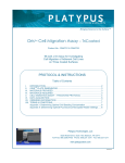
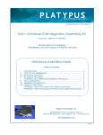
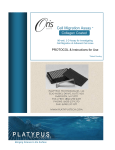
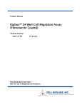
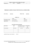
1
Bringing Science to the SurfaceTM
OrisTM Cell Invasion & Detection Assay
Product No.: CIA101DE & CIA200DE
96-well, 3-D Assay for Investigating
Cell Invasion of Adherent Cell Lines
PROTOCOL & INSTRUCTIONS
I.
II.
III.
IV.
V.
VI.
VII.
VIII.
IX.
INTRODUCTION ....................................................................................................................... 2
TM
ORIS PLATE DIMENSIONS (per well) ................................................................................... 2
MATERIALS PROVIDED ........................................................................................................... 3
MATERIALS REQUIRED ........................................................................................................... 3
PRECAUTIONS AND RECOMMENDATIONS .......................................................................... 4
CELL INVASION & DETECTION ASSAY PROTOCOL ............................................................. 5
DATA ACQUISITION ................................................................................................................. 8
ORDERING INFORMATION.................................................................................................... 10
TERMS & CONDITIONS ......................................................................................................... 10
APPENDIX I: Determining Optimal Cell Seeding Concentration .............................................. 11
APPENDIX II: Determining Optimal Fluorescence Microplate Reader Settings ....................... 11 Platypus Technologies, LLC
5520 Nobel Drive, Suite 100, Madison, WI 53711
Toll Free: 866.3296.4455 Phone: 608.237.1270 Fax: 608.237.1271
www.platypustech.com
RM0033.02
Oris™ CELL INVASION & DETECTION ASSAY
I.
INTRODUCTION
The Oris™ Cell Invasion & Detection Assay is a reproducible, sensitive, and flexible assay that can be used to monitor
cell invasion. Formatted for a 96-well plate, the assay utilizes Oris™ Cell Seeding Stoppers made from a medicalgrade silicone to restrict cell seeding to the outer annular regions of the wells. Removal of the stoppers reveals a 2 mm
diameter unseeded region in the center of each well, i.e., the detection zone, into which the seeded cells may then
invade. The Oris™ Detection Mask is applied to the plate bottom and restricts visualization to the detection zone, thus
allowing only invading cells to be detected (see Figure 1). The Oris™ Cell Invasion & Detection Assay is designed to be
used with any commercially available stain or labeling technique, but comes complete with a Calcein AM Staining
Reagent. Calcein AM is a fluorescent dye that passes through the membrane of live cells and is useful for short-term
labeling of cells at the end of the invasion period. Calcein AM is processed by esterases inside the cell allowing the dye
to bind calcium and be trapped inside the cell, and has an excitation/emission wavelength of 495/515 nm. Readout can
be performed by microscopy or use of a microplate reader.
The Oris™ Cell Invasion & Detection Assay kit has been uniquely designed to detect cellular invasion in vitro within a 3dimensional extracellular matrix comprised of a basement membrane extract (BME) of the Murine Engelbreth-HolmSwarm tumor. The Oris™ Cell Invasion & Detection Assay system has been designed for use with adherent cell
cultures. Performance of the Oris™ Cell Invasion & Detection Assay was optimized using the invasive HT-1080
fibrosarcoma and the non-invasive 3T3-Swiss albino fibroblast cell lines.
Using the Oris™ Cell Invasion & Detection Assay offers the following features & benefits:
•
•
•
Membrane-free Invasion - perform studies
without manipulating transmembrane inserts; no
membrane to restrict the ability to image cells.
Reproducible Results – obtain low well-to-well
CV's due to the unique assay design.
Preserve Cell Morphology – realize a more
native 3-D invasion environment since cells are
embedded in an extracellular matrix.
Apply BME Coating
& Populate Plates
with Oris™ Cell
Seeding Stoppers
Seed & Adhere
Cells onto Oris™
Plate
•
•
Remove Stoppers
to Create Detection
Zone & Apply BME
Overlay
Figure 1. Schematic of Oris™ Cell Invasion & Detection Assay
II.
Versatile - analyze data using multiple probes in
a single well by using a microscope, digital
imager, or fluorescence microplate reader.
Flexible - perform kinetic or endpoint cell
invasion assays without the use of special
instrumentation.
Incubate and Allow
Cells to Invade into
Detection Zone
Analyze Detection
Zone (Cells that HAVE
NOT Invaded into
Detection Zone are
Blocked from View
with Detection Mask
Attached)
ORISTM PLATE DIMENSIONS (per well)
Diameter of Well
6.5 mm
Diameter of Stopper Space (Detection Zone)
2 mm
Suggested Media Volume per Well (populated with Stoppers)
100 µl
Effective Area of Outer Annular Region (seeding region) per Well
30.03 mm2
Effective Area of Central Detection Zone per Well
3.14 mm2
Important: Read Instructions Before Performing any OrisTM Assay.
Platypus Technologies, LLC.
RM0033.02
5520 Nobel Drive, Suite 100
Madison WI 53711 USA
www.platypustech.com
Toll Free: 866.296.4455
Phone: 608.237.1270
Fax: 608.237.1271
pg. 2
III.
MATERIALS PROVIDED
Product No.: CIA101DE
Component
Oris™ 96-well Plate
Oris™ Cell Seeding Stoppers
Oris™ Detection Mask
Oris™ Stopper Tool
Oris™ Basement Membrane Extract (BME)
Stock Reagent
Calcein AM Reagent
Product No.: CIA200DE
Component
Oris™ 96-well Plates
Oris™ Cell Seeding Stoppers
Oris™ Detection Mask
Oris™ Stopper Tool
Oris™ Basement Membrane Extract (BME)
Stock Reagent
Calcein AM Reagent
Quantity
1
96
1
1
Storage
Room Temperature
Room Temperature
Room Temperature
Room Temperature
5 mL
< -20°C*
20 µL
-20°C with dessicant**
Quantity
2
2 x 96
2
2
Storage
Room Temperature
Room Temperature
Room Temperature
Room Temperature
2 x 5 mL
< -20°C*
2 x 20 µL
-20°C with dessicant**
* Oris™ BME Stock Reagent can be stored at -20°C in a manual defrost freezer if kit will be used within 3 months
of receipt. For long-term storage, maintain Oris™ BME Stock Reagent at -80°C.
** Calcein AM can be stored at -20°C in a manual defrost freezer with a dessicant for use within 6 months of
receipt.
IV.
MATERIALS REQUIRED
•
•
•
•
•
•
•
•
•
•
•
Biological Cells
Complete Cell Culture Growth Medium (containing serum)
Sterile PBS (containing both Calcium and Magnesium)
Hanks Balanced Salt Solution (HBSS)
Serum-Free Cell Culture Medium
Sterile Pipette Tips/Pipette or Multi-Channel Pipette
Trypsin or Cell Scraper
Inverted Microscope (optional)
Fluorescence Microplate Reader (optional)
Cell Culture Labeling Medium (phenol red-free/serum-free media)
Cell Labeling Fluorescent Agent (e.g., CellTracker™ Green) - required if performing staining in addition to or in
place of Calcein AM.
Platypus Technologies, LLC.
RM0033.02
5520 Nobel Drive, Suite 100
Madison WI 53711 USA
www.platypustech.com
Toll Free: 866.296.4455
Phone: 608.237.1270
Fax: 608.237.1271
pg. 3
V.
PRECAUTIONS AND RECOMMENDATIONS
For Research Use Only. Not for use in diagnostic procedures.
Handling and Use of the Oris™ BME Stock Reagent:
• Thaw on ice (2-8°C) overnight.
• Please note that there will be lot-to-lot variations in the concentration of the Oris™ BME Stock Reagent. Refer to the
BME concentration listed on the bottle label when preparing dilutions. It is crucial that the BME concentration be
optimized for cell line and experimental conditions, since different cell lines and different experimental conditions can
result in a range of cell invasiveness.
• A suggested starting concentration for the Oris™ BME Stock Reagent is 10 – 12 mg/mL. However, do not dilute
Oris™ BME Stock Reagent below 9 mg/mL, as this will preclude gel formation.
• Aliquot and freeze any remaining Oris™ BME Stock Reagent. Avoid repeated freeze-thaw cycles.
Recommendations for Preparation of Reference Wells:
• To establish t=0 pre-invasion reference wells by seeding both test and reference wells at the same time, it is necessary
to seed cells at different concentrations. Seed test wells at a density determined optimal in Appendix I, but seed
reference wells at sub-optimal density (50-75% confluency). Allow cells to adhere and remove stoppers from test wells
(overlay test wells with the Oris™ BME Stock Reagent and then incubate to allow for gel formation). Reference wells
will remain populated with Oris™ Cell Seeding Stoppers until the end of the assay. At that point, remove the Oris™
Cell Seeding Stoppers from the reference wells, overlay the cells with the Oris™ BME Stock Reagent, and incubate to
allow for gel formation. At this point, the entire plate can be stained and analyzed.
• To establish t=0 pre-invasion reference wells by seeding test and reference wells at the same concentration, it is
necessary to seed cells at different times during the assay. Seed test wells at density determined optimal in Appendix
I, allow cells to adhere, remove stoppers, overlay with the Oris™ BME Stock Reagent, and incubate to allow for gel
formation. Allow cells to invade for a set amount of time. At 4 - 18 hours prior to analyzing test wells, seed reference
wells at a density determined optimal in Appendix I, and allow cells to adhere. At the end of the assay, remove
stoppers from the reference wells, overlay them with the Oris™ BME Stock Reagent, and then incubate to allow for gel
formation. At this point, the entire plate can be stained and analyzed.
Oris™ is a trademark of Platypus Technologies, LLC.
CellTracker™ Green is a trademark of Invitrogen Corporation.
Platypus Technologies, LLC.
RM0033.02
5520 Nobel Drive, Suite 100
Madison WI 53711 USA
www.platypustech.com
Toll Free: 866.296.4455
Phone: 608.237.1270
Fax: 608.237.1271
pg. 4
VI.
CELL INVASION & DETECTION ASSAY PROTOCOL
The following steps should be performed in a biological hood using aseptic technique to prevent contamination.
1.
If desired, cells can be starved by incubating for 18 - 24 hours in serum-free medium prior to assay (0.5% fetal bovine serum
may be used if needed).
2.
Make 2 mL of a BME Coating Solution (final concentration of 3.5 mg/mL) by mixing thawed Oris™ BME Stock Reagent
with cold Hanks Balanced Salt Solution (HBSS) or serum-free media. For example, if you have a 14 mg/mL Oris™ BME
Stock Reagent, then mix 500 µL of the Oris BME Stock Reagent with 1.5 mL of HBSS or serum-free media.
NOTE: BME gels in 5-10 min above 15°C; therefore, keep the BME Coating Solution on ice while coating all wells. The use
of chilled pipette tips/reservoirs may be beneficial.
Also, keep the remaining Oris™ BME Stock Reagent on ice until ready to use (Oris™ BME Stock Reagent will be used
again in Step 17).
3.
Pipette 100 µl of the BME Coating Solution into the wells of Column 1. Avoid bubble formation in the BME Coating by not
fully expelling all contents of the pipette. Immediately remove the BME Coating Solution from the wells of Column 1 and
return contents back into the BME Coating Solution reservoir. Repeat procedure for the remainder of plate.
NOTE: The BME Coating Solution is only used in Step 3.
4.
After the BME Coating Solution has been applied to all
wells of the plate, check each well for excess solution. If
excess solution is apparent, remove via pipette.
5.
Incubate the plate in a humidified chamber (37°C, 5%
CO2) for 15 - 30 minutes.
6.
Under sterile conditions, populate the 96-well plate with
Oris™ Cell Seeding Stoppers:
• Vertically position the tip ends of two, 4-stopper strips
into one full column of 8 wells at a time (Figure 2A).
• Gently press down on the strip backbone to partially
insert the stoppers halfway into the well (Figure 2B).
• When both stopper strips have been partially inserted
in 1 column, ensure that the position of the stoppers is
vertical with respect to the well wall, making any
necessary adjustments (Figure 2C).
• Using the Oris™ Stopper Tool, firmly press down on
the strip backbone to fully insert the stoppers into
each well (Figure 2D and 2E). Repeat for all
remaining columns.
A)
B)
D)
C)
E)
Figure 2. Stopper Insertion Process. A) Placement of Stoppers into
Wells, B) Close-up of Stoppers Partially Inserted into
Wells, C) Proper Placement of Stoppers, D) Pressing of
Stoppers into Wells, and E) Fully Inserted Stoppers
NOTE: It is extremely important to ensure that the stoppers are inserted perpendicular to the well bottom and are fully
engaged with the bottom of the well. Failure to do so will increase the CV of your data set.
7.
Visually inspect the underside of the populated 96-well plate to ensure that the Oris™
Cell Seeding Stoppers are firmly sealed against the bottom of the plate. To inspect
the stoppers, turn the plate over and examine the stoppers for sealing (see Figure 3).
If incomplete sealing is observed, return the plate to the upright position and use a
sterile instrument to gently push the stopper back into the well until sealing is
observed.
A
B
C
NOTE: The sealing of the stoppers can be most easily observed if the plate is tipped
at an angle and viewed under indirect light to reveal the “bullseye” pattern at the
bottom of each well.
8.
Apply the Oris™ Detection Mask to the bottom of the 96-well plate if microplate
reader data is being collected. The Detection Mask is not necessary if collecting
imaging data.
Platypus Technologies, LLC.
RM0033.02
5520 Nobel Drive, Suite 100
Madison WI 53711 USA
www.platypustech.com
Figure 3. Stoppers that are:
A) Partially Sealed
B) Unsealed
C) Completely Sealed
Toll Free: 866.296.4455
Phone: 608.237.1270
Fax: 608.237.1271
pg. 5
VI.
CELL INVASION & DETECTION ASSAY PROTOCOL, continued
First Time Users: In order to prevent splashing of well contents, familiarize yourself with the attachment and removal of the
Detection Mask before any liquids are placed into the wells:
Aperture
Orientation A-1 Corner
Chamfer
Attachment Lugs
• Orient the chamfered corners of the mask with those of the 96-well plate,
ensuring that the A1 corner of the mask is aligned with the A1 well of the plate
(see Figure 4).
• Align the holes in the attachment lugs with the bosses on the bottom of the plate.
• Gently press the mask until it is flush with the bottom of the 96-well plate.
NOTE: It may be necessary to wash the mask with ethanol to remove dust and
debris since the mask is not sterile. The mask may be applied at any point during
the assay. For kinetic assays, it is often most convenient to apply the mask at the
beginning of the assay before any liquids are placed in the well. For endpoint
assays, using fixed and stained cells, it is often most convenient to apply the mask
just before reading assay results.
9.
Figure 4. Features of Detection Mask
If performing a kinetic analysis of cell invasion, pre-label cells with a fluorescent
stain now.
10. Collect cells and prepare a suspension that is 10-fold greater in density than the
optimal seeding concentration using complete cell culture growth medium
containing serum.
First Time Users: The optimum seeding density of cells must be determined as an
integral part of the design of the cell invasion assay. Please refer to Appendix I for
a discussion of this process.
IMPORTANT: For recommendations on designating ‘reference’ wells, please refer
to Section V: Precautions and Recommendations.
Figure 5. Media is Added with Single
or Multi-Channel Pipette
11. Pipette 100 µl of suspended cells into each test well through one of the side ports of the Oris™Cell Seeding Stopper.
NOTE: For best results, add or extract media by placing the pipette tip along the wall of the well (see Figure 5). Care should
be taken not to disturb the Oris™ Cell Seeding Stopper or the BME coating when introducing the pipette tip into the well. A
slender/elongated tip or a gel loading tip may be useful.
IMPORTANT: Lightly tap the plate on your work surface to evenly distribute well contents (extreme tapping may result in
splashing of well contents and lead to contamination).
12. Incubate the seeded plate containing the Oris™ Cell Seeding Stoppers in
a humidified chamber (37°C, 5% CO2) for 4 to 18 hours (cell line
dependent) to permit cell attachment.
A)
B)
C)
13. Remove plate from incubator.
14. Using the Oris™ Stopper Tool, remove stoppers (see Figure 6).
NOTE: It may be necessary to wash the Oris™ Stopper Tool with 70%
ethanol as the Stopper Tool is not sterile.
D)
E)
• Secure the 96-well plate by holding it firmly against the deck of your
work space. Slide the tines of the Oris™ Stopper Tool under the
backbone of the stopper strip, keeping the underside of the Oris™
Stopper Tool flush with the top surface of the plate.
• Lift the Oris™ Stopper Tool vertically to gently remove the
stoppers.
NOTE: DO NOT use the Oris™ Stopper Tool as a lever to pry the
stoppers from the well (see Figure 6E), as doing so may cause
displacement of seeded cells and may distort the detection zone area.
Platypus Technologies, LLC.
RM0033.02
5520 Nobel Drive, Suite 100
Madison WI 53711 USA
www.platypustech.com
Figure 6. Removal of Stoppers. Panels A, B, and C)
Position the Tines of the Stopper Tool
between the Stopper Tips, D) Lift Vertically,
and E) Do NOT Pry Stoppers
Toll Free: 866.296.4455
Phone: 608.237.1270
Fax: 608.237.1271
pg. 6
VI.
CELL INVASION & DETECTION ASSAY PROTOCOL, continued
15. Remove media with a pipette and gently wash wells with 100 µl of sterile PBS (or serum-free media) to remove any
unattached cells. Do not aspirate using an in-house vacuum.
16. Add 40 µl of the thawed Oris™ BME Stock Reagent to each well (supplements, such as FBS or growth factors, may be
mixed with BME) to create a 3-D BME overlay.
NOTE: Please note that there will be lot-to-lot variations in the concentration of the Oris™ BME Stock Reagent. Refer to
the BME concentration listed on the bottle label when preparing dilutions. It is crucial that the BME concentration be
optimized for cell line and experimental conditions, since different cell lines and different experimental conditions can result
in a range of cell invasiveness.
NOTE: BME gels in 5-10 min above 15°C, therefore, you must keep the thawed Oris™ BME Stock Reagent on ice until
ready to use. In addition, the use of chilled pipette tips/reservoirs might be beneficial.
17. Incubate plate in a humidified chamber (37°C, 5% CO2) for 30 - 60 minutes to permit polymerization of the 3-D BME overlay.
18. Add 100 µl of serum-free cell culture medium on top of the 3-D BME overlay. Optional: Invasion inhibitors or stimulants
may be added to the media.
19. Incubate plate in a humidified chamber (37°C, 5% CO2) to permit cell invasion (length of incubation is cell line dependent).
The 3-D BME overlay will remain gelled for up to 14 days. Refresh media or supplements, every 48 - 72 hours, as needed,
for the duration of the invasion experiment.
20. If performing an endpoint analysis of cell invasion, stain cells with a fluorescent stain after sufficient invasion has occurred.
Refer to Section VI and Appendix II for further information on data acquisition and fluorescence staining technique.
NOTE: Oris™ Cell Seeding Stoppers are for single use only; Platypus cannot guarantee the integrity of the stopper material
after a second sterilization procedure.
Platypus Technologies, LLC.
RM0033.02
5520 Nobel Drive, Suite 100
Madison WI 53711 USA
www.platypustech.com
Toll Free: 866.296.4455
Phone: 608.237.1270
Fax: 608.237.1271
pg. 7
VII. DATA ACQUISITION
The readout of the Oris™ Cell Invasion & Detection Assay can be conducted at any time, allowing the user to perform a kinetic
assay or an endpoint assay. The Oris™ Cell Invasion & Detection Assay is designed to be used with any commercially available
stain or labeling technique, but is supplied complete with the Calcein AM Reagent. The readout can be performed by using a
microscope, a microplate reader, or a High Content Screening or High Content Imaging Analysis platform.
Microscope Analysis
• Cell counting or image capture / analysis software, such as NIH ImageJ freeware, can be used.
• Note: Microscopic observations are possible using phase contrast or bright field microscopy with colorimetric stains.
• No need to attach the Oris™ Detection Mask to the Oris™ microplate.
• To set up reference controls, refer to Section V: Precautions and Recommendations.
Microplate Reader Analysis
• Attach the Oris™ Detection Mask to the bottom of the Oris™ microplate (see Step 8 of Protocol).
• Optimal settings will vary according to the microplate reader make and model. Consult Appendix II and the equipment
user manual for your particular instrument.
• The microplate reader MUST be set to read from the bottom of the plate.
• To set up reference controls, refer to Section V: Precautions and Recommendations.
Sample Data Obtained via Microscopy and Microplate Reader are shown in Figure 7.
5
• Wells were seeded with 50,000 HT-1080 cells (i.e., 100 µl of 5x10 cells/mL) that had been serum starved for 18 hours
and the plate was then incubated for 4 hours. The stoppers were removed from the wells and the OrisTM BME (final
concentration of [12 mg/mL]) with 10% FBS or without FBS was overlayed on the cells (four (4) wells were left stoppered
to represent t=0 reference). The plate was incubated in a humidified chamber for 48 hours to permit cell invasion. Cells
were labeled with Calcein AM and images were captured using a Zeiss Axiovert microscope (5X magnification).
Fluorescence in the detection zone was quantified by using a microplate reader. Each column represents the mean +/SD of at least 4 wells. A non-invasive cell line, 3T3-Swiss albino, served as the negative control. The images below,
captured without a detection mask in place, illustrate representative data from pre-invasion (t=0 hrs) and post-invasion
(t=48 hrs) wells. The graph depicts the average Relative Fluorescent Units (RFU’s) in the detection zones for each
condition, confirming the invasion augmenting effect of FBS on serum starved HT-1080 cells and the lack of invasion by
non-invasive 3T3-Swiss albino cells.
• As observed in Figure 7, HT-1080 cells formed invadopodial structures that projected into the central detection zone
(panel 1b) as compared to a time zero control (panel 1a).
• Invasion by serum-starved HT-1080 cells was augmented by including 10% v/v fetal bovine serum (FBS) in the BME
overlay (panel 1c).
• In contrast, as observed in images 2b and 2c, the non-invasive 3T3-Swiss albino cells did not form any invadopodial
structures and showed a nominal amount of FBS-independent migration into the central detection zone as compared to a
time zero control (image 2a).
Behavior of Invasive HT-1080 Cells in Oris
1a
1b
TM
Cell Invasion & Detection Assay
1c
Measurement
the OrisTM
Detection
ZoneDetection
Using a Plate
Reader
Analysis of
ofCells
Cell in
Invasion
into
the Oris™
Zone
t=0
t=48h (no FBS)
t=48h (10% FBS)
Behavior of Non-Invasive 3T3-Swiss albino Cells in
TM
Oris Cell Invasion & Detection Assay
2a
2b
2c
Invasive HT-1080 Cells
t=0
t=48h (no FBS)
Non-Invasive 3T3-Swiss albino Cells
t=48h (10% FBS)
Figure 7. Cell invasion data obtained via microscopy and microplate reader analysis
Platypus Technologies, LLC.
RM0033.02
5520 Nobel Drive, Suite 100
Madison WI 53711 USA
www.platypustech.com
Toll Free: 866.296.4455
Phone: 608.237.1270
Fax: 608.237.1271
pg. 8
Immunostaining Analysis
• As verified by immunostaining (shown in Figure 8), the structures projecting into the BME formed by the invasive HT-1080
cells exhibited classic hallmarks of invadopodia, namely colocalization of F-actin with cortactin (panel a), and the
expression of proteases such as MMP-9 and cathepsin B (panels b and c). These invadopodia could also be observed in
3-dimensions at different focal points along the z-axis. This is in contrast to any migratory activity observed by noninvasive cells that occurs only within the 2-dimensional space of the x and y axes (data not shown).
TM
Cell Invasion & Detection Assay were fixed, permeabilized, and pre-treated by sequential
TM
incubations in 3.7% formaldehyde, PBS, 0.5% Triton X-100, PBS, Image-iT FX signal enhancer, and PBS.
Immunostaining was performed by incubating with primary antibodies (2 hours, 37°C) and Alexa Fluor® 488 conjugated
secondary antibodies at 1:200 dilution (1 hour, 37°C) followed by an Alexa Fluor® 555 phalloidin counterstain (1 hour).
Images were collected using a Nikon TE300 inverted microscope equipped with a Photometrics Coolsnap fx CCD
camera, deconvolved using SlidebookTM v4.2 (Intelligent Imaging Innovations) and processed using Adobe® Photoshop®
CS2 (Adobe Systems).
• Cells from the Oris
Anti-Cortactin
a
b
F-actin is present in all eukaryotic cells, it is a major component of the cytoskeleton
and functions to define and maintain the cell shape. Cortactin is a cellular protein
that is present in and promotes the formation of lamellipodia and invadopodia.
These structures propel cells over surfaces as they move toward a target. Results
depicted in 8a demonstrate distinct areas of cortactin and F-actin colocalization
characteristic of invadopodia (Ref. 1 & 2).
Anti-Cathepsin B
Cathepsin B is a lysosomal protease that facilitates cell invasion by degrading the
surrounding extracellular matrix. Results depicted in 8b demonstrate distinct areas
of lysosomal and peripheral cellular expression of Cathepsin B characteristic of
protease expression observed in cancer cell invasion (Ref. 3).
Anti-MMP-9
c
MMP-9 is a protease that is involved in matrix degradation in cancer cell invasion.
Results depicted in 8c demonstrate evidence of MMP-9 expression and secretion
into surrounding areas of ECM, which are established characteristics of proteases
observed at invadopodia sites (Ref. 1& 2).
Figure 8. Confirmation of Cell Invasion using Immunostains
References:
1. Weaver, AM; Clin Exp Metastasis 2006. "Invadopodia: specialized cell structures for cancer invasion." 23:97-105.
2. Furmaniak-Kazmierczak et al; Circulation Research 2007. "Formation of extracellular matrix-digesting invadopodia by primary aortic
smooth muscle cells." 100:1328-1336.
3. Hulkower, KI et al; Eur. J. Biochem 2000. "Fluorescent microplate assay for cancer cell-associated cathepsin B." 267:4165-4170.
Platypus Technologies, LLC.
RM0033.02
5520 Nobel Drive, Suite 100
Madison WI 53711 USA
www.platypustech.com
Toll Free: 866.296.4455
Phone: 608.237.1270
Fax: 608.237.1271
pg. 9
VIII.
ORDERING INFORMATION
Product No.
Product Description
Package Size
CIA101DE
Oris™ Cell Invasion & Detection Assay, 1-pack:
Oris™ 96-well plate, 1
TM
Oris BME Stock Reagent, 5 mL
Oris™ Cell Seeding Stoppers, 96
Oris™ Detection Mask, 1 & Oris™ Stopper Tool, 1
Calcein AM Reagent, 20 µL
1-pack
CIA200DE
Oris™ Cell Invasion & Detection Assay, 2-pack:
Oris™ 96-well plates, 2
TM
Oris BME Stock Reagent, 2 x 5 mL
Oris™ Cell Seeding Stoppers, 2 x 96
Oris™ Detection Mask, 2 & Oris™ Stopper Tool, 2
Calcein AM Reagent, 2 x 20 µL
2-pack
CMA1.101
Oris™ Cell Migration Assay, 1-pack:
Oris™ 96-well, Tissue Culture Treated Plate (black, clear bottom) with Oris™
Cell Seeding Stoppers, 1
Oris™ Detection Mask, 1 & Oris™ Stopper Tool, 1
1-pack
CMA5.101
Oris™ Cell Migration Assay, 5-pack:
Oris™ 96-well, Tissue Culture Treated Plates (black, clear bottom) with Oris™
Cell Seeding Stoppers, 5
Oris™ Detection Mask, 1 & Oris™ Stopper Tool, 1
5-pack
CMATR1.101
CMATR5.101
Oris™ Cell Migration Assay - TriCoated, 1-pack:
Oris™ TriCoated, 96-well Plate with Oris™ Cell Seeding Stoppers, 1
32 wells, Uncoated, Tissue Culture Treated
32 wells, Collagen I coated
32 wells, Fibronectin coated
Oris™ Detection Mask, 1 & Oris™ Stopper Tool, 1
Oris™ Cell Migration Assay - TriCoated, 5-pack:
Oris™ TriCoated, 96-well Plates with Oris™ Cell Seeding Stoppers, 5
32 wells, Uncoated, Tissue Culture Treated
32 wells, Collagen I coated
32 wells, Fibronectin coated
Oris™ Detection Mask, 1 & Oris™ Stopper Tool, 1
1-pack
5-pack
To place an order, visit the Platypus Technologies website at: www.platypustech.com/order_main.html.
For technical assistance, contact Technical Support at (866) 296-4455 or [email protected].
IX.
TERMS & CONDITIONS
Certain uses of these products may be covered by U.S. Pat. No. 6,284,197, No. 7018838, No. 10/597,118, No. 11/342,413, and No. 60/836,109, licensed to PLATYPUS. Certain applications of
PLATYPUS products may require licenses from other parties. Determining the existence and scope of such third party intellectual property is the responsibility of the PURCHASER. Purchase of
the product provides the PURCHASER with a limited non-transferable license under any PLATYPUS patents or patent applications to use the product for internal research unless there is a
written limitation to this license in the product literature. PURCHASER is responsible for carefully reviewing the product literature and respecting any limitations to this license, e.g. limitations for
commercial use or research by for-profit institutions. These products may not be resold, modified for resale, used to manufacture commercial products, or used to develop commercial products
without the express written approval of PLATYPUS.
These products are intended for research or laboratory use only and are not to be used for any other purposes, including, but not limited to, unauthorized commercial purposes, in vitro
diagnostic purposes, ex vivo or in vivo therapeutic purposes, investigational use, in foods, drugs, devices or cosmetics of any kind, or for consumption by or use in connection with or
administration or application to humans or animals.
PLATYPUS warrants that its products shall conform substantially to the description of such goods as provided in product catalogues and literature accompanying the goods until their respective
expiration dates or, if no expiration date is provided, for 6 months from the date of receipt of such goods. PLATYPUS will replace, free of charge, any product that does not conform to the
specifications. This warranty limits PLATYPUS's liability only to the replacement of the nonconforming product.
THIS WARRANTY IS EXCLUSIVE AND PLATYPUS MAKES NO OTHER WARRANTY, EXPRESS OR IMPLIED, INCLUDING WITHOUT LIMITATION, ANY IMPLIED WARRANTY OF
MERCHANTABILITY OR FITNESS FOR A PARTICULAR PURPOSE. The stated express warranties, and the remedy provided for breach thereof, are in lieu of all other liability or obligations of
PLATYPUS for any damages whatsoever arising out of or in connection with the delivery, use, misuse, performance, or the inability to use any of its products. IN NO EVENT SHALL PLATYPUS
BE LIABLE UNDER ANY LEGAL THEORY (INCLUDING BUT NOT LIMITED TO CONTRACT, NEGLIGENCE, STRICT LIABILITY IN TORT, OR WARRANTY OF ANY KIND) FOR ANY
INDIRECT, SPECIAL, INCIDENTAL, CONSEQUENTIAL, OR EXEMPLARY DAMAGES (INCLUDING BUT NOT LIMITED TO LOST PROFITS) EVEN IF PLATYPUS HAD NOTICE OF THE
POSSIBILITY OF SUCH DAMAGES. Without limiting the effect of the preceding sentence, PLATYPUS's maximum liability, if any, shall not exceed the purchase price paid by PURCHASER for
the product.
This warranty shall not be effective if PLATYPUS determines, in its sole discretion that PURCHASER has altered or misused the goods or has failed to use or store them in accordance with
instructions furnished by PLATYPUS. PLATYPUS’s sole and exclusive liability and PURCHASER’s exclusive remedy with respect to goods proved to PLATYPUS’s satisfaction (applying
analytical methods reasonably selected by PLATYPUS) to be defective or nonconforming shall be the replacement of such goods free of charge, upon the return of such goods in accordance
with our instructions, although at its discretion, PLATYPUS may provide a credit or refund. If PLATYPUS manufactures custom goods for PURCHASER based on instructions, specifications, or
other directions provided by PURCHASER, PLATYPUS shall not be liable for the lack of sufficiency, fitness for purpose or quality of the goods to the extent attributable to such instructions,
specifications, or other directions. PLATYPUS shall not be liable for any loss, damage or penalty as a result of any delay in or failure to manufacture, deliver or otherwise perform hereunder due
to any cause beyond PLATYPUS’s reasonable control.
PLATYPUS shall not be liable for injury or damages resulting from the use or misuse of any of its products.
Platypus Technologies, LLC.
RM0033.02
5520 Nobel Drive, Suite 100
Madison WI 53711 USA
www.platypustech.com
Toll Free: 866.296.4455
Phone: 608.237.1270
Fax: 608.237.1271
pg. 10
APPENDIX I: Determining Optimal Cell Seeding Concentration
This procedure is intended to assist in determining the cell seeding density needed to achieve confluency of your cell line when
using the Oris™ Cell Invasion & Detection Assay. The intended goal is to achieve 90-95% confluencey of the monolayer
surrounding the Oris™ Cell Seeding Stoppers without overgrowth.
1.
2.
3.
4.
A suggested starting point is to evaluate three serial dilutions at the cell densities shown below. The cell seeding area of the
2
well with the stopper in place is ~ 0.3 cm . Based on the typical seeding density of your particular cell line, you can infer a
different cell number for your first serial dilution and adjust the numbers below accordingly.
Prepare a log-phase culture of the cell line to be tested. Collect cells and determine the total number of cells present.
6
6
6
Pellet cells by centrifugation. Prepare three serial dilutions at final concentrations of 1.0 x 10 , 0.5 x 10 and 0.25 x 10
cells/mL.
Dispense 100 µl of cell suspension per well into the 96-well plate to result in the following plate layout:
Column
Cells / well
Number of wells
1
100,000
8
2
50,000
8
3
25,000
8
Incubate the plate in a humidified chamber (37°C, 5% CO2) for 4 - 18 hours (cell line dependent) with cell seeding stoppers in
place to allow the cells to firmly attach to the well surface.
6. Following cell attachment, remove the Oris™ Cell Seeding Stoppers from each well (see Figure 6) and gently wash the wells
with PBS to remove non-attached cells.
•
Secure the 96-well plate by holding it firmly against the deck of your work space. Slide the tines of the Oris™ Stopper
Tool under the backbone of the stopper strip, keeping the underside of the tool flush with the top surface of the plate.
•
Lift the Oris™ Stopper Tool vertically to gently remove the stopper. Do not use the Oris™ Stopper Tool as a lever to
pry the stoppers from the well as doing so may cause displacement of the seeded cells.
7. Without a Detection Mask in place, use a microscope to visually inspect each well to determine the minimum cell seeding
concentration that yielded a confluent monolayer at the perimeter of the detection zone.
At this point, if you plan to obtain the results of the Oris™ Cell Invasion & Detection Assay via colorimetric analysis or microscopy,
you have successfully determined the optimal cell seeding concentration to be used in Step 11 of the Oris™ Cell Invasion &
Detection Assay Protocol.
5.
APPENDIX II: Determining Optimal Fluorescence Microplate Reader Settings
This procedure is intended to assist in optimizing your instrument settings when using a fluorescence microplate reader to capture
data from the Oris™ Cell Invasion & Detection Assay.
1.
2.
Using the optimal cell seeding concentration determined in Appendix I, perform a cell invasion assay per Section VI, Oris™
Cell Invasion & Detection Assay Protocol using culture conditions expected to result in robust cell invasion. Be sure to include
equal numbers of pre-invasion control wells (stoppers left in place until staining) and post-invasion test wells (stoppers
removed after cell attachment period). A minimum of 8 wells per condition are recommended.
Perform the desired fluorescent staining technique.
The Oris™ Cell Invasion & Detection Assay has been designed to work with all types of fluorescent stains and staining
techniques. The precise method for staining cells with fluorescent stains varies according to the nature of the individual stain.
It is important to stain cells using a fluorescent reagent that uniformly stains cells. Probes affected by experimental conditions
will increase variability of results and reduce correlation between fluorescence signal and cell invasion. Please consult the
manufacturer of your fluorescent stain for specific considerations.
The following is an example Fluorescent Staining Protocol for using Calcein AM:
a.
b.
c.
d.
e.
f.
To stain one fully-seeded 96-well plate, combine 5 μl of Calcein AM (1 mg/mL in dry DMSO) with 10 mL of phenol redfree and serum-free media or 1x PBS. Protect diluted Calcein AM solution from light until ready to use in step d.
Carefully remove culture medium from wells.
Wash wells with 100 μl of PBS (containing both Calcium and Magnesium).
Add 100 μl of diluted Calcein AM solution to each well.
Incubate plate at 37°C for 30 - 60 minutes.
Attach mask and read promptly with microplate reader using appropriate filter set and sensitivity/gain settings (for a
BioTek Synergy™ HT microplate reader, use 485/528 nm excitation/emission filters, sensitivity 55 nm).
3.
If not already in place, apply the Oris™ Detection Mask to the plate. Using the bottom probe of a fluorescence microplate
reader, obtain the fluorescence reading from each well. To achieve the optimal dynamic range, adjust the instrument settings
(e.g., gain) to result in the greatest difference in fluorescence signal between pre-invasion and post-invasion wells. Refer to
the instrument manual for your microplate reader for further guidance on instrument settings.
You have now successfully determined the optimal cell seeding concentration (to be used in Step 11 of the Oris™ Cell Invasion &
Detection Assay Protocol) and microplate reader settings for analysis of cell invasion using a fluorescence microplate reader.
Platypus Technologies, LLC.
RM0033.02
5520 Nobel Drive, Suite 100
Madison WI 53711 USA
www.platypustech.com
Toll Free: 866.296.4455
Phone: 608.237.1270
Fax: 608.237.1271
pg. 11