Download manual for the assessment of sperm counts
Transcript
MANUAL FOR THE ASSESSMENT OF
SPERM COUNTS
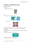
Leja 20 micron four chamber slide
General Information Leja® Standard
Count counting chambers
Manufacturer:
Leja Products B.V.
Luzernestraat 10
2153 GN Nieuw Vennep
The Netherlands
tel.:
fax.:
e-mail:
web:
+31 (0) 252 62 18 48
+31 (0) 252 62 18 05
[email protected]
www.leja.nl
Leja is certified ISO 9001:2000
Leja slides are in vitro diagnostic devices to be used by an authorized person.
Leja slides are designed for microscopic quantitative evaluation of cells in suspension, like blood and
semen. Leja slides are not made for self-testing. Authorized, qualified persons should handle Leja slides.
Leja slides should be stored in a closed box, protected from sunlight, and at room temperature. The shelf
life of Leja slides is years, but a haze can form inside the chamber. Filling will wash this away and does
not affect the functioning of the chamber.
Body fluids can be infectious. Do not touch the slides at the filling openings, and only touch the slide at
the outer left and right edges. Prevent spilling of body fluids.
Leja slides are made of glass. Glass is breakable and sharp edges will be formed after breaking.
Handle used Leja slides as infectious waste and discard used Leja slides according to the local
instructions for the handling of infectious waste.
General description.
The Leja slides are to be used for quantitative analysis of cells and particles in suspension. The slide has
to be used with a microscope. The microscope can be integrated in a Computer Assisted Semen Analysis
(CASA) system. They are mostly used for the analysis of semen. The Leja slides with chamber heights of
12 and 20 microns are suitable for the analysis of motile spermatozoa in combination with a 10x or 20x
objective lenses. The Leja slide with a chamber height of 100 microns is made for the analysis of small
numbers of cells. A separate instruction manual is available for the assessment of low numbers
(0-8.000/ml and 8.000 – 1.106/ml): manual 20 and 100 micron Leja slides.
The Leja slide is made of a standard microscopic object slide with the dimensions of 75x25x1 mm. It is
made of float glass and the dimensions of 75 x 25 mm are achieved by sawing, not by cutting and
breaking. The sawed, blunt edges prevent damage to the skin of the user.
On top of the Leja chamber is a cover slip. The dimensions of the cover slip of the Leja 4 chamber slide
are 32x 21x 0,7 mm.
Both glass plates are washed and coated. Clean glass has a very reactive surface; the coating prevents
sticking of the cells to the glass surface. The coating prevents the formation of air bubbles during the
filling process. However, small particles suspended in the semen sample can provoke the formation of air
bubbles.
Leja Products B.V.
Luzernestraat 10, 2153 GN Nieuw-Vennep - The Netherlands
2
The particulars of the Leja slides are printed at the left and right sides of the object slide.
Leja slides are available with different chamber numbers and different chamber heights. There are slides
with 2, 4 and 8 chambers and chamber heights are available in 12, 20 and 100 microns. The content of
the chambers varies between 1 µl and 25 µl. The content of the chamber is indicated on the slide. The
effective surface of the chamber to be used for analytical purposes varies between 20 mm2 (eight
chamber slide), 66 mm2 (4 chamber slide), and 250 mm2 (100 micron chamber height, two chamber
slide).
The two glass plates of the Leja slide are at a well-defined distance from each other. A resin is printed on
the object slide. In this resin spacers of a well-defined diameter are present. A cover slip is placed on the
printed pattern and the two glass plates are pressed together in such a way that the two plates stay
parallel. The spacers define the distance between the two glass plates. During this process the resin is
cured. The production of the Leja slides takes place in a clean room, preventing the settlement of dust
particles inside the chamber during the production process.
Patents protect the production process and the chamber designs.
Chamber height
The tolerance of the chamber height is maximum ± 5%. The 100-micron chamber has a chamber height
of 100 ± 5 micron. The actual average height, the upper and lower limits and the 95% confidence interval
of each lot is published at our web side.
Segre-Silberberg effect compensation factor
In principle, all capillary filled slides are affected by this phenomenon. It causes transport of cells to the
filling front during the flow of the sample into the chamber. The Segre-Silberberg effect causes
underestimation of the concentrations assessed in the central area of the analysis chamber. The
dimension of the correction factor of the Segre-Silberberg effect depends on many variables, like the
development of a full Poiseuille flow, chamber height, surface properties of the counting chamber, surface
tension, flow velocity and viscosity of the sample. For sperm cells suspended in watery solutions the
compensation factor will be 1.30. For very viscous samples the correction factor will be near 1.00. Since
all variables are kept constant the only variable that affects the dimension of the Segre Silberberg effect is
the viscosity of the sample. Filling time and viscosity are closely associated. By measuring the filling time
of the Leja chamber with a capillary length of 21 mm, the Segre Silberberg compensation factor for that
specific sample is known.
Due to the chamber height of 100 microns, the Segre Silberberg effect is negligible.
Quality control
1.
Chamber height.
After the production process each slide must pass quality control. Printing quality and resin tracks are
checked visually. Chamber height is checked with two methods. 1) All chambers are checked with
interference patterns of monochromatic light. 2) Out of each box of 25 slides 3 slides are tested with a
specific device, specially constructed for Leja, which measures the chamber height in microns. This
device works according to the principle of interference of the complete spectrum of visible light. The
results of the assessments of each lot are published at the website of Leja.
Leja Products B.V.
Luzernestraat 10, 2153 GN Nieuw-Vennep - The Netherlands
3
2.
Toxicity.
Each lot is tested for toxicity with diluted boar semen; boar semen is much more sensitive to toxic effects
than human spermatozoa. The motility of the spermatozoa is tested in the central area (at least 0.8 mm
from the resin border). The motility may decrease with a maximum of 10 % within a test period of 6
minutes.
The results of the toxicity assay are published at the website of Leja. 100 micron chambers are not tested
for toxicity; sharpness of vision of 10x and 20x objective lenses is much less than 100 micron.
3.
Air bubble formation.
Each lot is tested for air bubble formation during the filling process. Each day slides are randomly
selected and filled with water. Lots pass quality control when no air bubble formation is observed.
Waste handling.
Unused Leja chambers can be discarded as household waste. Leja chambers filled with human bodily
fluids have to be treated as infectious and should be treated accordingly. Follow the instructions of local
health authorities how to treat used, filled Leja chambers.
Leja Products B.V.
Luzernestraat 10, 2153 GN Nieuw-Vennep - The Netherlands
4
Table of contents of different procedures
Determination of sperm concentration using CASA system
Variables and formulas
Laboratory manual (procedure)
Page
Page
Page
6
6
7
Determination of sperm concentration manually
Variables and formulas
Determination of Sx
Laboratory manual (procedure)
Page
9
Page
9
Page 10
Page 11
First time use of Leja slides, calibration of microscope
Page 13
Leja Products B.V.
Luzernestraat 10, 2153 GN Nieuw-Vennep - The Netherlands
5
Computer Automated Semen Analysis.
Be sure that the CASA system has been properly set and calibrated. See CASA instruction manual
provided by the manufacturer of the CASA system.
Follow the instructions of the WHO laboratory manual for the examination of human semen and spermcervical mucus interaction (see reference list) for collection and handling of human semen.
Introduction
Motility patterns of sperm cells are sensitive to temperature. To get repeatable and comparable results
the temperature regime during storage and assessments have to be well controlled. It is recommended to
prevent temperature shocks. If sperm samples are cooled down to room temperature or to lower
temperatures, the motion patterns can be impeded or completely blocked. The restoration of the motion
patterns needs time. It is advisable to treat samples according to a standard protocol and to measure
motility patterns at a fixed time interval after collection of the sperm after liquefaction.
It is advisable to keep the sample at 37° C for an appropriate time (30 minutes).
It is advisable to keep the slides on a hot plate of 37° C during a few minutes before filling.
Variables and formulas
TC1
=
total number of counted cells first round
TC2
=
total number of counted cells second round
Ci1
=
initial concentration first round, not corrected for Segre Silberberg effect.
Ci2
=
initial concentration second round, not corrected for Segre Silberberg effect
Ct1
=
true concentration first round (corrected for the Segre Silberberg effect)
Ct2
=
true concentration second round (corrected for the Segre Silberberg effect)
Sx
=
Segre Silberberg compensation factor, see Appendix 1
Cf
=
final true concentration (Ct1 + Ct2) / 2
SRn
=
√ (TC1 +TC2); square root of the sum of the number of spermatozoa counted
in the first and in the second round;
ADn
=
|TC1 – TC2|; absolute difference between the number of spermatozoa
counted in the first and in the second round.
Leja Products B.V.
Luzernestraat 10, 2153 GN Nieuw-Vennep - The Netherlands
6
Procedure CASA
Needed:
•
•
•
•
37° C hotplate
Pipette to pipette 8 µl (2 chamber slide) or 4 µl (4 chamber slide)
Stopwatch
CASA system
1. Loading of the Leja slide and the assessment of filling time of the chamber
2. Have a stopwatch ready.
3. Set the stopwatch at zero.
4.
Mix the semen sample gently; be convinced that the sample has been liquefied.
5. Load the pipette with some more volume than indicated at the chamber.
6. Place the tip of the pipette on the filling spot of the slide.
7. Push the button of the pipette and the start button of the stopwatch at the same time.
8. Observe the filling of the chamber and push the stop button of the stopwatch as the filling front
has reached the out stream opening of the chamber**.
9. Take away excess fluid
10. Note the filling time; write it at the appropriate field of your computer screen.
11. Place the loaded slide at the stage of the CASA system. Count at least 4 different fields (stay
away at least 2 fields from the chamber resin track).
12. Store the number of counted cells = TC1.
13. Store the assessed concentration = Ci1.
14. Go to appendix 1; take the filling time that you obtained under 1 and read the matching Sx (the
correction factor).
15. Get the accurate concentration Ct1 = Ci1 * Sx.
16. If possible, count at least 200 cells. The precision of the assessment will increase when the
number of assessed cells increases. Normally we count 10 fields automatically with the CASA
system.
17. Repeat the assessment of the same sample in a new chamber, obtain TC2 and Ct2.
18. Verify whether the assessments are representative of semen sample.
19. Take the sum of the number of counted cells of the two assessments (TC1 + TC2).
Leja Products B.V.
Luzernestraat 10, 2153 GN Nieuw-Vennep - The Netherlands
7
20. Multiply by 2 the square root of the sum of TC1 and TC2: 2 * √ (TC1 +TC2).
21. Take the absolute difference of the two assessments: |TC1 – TC2|.
22. If |TC1 – TC2| ≤ 2 * √ (TC1 + TC2), the assessments have resulted in two observations within
the limits of 95% confidence interval and the assessments are accepted.
23. If |TC1 – TC2| > 2*(TC1 + TC2), the assessments are rejected and one has to start over completely
24. Calculate the true sperm concentration. This is the average value of the two assessed
concentrations Cf: Cf = (Ct1 + Ct2) / 2
NB.
If your CASA system lacks the facility to correct for the Segre-Silberberg effect, write the filling time
on the patient’s file and perform the corrections yourself by using the values as depicted in
Appendix 1.
NB.
If it is not possible to count 200 cells, the concentration will be low and it is advised to assess the
concentration in a different way (see Leja manual for the assessment of low sperm count and
azoospermia).
Leja Products B.V.
Luzernestraat 10, 2153 GN Nieuw-Vennep - The Netherlands
8
Manual Analysis.
Follow the instructions of the WHO laboratory manual for the examination of human semen and spermcervical mucus interaction (see reference list) for collection and handling of human semen. It is also
advised to follow this manual for the assessment of the motility of human semen.
Variables and formulas
TC1
=
total number of counted cells in the first round
TC2
=
total number of counted cells in the second round
AF
=
number of assessed fields
SB1
=
number of assessed small boxes in the first round
SB2
=
number of assessed small boxes in the second round
N1
=
TC1/SB1; average number of cells per small box of the reticle, first round
N2
=
TC2/SB2; average number of cells per small box of the reticle, second round
Ci1
=
initial concentration first round (not corrected for the Segre Silberberg effect)
Ci2
=
initial concentration second round (not corrected for the Segre Silberberg effect)
Ct1
=
true concentration first round (corrected for the Segre Silberberg effect)
Ct2
=
true concentration second round (corrected for the Segre Silberberg effect)
Sx
=
Segre Silberberg compensation factor, see Appendix 1
F
=
the figure by which the content of a small box projected by the eyepiece reticle on the
Leja chamber has to be multiplied to get 1 nl (one nano litre).
D
=
the seize in microns of an arm of a small box of a 10 x 10 mm (10 x 10 small blocks) eye
piece reticle
Ci1
=
N1 * F
F = ~5 using a 10x objective lens and the linear magnification is 10x indeed.
F = ~20 using a 20x objective lens and the linear magnification is 20x indeed
(the correction factors depicted above in bold are estimates and depending on your microscope,
see page 13 to calibrate your microscope)
Ct1
=
Ci1 * S1
Ct2
=
Ci2 * S2
Cf
=
final true concentration (Ct1 + Ct2)/2
SRn
=
ADn
=
√ (TC1 +TC2); square root of the sum of the number of spermatozoa counted in the first
and in the second round;
|TC1 – TC2|; absolute difference between the number of spermatozoa counted in the first
and in the second round.
Leja Products B.V.
Luzernestraat 10, 2153 GN Nieuw-Vennep - The Netherlands
9
Procedure of determination of Sx
Needed:
•
•
•
•
•
Leja 2 or 4 chamber slides with side openings (chamber length 21 mm)
Calibrated microscope
Stopwatch
Pipette to pipette 4 or 8 µl
Appendix 1 of this manual
If you already use Leja 20 micron slides and you have calibrated your microscope, you need to determine
Sx, the correction factor needed to compensate for the Segre Silberberg effect. If you have not yet used
Leja slides before or calibrated your microscope, please refer to page 13. This is the description of the
determination of Sx only. This procedure is incorporated the manual to determine sperm concentration as
well.
1. Assess the filling time of the Leja chamber with a stopwatch, and note the number of seconds.
2. Take away the excess of fluid from the loading area.
3. Take some time to study the sample under the microscope. Assess the motility according to the
WHO laboratory manual for the examination of human semen and sperm-cervical mucus
interaction (see reference list).
4. Calculate Ci1
=
N1 x F
:
the initial concentration 1.
5. Go to appendix 1. Go to column 1, filling times, and look up the corresponding compensation
factor Sx.
6. Multiply Sx with Ci, to get the Cf, the final true concentration.
By using Sx , the outcome will be fully comparable with hemocytometer assessments.
There is a rule of thumb:
Diluted sperm
Sx= 1.23
Normal viscose sperm
Sx= 1.1
Very viscose sperm
Sx= 1.0 – 1,05
Leja Products B.V.
Luzernestraat 10, 2153 GN Nieuw-Vennep - The Netherlands
10
Manual to determine sperm concentration
For manual analysis you need
•
•
•
•
•
Leja 2 or 4 chamber slides with side openings (chamber length 21 mm)
Stopwatch
Pipette to pipette 8 µl or 4 µl
Microscope with 10x or 20x positive or negative phase contrast
Eyepiece reticle with 10x10 mm grid
This is only suitable if you are convinced that the linear magnification of your microscope 10x objective
lens is 10x indeed. If you have a 20x objective lens, you have to be convinced that the linear
magnification of your 20x objective lens is 20x indeed. Otherwise, calibrate your microscope (see next
section).
Getting started
1. Mix the completely liquefied semen specimen gently and take a sample with the 8µl pipette (2
chamber slide; for the 4 chamber slide take 7µl). One has to use more semen than the content of
the chamber; otherwise it will be not possible to assess properly the filling time.
2. Place the tip of the pipette at the loading place of the Leja slide.
3. Push the start button of the stop watch and push to empty the pipette at the very same moment;
4. Observe the filling of the chamber and push the stop button of the stopwatch as the filling front
reaches the out stream opening.
5. Note the filling time at the patient file;
6. Wipe away the excess of fluid on the filling area.
7. Place the loaded slide at the stage of the microscope and observe the sample for particulars.
Perform the motility analysis at least in 5 different fields according to the WHO laboratory manual
for the examination of human semen and sperm-cervical mucus interaction (see reference list).
8. Select at least five different fields for counting the sperm cells. Stay away two full fields from the
resin edge of the chamber.
9. Count the cells per small box of the eyepiece reticle, note SB1 = the number of assessed small
boxes.
If you use a 10x objective lens it could be convenient to count 5 boxes or multiples of 5 boxes at the time
because the count equals the concentration in millions per ml. If you use a 20x objective lens it is handy
to count 20 small boxes at the time, because this equals the concentration in millions per ml. Count, if
possible, at least 200 cells per chamber.
Leja Products B.V.
Luzernestraat 10, 2153 GN Nieuw-Vennep - The Netherlands
11
10. Note TC1 = total number of counted cells.
11. Note AF = number of analyzed fields.
12. Calculate N1 = (TC1 / SB1) = the average number of cells per small box.
13. Calculate Ci1 = N1 x 5 (using a 10x objective lens) the initial concentration 1.
14. Calculate Ci1 = N1 x 20 (using a 20x objective lens) the initial concentration 1.
15. Calculate Ct1 = C11 * Sx
Go to Appendix 1.
In the first column the chamber filling time is depicted.
In the second column Sx, the compensation factor of Segre Silberberg effect is depicted.
16. Repeat the procedure. Load a new chamber. Assess the chamber filling time and get initial
concentration 2 (Ci2). Be sure that the same numbers of fields and the same number of small
boxes have been assessed. Write down the total number of assessed cells (TC2) in this second
round. Calculate the initial second concentration Ci2, and calculate the true concentration of the
second assessment, Ct2.
17. Multiply the square root SRn of the sum by 2: 2* √ (TC1 + TC2).
18. Take the absolute difference ADn: |TC1 – TC2|.
19. If |TC1 – TC2| ≤ 2 * √ (TC1 + TC2), the assessment have resulted in two observations within
the limits of 95% confidence interval and the assessments are accepted.
20. If |TC1 – TC2| > 2*(TC1 + TC2), the assessments are rejected and one has to start over
completely.
21. If the two assessments are accepted, calculate the true final concentration Cf by taking the
average value of Ct1 and Ct2; Cf = (Ct1 + Ct2 ) / 2.
Leja Products B.V.
Luzernestraat 10, 2153 GN Nieuw-Vennep - The Netherlands
12
First time use: Calibration of your microscope.
Assessment of D
To calibrate your microscope you need an eyepiece reticle and a stage micrometer. The dimensions of
the eyepiece reticle are 10 mm x 10 mm, divided in blocks of 1 x 1 mm. The dimension of the stage
micrometer is 1 mm. The smallest distance between two lines is 10 micron. The distance between a
larger line and the nearest largest line is 50 microns. The distance between two nearest largest lines is
100 microns.
Be sure that the eyepiece reticle has been placed inside the microscope eyepiece at the proper position.
1. Use the objective lens intended to use for the assessment of the cell concentrations (normally a
10x or a 20x objective lens).
2. Place the stage micrometer on the stage of the microscope. Focus both views; the view of the
micrometer and of the eyepiece reticle. Turn the eyepiece to align the stage micrometer and the
lines of the eyepiece reticle.
3. Read the stage micrometer to get the distance between the two outer lines of the eyepiece
reticle. The distance can vary and depends on the type of microscope and the presence of inbetween-lenses. Using a microscope without in-between-lenses and a 10x objective it should be
near 1000 microns (1 mm). Divide this figure by 10 to get the dimension the side of a small box,
D. Using a 10x objective lens and a microscope without in-between-lenses this figure should be
near 100 microns.
The calculation of F
106 / D2 x T
F
=
T
=
chamber height, 20 microns
D
=
length of the side of a small box of the eye piece reticle
D2 * T =
volume of a small box projected in a Leja slide with a chamber height of 20 microns, in µ3
106
figure needed to get the proper dimension (1 nl = 106 µ3)
=
F is the figure by which the content of a small box projected by the eyepiece reticle on the Leja chamber
has to be multiplied to get 1 nl (one nano litre).
F should be near 5 using a microscope without in between lenses and a 10x objective lens; F should be
near 20 using a microscope without in between lenses and a 20x objective lens.
Each objective lens and each microscope will have its own F factor.
You need to calculate the factor F only once, on the first occasion of using the Leja chambers.
However, if you change to another microscope you have to assess F again.
Leja Products B.V.
Luzernestraat 10, 2153 GN Nieuw-Vennep - The Netherlands
13
Reference list
Douglas-Hamilton DH, Smith NG, Kuster CE, Vermeiden JP, Althouse GC.Particle distribution in lowvolume capillary-loaded chambers. J Androl. 2005 Jan-Feb; 26(1): 107-14.
Douglas-Hamilton DH, Smith NG, Kuster CE, Vermeiden JP, Althouse GC.Capillary-loaded particle fluid
dynamics: effect on estimation of sperm concentration. J Androl. 2005 Jan-Feb;26(1):115-22.
WHO Laboratory manual for the examination of human semen and sperm-cervical mucus interaction,
fourth edition, Cambridge University Press, 1999
ISBN 0 521 64599 9
Leja Products B.V.
Luzernestraat 10, 2153 GN Nieuw-Vennep - The Netherlands
14
Appendix 1
Filling time in
seconds
2,0
2,1
2,2
2,3
2,4
2,5
2,6
2,8
2,9
3,2
3,4
3,6
3,8
4,0
4,2
4,5
5,0
5,3
5,5
6,0
7,0
8,0
Sx
1,32
1,31
1,30
1,29
1,28
1,27
1,26
1,25
1,24
1,23
1,22
1,21
1,20
1,19
1,18
1,17
1,16
1,15
1,14
1,13
1,11
1,10
Filling time in
seconds
9,0
10,0
11,0
12,0
13,0
14,0
15,0
16,0
17,0
18,0
19,0
20,0
21,0
22,0
23,0
24,0
25,0
30,0
60,0
120,0
180,0
240,0
Sx
1,09
1,08
1,08
1,07
1,06
1,06
1,06
1,05
1,05
1,05
1,04
1,04
1,04
1,04
1,04
1,04
1,03
1,03
1,01
1,01
1,00
1,00
In this table the relationship between filling time and Sx, the compensation factor of the Segre
Silberberg effect is depicted. The figures are only valid for Leja chambers with a capillary length of 21
mm and a chamber height of 20 µm and for watery solutions, like semen or plasma. Filling time 2,2
seconds equals the filling time of water (viscosity 1 cP).
Leja Products B.V.
Luzernestraat 10, 2153 GN Nieuw-Vennep - The Netherlands
15


