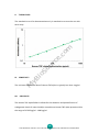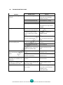
1
Human TNF alpha ELISA Kit
Catalog # MBS824724
M
yB
io
So
ur
ce
.c
om
User Manual
Sandwich Enzyme-Linked Immunosorbent Assay for Quantitative
Detection of Human TNF alpha Concentrations in Cell Culture
Supernatants, Serum, Plasma, Urine, Tissue Homogenates.
For research use only. Not for diagnostic or therapeutic procedures.
FOR RESEARCH USE ONLY. NOT FOR USE IN DIAGNOSTIC OR THERAPEUTIC PROCEDURES.
I. INTRODUCTION.………………...............................................................……………………….2
II. ASSAY PRINCIPLES.....................................................................................................3
III. KIT COMPONENTS.………..............................................................……………………….…4
IV. STORAGE AND STABILITY.………........................………...................……………………….….4
V. MATERIALS REQUIRED BUT NOT PROVIDED.………………........................……….…….….5
VI. HEALTH AND SAFETY PRECAUTIONS…………......................................………….………..5
om
VII. REAGENT PREPARATION………….......…………….............................................………....6
VIII. ASSAY PROCEDURE................................…….......................................………………...9
.c
IX. ASSAY PROCEDURE SUMMARY…..……………...…......................................................11
ce
X. TYPICAL DATA……………..………...............……....................................................…….…12
ur
XI. SENSITIVITY……………………................................................................…..….……...…12
XII. SPECIFICITY……………......................................................................………………….…12
So
XIII. CROSS REACTIVITY………………..............................................................………………13
M
yB
io
XIV. REFERENCES……………................…………...……................……................................13
XV. TROUBLESHOOTING GUIDE……………….………..........................................…………….14
XVI. TECHNICAL SUPPORT.…….............................................................…………..…..……15
XVII. NOTES.……...................................................................................…………..…..……15
1
FOR RESEARCH USE ONLY. NOT FOR USE IN DIAGNOSTIC OR THERAPEUTIC PROCEDURES.
I.
INTRODUCTION
Tumor necrosis factor-alpha (TNF-alpha, or TNF) is secreted by macrophages in
response to inflammation, infection and cancer. Human Tumor Necrosis Factor (TNF)
and Lymphotoxin (TNF-beta) are cytotoxic proteins which have similar biological
activities and share 30% amino acid homology. TNF-alpha is produced by monocytes,
which can stimulate endothelial cells to produce the multilineage growth factor
granulocyte-macrophage colony-stimulating factor and extend the role of this
immunoregulatory protein to the regulation of hematopoiesis in vitro. TNF is a
om
soluble protein that causes damage to tumor cells but has no effect on normal cells.
Human TNF has been purified to apparent homogeneity as a 17.3-kilodalton protein
.c
from HL-60 leukemia cells and has showed cytotoxic and cytostatic activities against
ce
various human tumor cell lines. The human TNF cDNA is 1585 base pairs in length
ur
and encodes a protein of 233 amino acids. The mature protein begins at residue 77,
M
yB
io
human chromosome 6.2
So
leaving a long leader sequence of 76 amino acids. TNF-alpha has been mapped to
2
FOR RESEARCH USE ONLY. NOT FOR USE IN DIAGNOSTIC OR THERAPEUTIC PROCEDURES.
II.
ASSAY PRINCIPLES
The Human TNF alpha ELISA (Enzyme-Linked Immunosorbent Assay) kit is an in vitro
enzyme-linked immunosorbent assay for the quantitative measurement of Human
TNF alpha in Cell Culture Supernatants, Serum, Plasma, Urine, Tissue Homogenates.
This assay employs an antibody specific for Human TNF alpha coated on a 96-well
plate. Standards and samples are pipetted into the wells and TNF alpha present in a
sample is bound to the wells by the immobilized antibody. The wells are washed
and biotinylated anti-Human TNF alpha antibody is added. After washing away
om
unbound biotinylated antibody, HRP-conjugated streptavidin is pipetted to the
wells. The wells are again washed, a TMB substrate solution is added to the wells
.c
and color develops in proportion to the amount of TNF alpha bound. The Stop
ce
Solution changes the color from blue to yellow, and the intensity of the color is
M
yB
io
So
ur
measured at 450 nm.
3
FOR RESEARCH USE ONLY. NOT FOR USE IN DIAGNOSTIC OR THERAPEUTIC PROCEDURES.
III.
KIT COMPONENTS
Component
Volume
12 x 8 Strips
Human TNF alpha Standard
1 ng x 2
Biotin-Labeled Detection Antibody (100X)
120 µl
Streptavidin-HRP (100X)
120 µl
Standard/Sample Diluent
30 ml
Detection Antibody Diluent
12 ml
om
96-well Plate Coated With Anti-Human TNF alpha Antibody
Streptavidin-HRP Diluent
12 ml
30 ml
.c
Wash Buffer (20X)
ce
TMB Substrate Solution
Stop Solution
IV.
So
12 ml
3 Strips
1 Manual
M
yB
io
Technical Manual
ur
Plate Adhesive Strips
12 ml
STORAGE AND STABILITY
All kit components are stable at 2 to 8 °C. Standard (recombinant protein) should be
stored at -20 °C or -80 °C (recommended at -80 °C) after reconstitution. Opened
Microplate Wells or reagents may be store for up to 1 month at 2 to 8 °C. Return
unused wells to the pouch containing desiccant pack, reseal along entire edge.
Note: the kit can be used within one year if the whole kit is stored at -20 °C. Avoid
repeated freeze-thaw cycles.
4
FOR RESEARCH USE ONLY. NOT FOR USE IN DIAGNOSTIC OR THERAPEUTIC PROCEDURES.
V.
MATERIALS REQUIRED BUT NOT PROVIDED
1. Microplate reader capable of measuring absorbance at 450 nm.
2. Adjustable pipettes and pipette tips to deliver 2 µl to 1 ml volumes.
3. Adjustable 1-25 ml pipettes for reagent preparation.
4. 100 ml and 1 liter graduated cylinders.
5. Absorbent paper.
6. Distilled or deionized water.
7. Computer and software for ELISA data analysis.
.c
ce
HEALTH AND SAFETY PRECAUTIONS
ur
VI.
om
8. Tubes to prepare standard or sample dilutions.
1. Reagents provided in this kit may be harmful if ingested, inhaled or absorbed
M
yB
io
conducting the experiment.
So
through the skin. Please carefully review the MSDS for each reagent before
2. Stop Solution contains 2 N Sulfuric Acid (H 2 SO 4 ) and is an extremely corrosive
agent. Please wear proper eye, hand and face protection when handling this material.
When the experiment is finished, be sure to rinse the plate with copious amounts of
running water to dilute the Stop Solution prior to disposing the plate.
5
FOR RESEARCH USE ONLY. NOT FOR USE IN DIAGNOSTIC OR THERAPEUTIC PROCEDURES.
VII.
REAGENT PREPARATION
1. Sample Preparation
Store samples to be assayed within 24 hours at 2-8°C. For long-term storage, aliquot
and freeze samples at -20°C. Avoid repeated freeze-thaw cycles.
Cell culture supernates: Remove particulates by centrifugation, assay immediately or
aliquot and store samples at -20°C.
Serum: Allow the serum to clot in a serum separator tube (about 4 hours) at room
temperature. Centrifuge at approximately 1000 X g for 15 minutes. Analyze the
om
serum immediately or aliquot and store samples at -20°C.
Plasma: Collect plasma using heparin or EDTA as an anticoagulant. Centrifuge for 15
.c
minutes at 1500 X g within 30 minutes of collection. Assay immediately or aliquot
ce
and store samples at -20°C.
ur
Cell Lysates: Collect cells and rinse cells with PBS. Homogenize and lyse cells
throughly in lysate solution. Centrifuge cell lysates at approximately 10000 X g for 5
So
minutes to remove debris. Aliquots of the cell lysates were removed and assayed.
M
yB
io
Bone Tissue: Extract demineralized bone samples in 4 M Guanidine-HCl and protease
inhibitors. Dissolve the final sample in 2 M Guanidine-HCl.
Tissue Homogenates: Rinse tissue with PBS to remove excess blood, chopped into
1-2 mm pieces, and homogenize with a tissue homogenizer in PBS or in lysate
solution, lysate solution: tissue net weight = 10ml : 1g (i.e. Add 10ml lysate solution
to 1g tissue). Centrifuge at approximately 5000 X g for 5 minutes. Assay immediately
or aliquot and store homogenates at -20°C. Avoid repeated freeze-thaw cycles.
Urine: Urinary samples should be cleared by centrifugation and then can be used
directly without dilution. Storage at -20°C.
2. Human TNF alpha Standard Preparation
Reconstitute the lyophilized Human TNF alpha Standard by adding 1 ml of
Standard/Sample Diluent to make the 1000 pg/ml standard stock solution. Allow
6
FOR RESEARCH USE ONLY. NOT FOR USE IN DIAGNOSTIC OR THERAPEUTIC PROCEDURES.
solution to sit at room temperature for 5 minutes, then gently vortex to mix
completely. Use within one hour of reconstituting. Two tubes of the standard (1 ng
per tube) are included in each kit. Use one tube for each experiment.
Perform 2-fold serial dilutions of the top standards to make the standard curve
within the range of this assay (15.625 pg/ml - 1000 pg/ml) as below.
Standard/Sample Dilution Buffer serves as the zero standard (0 pg/ml).
Standard
Add
Into
1000 pg/ml
500 µl of the Standard (1000 pg/ml)
500 µl of the Standard/Sample Diluent
250 pg/ml
500 µl of the Standard (500 pg/ml)
500 µl of the Standard/Sample Diluent
125 pg/ml
500 µl of the Standard (250 pg/ml)
500 µl of the Standard/Sample Diluent
62.5 pg/ml
500 µl of the Standard (125 pg/ml)
31.25 pg/ml
500 µl of the Standard (62.5 pg/ml)
500 µl of the Standard/Sample Diluent
15.625 pg/ml
500 µl of the Standard (31.25 pg/ml)
500 µl of the Standard/Sample Diluent
0 ng/ml
1 ml of the Standard/Sample Diluent
.c
om
500 pg/ml
So
ur
ce
500 µl of the Standard/Sample Diluent
M
yB
io
Note: The standard solutions are best used within 2 hours. The 1000 pg/ml standard
solution should be stored at 4°C for up to 12 hours, or at -20°C for up to 48 hours.
Avoid repeated freeze-thaw cycles.
3. Biotin-Labeled Detection Antibody Working Solution Preparation
The Biotin-Labeled Detection Antibody should be diluted in 1:100 with the Detection
Antibody Diluent and mixed thoroughly. The solution should be prepared no more
than 2 hours prior to the experiment.
4. Streptavidin-HRP Working Solution Preparation
The Streptavidin-HRP should be diluted in 1:100 with the Streptavidin-HRP Diluent
and mixed thoroughly. The solution should be prepared no more than 1 hour prior to
the experiment.
7
FOR RESEARCH USE ONLY. NOT FOR USE IN DIAGNOSTIC OR THERAPEUTIC PROCEDURES.
5. Wash Buffer Working Solution Preparation
Pour entire contents (30 ml) of the Wash Buffer Concentrate into a clean 1,000 ml
graduated cylinder. Bring final volume to 600 ml with glass-distilled or deionized
M
yB
io
So
ur
ce
.c
om
water (1:20).
8
FOR RESEARCH USE ONLY. NOT FOR USE IN DIAGNOSTIC OR THERAPEUTIC PROCEDURES.
VIII.
ASSAY PROCEDURE
The Streptavidin-HRP Working Solution and TMB Substrate Solution must be kept
warm at 37°C for 30 minutes before use. When diluting samples and reagents, they
must be mixed completely and evenly. Standard detection curve should be prepared
for each experiment. The user will decide sample dilution fold by crude estimation of
protein amount in samples.
1. Add 100 µl of each standard and sample into appropriate wells.
om
2. Cover well and incubate for 90 minutes at room temperature or over night at 4°C
with gentle shaking.
.c
3. Remove the cover, discard the solution and wash plate 3 times with Wash Buffer
ce
Working Solution, and each time let Wash Buffer Working Solution stay in the wells
ur
for 1 - 2 minutes. Blot the plate onto paper towels or other absorbent material. Do
NOT let the wells completely dry at any time.
So
4. Add 100 µl of Biotin-Labeled Detection Antibody Working Solution into each well
M
yB
io
and incubate the plate at 37°C for 60 minutes.
5. Wash plate 3 times with Wash Buffer Working Solution, and each time let Wash
Buffer Working Solution stay in the wells for 1 - 2 minutes. Discard the Wash Buffer
Working Solution and blot the plate onto paper towels or other absorbent material.
6. Add 100 µl of Streptavidin-HRP Working Solution into each well and incubate the
plate at 37°C for 45 minutes.
7. Wash plate 5 times with Wash Buffer Working Solution, and each time let wash
buffer stay in the wells for 1 - 2 minutes. Discard the wash buffer and blot the plate
onto paper towels or other absorbent material.
8. Add 100 µl of TMB Substrate Solution into each well and incubate plate at 37°C in
dark for 30 minutes.
9. Add 100 µl of Stop Solution into each well. The color changes into yellow
immediately.
9
FOR RESEARCH USE ONLY. NOT FOR USE IN DIAGNOSTIC OR THERAPEUTIC PROCEDURES.
10. Read the O.D. absorbance at 450nm in a microplate reader within 30 minutes
after adding the Stop Solution.
For calculation, (the relative O.D.450) = (the O.D.450 of each well) - (the O.D.450 of
Zero well). The standard curve can be plotted as the relative O.D.450 of each
standard solution (Y) vs. the respective concentration of the standard solution (X).
The concentration of the samples can be interpolated from the standard curve.
om
Note: If the samples measured were diluted, multiply the dilution factor to the
M
yB
io
So
ur
ce
.c
concentrations from interpolation to obtain the concentration before dilution.
10
FOR RESEARCH USE ONLY. NOT FOR USE IN DIAGNOSTIC OR THERAPEUTIC PROCEDURES.
So
ur
ce
.c
om
ASSAY PROCEDURE SUMMARY
M
yB
io
IX.
11
FOR RESEARCH USE ONLY. NOT FOR USE IN DIAGNOSTIC OR THERAPEUTIC PROCEDURES.
X.
TYPICAL DATA
The standard curve is for demonstration only. A standard curve must be run with
XI.
M
yB
io
So
ur
ce
.c
om
each assay.
SENSITIVITY
The minimum detectable dose of Human TNF alpha is typically less than 4 pg/ml.
XII.
SPECIFICITY
The Human TNF alpha ELISA Kit allows for the detection and quantification of
endogenous levels of natural and/or recombinant Human TNF alpha proteins within
the range of 15.625 pg/ml - 1000 pg/ml.
12
FOR RESEARCH USE ONLY. NOT FOR USE IN DIAGNOSTIC OR THERAPEUTIC PROCEDURES.
XIII.
CROSS REACTIVITY
No detectable cross-reactivity with other relevant proteins.
XIV.
REFERENCES
1. Brenner, D. A.; O'Hara, M.; Angel, P.; Chojkier, M.; Karin, M. Prolonged activation of
JUN and collagenase genes by tumour necrosis factor-alpha. Nature 337: 661-663,
om
1989.
2. Nedwin, G. E.; Naylor, S. L.; Sakaguchi, A. Y.; Smith, D.; Jarrett-Nedwin, J.; Pennica,
.c
D.; Goeddel, D. V.; Gray, P. W. Human lymphotoxin and tumor necrosis factor genes:
ce
structure, homology and chromosomal localization. Nucleic Acids Res. 13: 6361-6373,
ur
1985.
3. Broudy, V. C.; Kaushansky, K.; Segal, G. M.; Harlan, J. M.; Adamson, J. W. Tumor
So
necrosis factor type alpha stimulates human endothelial cells to produce
M
yB
io
granulocyte/macrophage colony-stimulating factor. Proc. Nat. Acad. Sci. 83:
7467-7471, 1986.
4. Wang, A. M.; Creasey, A. A.; Ladner, M. B.; Lin, L. S.; Strickler, J.; Van Arsdell, J. N.;
Yamamoto, R.; Mark, D. F. Molecular cloning of the complementary DNA for human
tumor necrosis factor. Science 228: 149-154, 1985.
13
FOR RESEARCH USE ONLY. NOT FOR USE IN DIAGNOSTIC OR THERAPEUTIC PROCEDURES.
XV.
TROUBLESHOOTING GUIDE
Problem
High signal and background in
all wells
Solution
• Insufficient washing
• Increase number of washes
• Increase time of soaking
between in wash
• Too much Streptavidin-HRP
• Check dilution, titration
• Incubation time too long
• Reduce incubation time
• Development time too long
• Decrease the incubation
time before the stop solution
is added
• Reagent added in incorrect
order, or incorrectly prepared
• Review protocol
• Standard has gone bad (If
there is a signal in the sample
wells)
• Check the condition of
stored standard
• Assay was conducted from an
incorrect starting point
• Reagents allows to come to
20 - 30 °C before performing
assay
• Insufficient washing-unbound
Streptavidin-HRP remaining
• Increase number of washes
Carefully
• Too much Streptavidin-HRP
• Check dilution
• Plate sealer or reservoir
reused, resulting in presence of
residual Streptavidin-HRP
• Use fresh plate sealer and
reagent reservoir for each
step
Standard curve achieved but
poor discrimination between
point
• Plate not developed long
enough
• Increase substrate solution
incubation time
• Improper calculation of
standard curve dilution
• Check dilution, make new
standard curve
No signal when a signal is
expected, but standard curve
looks fine
• Sample matrix is masking
detection
• More diluted sample
Recommended
Samples are reading too high,
but standard curve is fine
• Samples contain protein levels
above assay range
• Dilute samples and run
Again
Edge effect
• Uneven temperature around
work surface
• Avoid incubating plate in
areas where environmental
conditions vary
• Use plate sealer
So
M
yB
io
Too much signal-whole plate
turned uniformly blue
ur
ce
.c
om
No signal
Possible Cause
14
FOR RESEARCH USE ONLY. NOT FOR USE IN DIAGNOSTIC OR THERAPEUTIC PROCEDURES.
XVI.
TECHNICAL SUPPORT
NOTES
M
yB
io
XVII.
So
ur
ce
.c
om
For troubleshooting, information or assistance, please go online
15
FOR RESEARCH USE ONLY. NOT FOR USE IN DIAGNOSTIC OR THERAPEUTIC PROCEDURES.