1
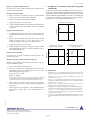
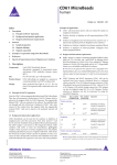
CD326 (EpCAM)
MicroBeads
human
Order no. 130-061-101
Index
Example applications
1.Description
●
Enrichment of disseminated carcinoma cells from peripheral
blood, bone marrow, and lymphoid tissue, for example, of
patients with epithelial cancer for subsequent analysis, e.g.,
enumeration, cultivation, or RT-PCR.
●
Purification of CD326 (EpCAM)+ cells from epithelial tissues
(e.g. carcinomas).
1.1 Principle of MACS® Separation
1.2 Background and product applications
1.3 Reagent and instrument requirements
2.Protocol
2.1 Sample preparation
2.2 Magnetic labeling
2.3 Magnetic separation
1.3 Reagent and instrument requirements
●
Buffer: Prepare a solution containing phosphate-buffered saline
(PBS) pH 7.2, 0.5% bovine serum albumin (BSA), and 2 mM
EDTA by diluting MACS BSA Stock Solution (# 130-091-376)
1:20 with autoMACS™ Rinsing Solution (# 130-091-222). Keep
buffer cold (4−8 °C). Degas buffer before use, as air bubbles
could block the column.
▲ Note: EDTA can be replaced by other supplements such as anticoagulant citrate
dextrose formula-A (ACD-A) or citrate phosphate dextrose (CPD). BSA can be
replaced by other proteins such as human serum albumin, human serum, or fetal
calf serum. Buffers or media containing Ca 2+ or Mg2+ are not recommended for
use.
●
MACS Columns and MACS Separators: CD326 (EpCAM)+
cells can be enriched by using MS, LS or XS Columns (positive
selection) or depleted with the use of LD, CS, or D Columns.
Cells which strongly express the CD326 (EpCAM) antigen
can also be depleted using MS, LS, or XS Columns. Positive
selection or depletion can also be performed by using the
autoMACS Separator.
3. Example of a separation using CD326 (EpCAM)
MicroBeads
4.References
5.Appendix
1.Description
Components
Size
2 mL CD326 (EpCAM) MicroBeads, human:
MicroBeads conjugated to monoclonal antibody
(isotype: mouse IgG1).
For 10⁹ total cells, up to 20 separations.
Product format CD326 (EpCAM) MicroBeads are supplied in
buffer containing stabilizer and 0.05% sodium
azide.
Storage
Store protected from light at 2−8 °C. Do not
freeze. The expiration date is indicated on the
vial label.
1.1 Principle of MACS® Separation
First, the CD326 (EpCAM)+ cells are magnetically labeled with
CD326 (EpCAM) MicroBeads. Then the cell suspension is loaded
onto a MACS® Column which is placed in the magnetic field of a
MACS Separator. The magnetically labeled CD326 (EpCAM)+ cells
are retained within the column. The unlabeled cells run through;
this cell fraction is depleted of CD326 (EpCAM)+ cells. After
removing the column from the magnetic field, the magnetically
retained CD326 (EpCAM)+ cells can be eluted as the positively
selected cell fraction.
Column
Max. number Separator
of total cells
Positive selection
2×10⁸
MS10⁷
MiniMACS, OctoMACS, VarioMACS, SuperMACS
LS10⁸
2×10⁹
MidiMACS, QuadroMACS,
VarioMACS, SuperMACS
XS10⁹
Depletion
2×10¹⁰
SuperMACS
5 ×10⁸
LD10⁸
CS
2×10⁸
MidiMACS, QuadroMACS,
VarioMACS, SuperMACS
VarioMACS, SuperMACS
SuperMACS
D10⁹
1.2 Background and product applications
140-000-108.09
Epithelial cell adhesion molecule (EpCAM), which is also known as
CD326 and human epithelial antigen (HEA), is broadly expressed
on cells of epithelial origin and on epithelial derived tumor cells¹.
CD326 (EpCAM) MicroBeads are used for the positive selection of
viable epithelial tumor cells from peripheral blood, bone marrow,
lymphoid tissue and serous effusions of patients with carcinomas.
In order to prevent FcR-mediated non-specific labeling of nonepithelial cells it is strongly recommended to use FcR Blocking
Reagent before magnetically labeling.
Max. number
of labeled cells
Positive selection or depletion
autoMACS2×10⁸
4×10⁹
autoMACS
▲ Note: Column adapters are required to insert certain columns into the
VarioMACS™ or SuperMACS™ Separators. For details see the respective MACS
Separator data sheet.
●
FcR Blocking Reagent containing human IgG (# 130-059-901)
to avoid Fc receptor-mediated staining.
www.miltenyibiotec.com
Miltenyi Biotec GmbH
Friedrich-Ebert-Str. 68
51429 Bergisch Gladbach, Germany
Phone +49 2204 8306 0 Fax +49 2204 85197
Miltenyi Biotec Inc.
2303 Lindbergh Street, Auburn, CA 95602, USA
Phone 800 FOR MACS, +1 530 888 8871 Fax +1 530 888 8925
page 1/4
Order no. 130-061-101
●
(Optional) Fluorochrome-conjugated CD326 antibody for
flow cytometric analysis, for example, CD326 (EpCAM)-FITC
(#
130-080-301), CD326 (EpCAM)-PE (#
130-091-253), or
CD326 (EpCAM)-APC (# 130-091-254) and CD45 conjugated
to a different fluorochrome, for example, CD45-FITC
(# 130-080-202), CD45-PE (# 130-080-201), or CD45-APC
(# 130-091-230).
1.
Determine cell number.
2. Centrifuge cell suspension at 300×g for 10 minutes. Aspirate
supernatant completely.
3. Resuspend cell pellet in 300 µL of buffer per 5×10⁷ total cells.
4. Add 100 µL of FcR Blocking Reagent per 5×10⁷ total cells and
mix well.
●
(Optional) Propidium iodide (PI) or 7-AAD for flow-cytometric
exclusion of dead cells.
●
(Optional) Pre-Separation Filters (# 130-041-407) to remove
cell clumps.
5. Add 100 µL of CD326 (EpCAM) MicroBeads per 5×10⁷ total
cells.
●
(Optional) for bone marrow: HEPES buffered cell culture
medium (e.g. Iscove’s modified Dulbecco medium [IMDM]).
●
(Optional) for bone marrow: Heparin.
●
(Optional) for bone marrow and cryopreserved cells: DNaseI.
●
(Optional) for cryopreserved cells: Dulbecco’s PBS (pH 7.0−7.2),
containing 0.5 mM MgCl2 and 1 mM CaCl2, 0.5% BSA.
2.Protocol
2.1 Sample preparation
▲ Note: FcR Blocking Reagent should be used to block Fc receptor-mediated
labeling of non-epithelial cells.
6. Mix well and refrigerate for 30 minutes (4−8 °C).
▲ Note: Working on ice may require increased incubation times. Higher
temperatures and/or longer incubation times may lead to non-specific cell
labeling.
7. (Optional) Add staining antibodies, for example, 50 µL of
CD326 (EpCAM)-FITC (# 130-080-301) and CD45 conjugated
to another fluorochrome (e.g. CD45-PE, # 130-080-201), and
refrigerate for 5 minutes in the dark (4−8 °C).
8. Wash cells by adding 5−10 mL of buffer per 5×10⁷ cells and
centrifuge at 300×g for 10 minutes. Aspirate supernatant
completely.
When working with anticoagulated peripheral blood or buffy coat,
peripheral blood mononuclear cells (PBMCs) should be isolated
by density gradient centrifugation, e.g. using Ficoll-Paque™. For
details see section General Protocols in the user manuals or visit
www.miltenyibiotec.com/protocols.
▲ Note: For higher cell numbers, scale up buffer volume accordingly.
▲ Note: For depletion with LD Columns, resuspend for up to 1.25×10⁸ cells in
500 µL of buffer.
10. Proceed to magnetic separation (2.3).
▲ Note: To remove platelets after density gradient separation, resuspend cell pellet
in buffer and centrifuge at 200×g for 10−15 minutes at 20 °C. Carefully aspirate
supernatant. Repeat washing step.
When working with tissues, prepare a single-cell suspension by
a standard preparation method. For details see section General
Protocols in the user manuals or visit www.miltenyibiotec.com/
protocols.
▲ Note: Dead cells may bind non-specifically to MACS MicroBeads. To remove
dead cells, we recommend using density gradient centrifugation.
2.2 Magnetic labeling
▲ Work fast, keep cells cold, and use pre-cooled solutions. This will
prevent capping of antibodies on the cell surface and non-specific
cell labeling.
▲ Volumes for magnetic labeling given below are for up to 5×10⁷
total cells. When working with fewer than 5×10⁷ cells, use the same
volumes as indicated. When working with higher cell numbers,
scale up all reagent volumes and total volumes accordingly (e.g. for
10⁸ total cells, use twice the volume of all indicated reagent volumes
and total volumes).
▲ For optimal performance it is important to obtain a singlecell suspension before magnetic separation. Pass cells through
30 µm nylon mesh (Pre-Separation Filters, # 130-041-407) to remove
cell clumps which may clog the column.
▲ Always include positive and negative controls when working with
patient samples. For a positive control, PBMCs spiked with tumor
cells should be used. For a negative control, simply use PBMCs from
a healthy donor.
9. Resuspend up to 10⁸ cells in 500 µL of buffer.
2.3 Magnetic separation
▲ Choose an appropriate MACS Column and MACS Separator
according to the number of total cells and the number of
CD326 (EpCAM)+ cells. For details see table in section 1.3.
Magnetic separation with MS or LS Columns
1.
Place column in the magnetic field of a suitable MACS Separator.
For details see respective MACS Column data sheet.
2. Prepare column by rinsing with appropriate amount of buffer:
MS: 500 µL
LS: 3 mL
3. Apply cell suspension onto the column.
4. Collect unlabeled cells that pass through and wash column
with appropriate amount of buffer. Perform washing steps
by adding buffer three times. Only add new buffer when the
column reservoir is empty.
MS: 4×500 µL
LS: 4×3 mL
Collect total effluent; this is the unlabeled cell fraction.
5. Remove column from the separator and place it on a suitable
collection tube.
6. Pipette an appropriate amount of buffer onto the column.
Immediately flush out the magnetically labeled cells by firmly
pushing the plunger into the column.
MS: 1 mL
LS: 5 mL
140-000-108.09
▲ Note: To increase the purity of the magnetically labeled fraction, pass the
cells over a new, freshly prepared column.
www.miltenyibiotec.com
Unless otherwise specifically indicated, Miltenyi Biotec
products and services are for research use only and not for
diagnostic or therapeutic use.
page 2/4
Order no. 130-061-101
Magnetic separation with XS Columns
For instructions on the column assembly and the separation, refer
to the XS Column data sheet.
Depletion with LD Columns
1. Place LD Column in the magnetic field of a suitable MACS
Separator. For details see LD Column data sheet.
2. Prepare column by rinsing with 2 mL of buffer.
3. Example of a separation using CD326 (EpCAM)
MicroBeads
Enrichment of tumor cells from PBMCs spiked with cells from a
breast cancer cell line (BT474) using CD326 (EpCAM) MicroBeads,
a MiniMACS™ Separator, and an MS Column. Cells are fluorescently
stained with CD326 (EpCAM)-FITC (# 130-080-301) and CD45-PE
(# 130-080-201). Cell debris and dead cells are excluded from the
analysis based on scatter signals and PI fluorescence.
3. Apply cell suspension onto the column.
Collect unlabeled cells that pass through and wash column with
2×1 mL of buffer. Collect total effluent. This is the unlabeled
cell fraction.
CD45-PE
4.
Cell mixture before separation
Depletion with CS Columns
1. Assemble CS Column and place it in the magnetic field of a
suitable MACS Separator. For details see CS Column data
sheet.
2. Prepare column by filling and rinsing with 60 mL of buffer.
Attach a 22G flow resistor to the 3-way-stopcock of the
assembled column. For details see CS Column data sheet.
CD326 (EpCAM)-FITC
3. Apply cell suspension onto the column.
Negative fraction with cells
depleted of breast cancer cells
For instructions on column assembly and separation refer to the
D Column data sheet.
Positive fraction with enriched
breast cancer cells
CD45-PE
Depletion with D Columns
CD45-PE
4. Collect unlabeled cells that pass through and wash column
with 30 mL buffer from the top. Collect total effluent. This is
the unlabeled cell fraction.
Magnetic separation with the autoMACS™ Separator
▲ Refer to the autoMACS™ User Manual for instructions on how to
use the autoMACS Separator.
1.
2. Place tube containing the magnetically labeled cells in the
autoMACS Separator. For a standard separation, choose one of
the following separation programs:
Positive selection: "Posseld"
Depletion: "Depletes"
▲ Note: Program choice depends on the isolation strategy, the strength of
magnetic labeling and the frequency of magnetically labeled cells. For details
see autoMACS user manual, section autoMACS Cell Separation Programs.
3. When using the program "Posseld", collect positive fraction
from outlet port pos2. This is the purified CD326 (EpCAM)+
cell fraction.
CD326 (EpCAM)-FITC
Prepare and prime autoMACS Separator.
When using the program "Depletes", collect unlabeled fraction
from outlet port neg1. This is the CD326 (EpCAM)- cell
fraction.
CD326 (EpCAM)-FITC
4.References
1.
Moldenhauer, G. et al. (1987) Epithelium-specific surface glycoprotein of Mr
34,000 is a widely distributed human carcinoma marker. Br. J. Cancer 56: 714–
721.
2.
Coligan, J. E. et al. (eds.): Current Protocols in Immunology. New York, John
Wiley & Sons, Inc. (1994).
3.
Garau, D. et al. (1997) Detection of Breast Cancer Cells using the Magnetic
Cell Separation (MACS) System: Implications for Stem cell Purging. Europ. J.
Histochem. 41(S2): 18–19. [378]
4.
Krüger, W. et al. (1999) Improvement of breast cancer cell detection by
immunomagnetic enrichment. Cytotherapy 1: 135–139. [568]
5.
Krüger, W. et al. (2000) Immunomagnetic tumor cell selection-implications for
the detection of disseminated cancer cells. Transfusion 40: 1489–1493. [984]
6.
Leinung, S. et al. (2000) Cytokeratin-positive cells in bone marrow in
comparison with other prognostic factors in colon carcinoma. Langenbeck's
Arch. Surg. 385: 337–343. [986]
7.
Tögel, F. et al. (2001) Urokinase-like Plasminogen Activator Receptor Expression
on Disseminated Breast Cancer Cells. J. Hematother. Stem Cell Res. 10: 141–145.
[985]
140-000-108.09
www.miltenyibiotec.com
Unless otherwise specifically indicated, Miltenyi Biotec
products and services are for research use only and not for
diagnostic or therapeutic use.
page 3/4
Order no. 130-061-101
5.Appendix
Preparation of peripheral blood mononuclear cells (PBMCs)
using Ficoll-Paque™
▲ The peripheral blood or buffy coat should not be older than
8 hours and supplemented with anticoagulants (e.g. heparin, EDTA,
citrate, ACD-A or citrate phosphate dextrose (CPD)).
▲ PBMCs may be stored in the refrigerator overnight in PBS
containing 0.5% BSA supplemented with autologous serum after
the last washing step.
1. Dilute cells with 2−4× the volume of PBS containing 2 mM
EDTA (PBS/EDTA) or 0.6% ACD-A.
▲ Note: The more diluted the blood sample, the better the purity of the
mononuclear cells.
2. Carefully layer 35 mL of diluted cell suspension over 15 mL
Ficoll-Paque™ (ρ = 1.077) in a 50 mL conical tube.
3. Centrifuge at 400×g for 30−40 minutes at 20 °C in a swingingbucket rotor without brake.
4. Aspirate the upper layer leaving the mononuclear cell layer
undisturbed at the interphase.
8. Fill the conical tube with PBS containing 2 mM EDTA or 0.6%
ACD-A, mix, and centrifuge at 300×g for 10 minutes at 20 °C.
Carefully aspirate supernatant completely.
9. Repeat step 8.
▲ Note: This step increases the purity of the CD326 (EpCAM) cell separation.
10. Resuspend cell pellet in a final volume of 300 µL of buffer for
up to 10⁸ total cells. Proceed to magnetic labeling (2.2).
Preparation of cryopreserved cells
1. Thaw frozen cells (usually in 1−2 mL vials containing 10⁷−10⁸
cells) quickly at 40 °C in a waterbath by shaking the tube. Take
the tube from the waterbath when there is still a very small
piece of ice in the suspension and continue shaking for some
seconds until the ice has disappeared.
2. Place cells immediately on ice.
3. Transfer cells to a 50 mL conical tube and dilute the cells
(1−2 mL) slowly with cold (4 °C) Dulbecco's PBS (pH 7.0−7.2),
containing 0.5 mM MgCl2 and 1 mM CaCl2, 0.5% BSA,
100 U/mL DNase to a final volume of 20 mL.
4. Incubate cells for 10 minutes on ice (4 °C).
5. Centrifuge cell suspension at 300×g for 10 minutes at 4 °C.
5.Carefully transfer the interphase cells (lymphocytes,
monocytes, and thrombocytes) to a new 50 mL conical tube.
6. Carefully remove supernatant.
6. Fill the conical tube with PBS containing 2 mM EDTA or 0.6%
ACD-A, mix and centrifuge at 300×g for 10 minutes at 20 °C.
Carefully aspirate supernatant completely.
7. Resuspend cell pellet in 15 mL of Dulbecco's PBS (pH 7.0−7.2),
containing 0.5 mM Mg2+ and 1 mM Ca 2+, 0.5% BSA, and
100 U/mL DNase and centrifuge at 300×g for 10 minutes at 4 °C.
7. For removal of platelets, resuspend cell pellet in 50 mL of PBS
containing 2 mM EDTA or 0.6% ACD-A and wash twice:
centrifuge at 200×g for 10−15 minutes at 20 °C. Remove
supernatant completely.
8. Carefully remove supernatant completely.
▲ Note: This step increases the purity of the CD326 (EpCAM) cell separation.
8.
Repeat step 7. Most of the platelets will remain in the supernatant
upon centrifugation at 200×g.
9. Resuspend cell pellet in a final volume of 300 µL of buffer for
up to 5×10⁷ total cells. Proceed to magnetic labeling (2.2).
Preparation of bone marrow cells
1. Collect bone marrow in 50 mL tubes containing 5 mL PBS,
pH 7.2, containing 2 mM EDTA, or 0.6% ACD-A, or 200 U/mL
heparin.
▲ Note: Store cells at 4 °C if the cells cannot be processed immediately.
2. For preparation of a single-cell suspension of bone marrow
cells dilute with 10 × the volume of RPMI 1640 containing
0.02% collagenase B and 100 U/mL DNase and shake gently at
room temperature for 45 minutes.
3. Pass cells through 30 µm nylon mesh (Pre-Separation Filters,
# 130-041-407), in order to remove cells clumps. Wet filter with
buffer before use.
4. Carefully layer 35 mL of diluted cell suspension over 15 mL
Ficoll-Paque in a 50 mL conical tube.
9. Resuspend cell pellet in a final volume of 300 µL of buffer for
up to 5×10⁷ total cells. Proceed to magnetic labeling (2.2).
Preparation of cells from lymphoid tissue
1. Isolate single-cell suspension from lymphoid tissue by a
standard preparation method.
2. Centrifuge cells at 300×g for 10 minutes in a 15 mL conical tube.
3. Carefully aspirate supernatant completely.
4. Resuspend cell pellet in a final volume of 300 µL of buffer for
up to 5×10⁷ total cells. Proceed to magnetic labeling (2.2).
Warnings
Reagents contain sodium azide. Under acidic conditions sodium azide yields
hydrazoic acid, which is extremely toxic. Azide compounds should be diluted with
running water before discarding. These precautions are recommended to avoid
deposits in plumbing where explosive conditions may develop.
Warranty
The products sold hereunder are warranted only to be free from defects in workmanship
and material at the time of delivery to the customer. Miltenyi Biotec GmbH
makes no warranty or representation, either expressed or implied, with respect to
the fitness of a product for a particular purpose. There are no warranties, expressed
or implied, which extend beyond the technical specifications of the products.
Miltenyi Biotec GmbH’s liability is limited to either replacement of the products or refund
of the purchase price. Miltenyi Biotec GmbH is not liable for any property damage,
personal injury or economic loss caused by the product.
140-000-108.09
5. Centrifuge at 400×g for 35 minutes at 20 °C in a swingingbucket rotor without brake.
MACS is a registered trademark and autoMACS, MidiMACS, MiniMACS, OctoMACS,
QuadroMACS, SuperMACS, and VarioMACS are trademarks of Miltenyi Biotec
GmbH.
6. Aspirate the upper layer leaving the mononuclear cell layer
undisturbed at the interphase.
Ficoll-Paque is a trademark of GE Healthcare companies.
7. Carefully transfer the interphase cells (lymphocytes,
monocytes, and thrombocytes) to a new 50 mL conical tube.
© 2007 Miltenyi Biotec GmbH.
www.miltenyibiotec.com
Unless otherwise specifically indicated, Miltenyi Biotec
products and services are for research use only and not for
diagnostic or therapeutic use.
page 4/4