1
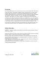
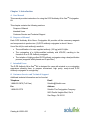
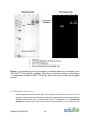
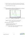
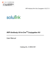
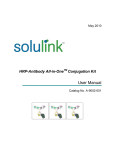
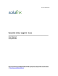
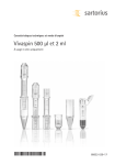
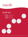
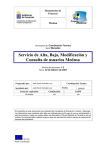
Version 08.31.2012 R-PE Antibody All-In-One Conjugation Kit Technical Manual Catalog # A-9001-006 Note: This protocol and any documents linked below can be downloaded from the appropriate category in the Solulink Library at http://www.solulink.com/library. Catalog # A-9001-006 1 Disclaimer The products offered here are for research use only. Any commercial application will require a license from Solulink. The Solulink Conjugation System is patented and has multiple patents pending. Please contact Solulink for information regarding licensing information. Solulink products and methods may be covered by one or more of the following United States patents Nos. 6,686,461, 6,800,728, 7,102,024, 7,173,125, 7,462,689 and other pending patent applications. Information in this manual is subject to change without notice and does not constitute a commitment on the part of Solulink, Inc. It is supplied on an “as is” basis without any warranty of any kind, either explicit or implied. Information may be changed or updated in this manual at any time. This document may not be copied, transferred, reproduced, disclosed, or duplicated, in whole or in part, without the prior written consent of Solulink, Inc. This documentation is proprietary information and protected by the copyright laws of the United States and international treaties. The manufacturer of this documentation is Solulink, Inc Safety Information WARNING – CHEMICAL HAZARD. Some chemicals used can be potentially hazardous, and can cause injury or illness. • Read and understand the Material Safety Data Sheets (MSDS) available at Solulink.com before you store, handle, or work with any chemicals or hazardous materials. • Minimize contact with and inhalation of chemicals. Wear appropriate personal protective equipment when handling chemicals (e.g. safety glasses, gloves, or clothing). For additional safety guidelines consult the MSDS. • Check regularly for chemical leaks or spills. If a leak or spill occurs, follow the manufacturer’s clean-up procedures as recommended in the MSDS. • Comply with all local, state/provincial, or national laws and regulations related to chemical storage, handling and disposal. Catalog # A-9001-006 2 Table of Contents Chapter 1: Introduction...................................................................................................................4 A. B. C. D. User Manual...................................................................................................................................... 4 Purpose of Manual............................................................................................................................ 4 Intended Users.................................................................................................................................. 4 Customer Service and Technical Support ......................................................................................... 4 Chapter 2: Overview of Conjugation ..............................................................................................5 A. B. C. D. E. Product Description .......................................................................................................................... 5 All-in-OneTM Conjugation Technology............................................................................................... 5 All-in-OneTM Conjugation Process Summary..................................................................................... 8 Materials Provided and Storage Conditions ..................................................................................... 9 Additional Materials Required But Not Provided ............................................................................. 9 Chapter 3: All-in-OneTM Conjugation Protocol...............................................................................9 A. B. C. D. E. F. G. H. I. IgG Sample Preparation (5 minutes)............................................................................................... 10 Buffer Exchange IgG (3 minutes)..................................................................................................... 10 HyNic Modify IgG (2 h).................................................................................................................... 12 Buffer Exchange IgG (3 minutes)..................................................................................................... 12 Conjugate Formation (2 h).............................................................................................................. 13 Conjugate Purification Stage 1: Affinity Isolation (1 ¾ hours) ........................................................ 13 Buffer Exchange Conjugate (3 minutes) ......................................................................................... 15 Conjugate Purification Stage 2: Q Spin Filter (35 min) ................................................................... 16 Buffer Exchange Conjugate (3 minutes) ......................................................................................... 17 Chapter 4: Appendix......................................................................................................................18 A. B. C. D. E. F. G. H. Human IgG-R-PE Conjugate (Monoclonal): An example................................................................ 18 Bradford Protein Assay ................................................................................................................... 19 Using a NanoDropTM to Measure Antibody Concentration ............................................................ 21 R-PE Absorption Spectrum (Unmodified R-PE)............................................................................... 24 R-PE Absorption Spectrum (4FB-modified)..................................................................................... 25 Concentration of Dilute Antibody Solutions ................................................................................... 25 Troubleshooting Guide ................................................................................................................... 27 References ...................................................................................................................................... 28 Catalog # A-9001-006 3 Chapter 1: Introduction A. User Manual This manual provides instructions for using the R-PE Antibody All-in-OneTM Conjugation Kit. This chapter contains the following sections: Purpose of Manual Intended Users Customer Service and Technical Support B. Purpose of Manual Each R-PE Antibody All-in-OneTM Conjugation Kit provides all the necessary reagents and components to produce two (2) R-PE antibody conjugates in about 6 hours. Use of the kit (for each antibody) results in: The modification of a user-supplied antibody (100 µg) with S-HyNic. The conjugation of a HyNic-modified antibody with 4FB-PE, resulting in the formation of an R-PE Antibody conjugate. The isolation of highly purified R-PE Antibody conjugates using a dual purification process (magnetic affinity beads and Q spin filter) C. Intended Users The R-PE Antibody All-in-OneTM Kit is designed for users with minimal or no conjugation experience allowing them to prepare customized, high purity, ready-to-use R-PE Antibody conjugates in a single day. D. Customer Service and Technical Support Additional technical information can be found at: Telephone Email 1-888-625-0670 (Toll Free) [email protected] Fax Address 1-858-625-0770 Solulink-The Conjugation Company 9853 Pacific Heights Blvd, Ste H San Diego, CA 92121 Catalog # A-9001-006 4 Chapter 2: Overview of Conjugation A. Product Description Each R-PE Antibody All-in-OneTM Conjugation kit is designed to produce two high quality ready-to-use R-PE antibody conjugates. Each conjugation requires 100 µg of user supplied mammalian IgG from any species. The kit is designed to produce heterodimer conjugates (1:1 R-PE to IgG ratio) devoid of virtually all free R-PE or residual IgG. Any suitably purified monoclonal or polyclonal antibody free of protein carriers or stabilizers (e.g. BSA or gelatin) can be used to form the conjugate. The kit employs a dual purification strategy using magnetic affinity beads and a Q spin filter. Conjugates produced are fully compatible with all demanding downstream applications including flow-cytometry and/or sensitive microarray detection. Each kit contains sufficient reagents to produce two ready-to-use R-PE antibody conjugates (~125 µg each). B. All-in-OneTM Conjugation Technology Conjugation Chemistry HydraLinKTM chemistry is based on the use of two complementary heterobifunctional linkers; S-HyNic and Sulfo-S-4FB (Figure 1). S-HyNic (Succinimidyl-6-hydrazinonicotinamide) is first used to modify and incorporate protected aromatic hydrazines (HyNic groups) into the antibody via acylation of lysine residues. In a similar fashion the second linker, Sulfo-S-4FB (Sulfo-N-succinimidyl-4-formylbenzamide) is used by Solulink to form a pre-activated form of R-PE called 4FB-PE (provided in the kit). Incubation of the HyNic-modified antibody with pre-activated 4FB-PE in the presence of aniline as catalyst leads to rapid and efficient conversion of the antibody to conjugate through formation of stable bis-arylhydrazone bonds (Figure 2). Figure 1. Structure of S-HyNic and Sulfo-S-4FB linkers used to conjugate R-PE to antibody. Catalog # A-9001-006 5 Figure 2. Conjugation chemistry used in linking IgG to R-PE. Purification Technology This All-in-OneTM kit uses a two-stage purification process to generate highly purified R-PE/antibody conjugates as illustrated in Figure 3. Kits have been engineered to consistently produce a 1:1 heterodimer product starting with any mammalian antibody. The perfect heterodimer ratio (1:1) is made possible by the recent discovery that hydrazone bond formation is rapidly and efficiently catalyzed in the presence of aniline1,2,3. Aniline facilitates hydrazone bond formation between biomolecules at low protein concentrations and equivalent mole-ratios, both of which are necessary for the consistent formation of a heterodimer. In the first stage of the purification process the 1:1 heterodimer and any excess un-conjugated IgG are purified away from excess 4FB-PE using a magnetic affinity matrix or bead. In a second purification stage, a novel Q spin filter selectively binds conjugate but excludes free IgG from binding based on well-known biophysical properties of nearly all antibodies4,5. This novel dual purification process completely avoids costly and time consuming column chromatography procedures while removing all traces of free IgG and virtually all free R-PE. The result is a high activity conjugate consisting primarily of 1:1 heterodimer (>80%) with minor residual quantities of 1:2 heterodimer (~20%) in Catalog # A-9001-006 6 high yield (see Appendix). All purified conjugates are ready for the most demanding downstream applications Figure 3. Two-stage purification of IgG-R-PE conjugate. Catalog # A-9001-006 7 C. All-in-OneTM Conjugation Process Summary Figure 4. All-in-OneTM conjugation process summary. Catalog # A-9001-006 8 D. Materials Provided and Storage Conditions Components S-HyNic Amount 2 x 100 µg 4FB – R-PE Buffer A Buffer B Buffer C Buffer D Buffer E Buffer F Anhydrous DMF Affinity Magnetic Beads Red Cap Spin Column Yellow Cap Spin Column Brown Cap Spin Column Blue Cap Spin Column Q Spin Column & Q Collection Tubes Collection Tubes 2 x 48 µl 5 mL 5 mL 5 mL 0.5 mL 5 mL 0.5 mL 500 µL 2 x 100 µL 2 2 2 2 2/4 16 Storage conditions Keep desiccated at or below room temperature within desiccated foil pouch Keep refrigerated (2-8oC) Keep refrigerated (2-8oC) Keep refrigerated (2-8oC) Keep refrigerated (2-8oC) Keep refrigerated (2-8oC) Keep refrigerated (2-8oC) Keep refrigerated (2-8oC) Keep refrigerated (2-8oC) Keep refrigerated (2-8oC) Keep refrigerated (2-8oC) Keep refrigerated (2-8oC) Keep refrigerated (2-8oC) Keep refrigerated (2-8oC) Room temperature or refrigerated (2-8oC) Room temperature E. Additional Materials Required But Not Provided Bradford Protein Assay Reagents (verification of final conjugate concentration) Standard UV-VIS or NanoDropTM Spectrophotometer Pipettes (P-10, P-100, P-1000) and tips Microcentrifuge (e.g. Eppendorf or MicroMax) 1.5 ml microfuge tubes Chapter 3: All-in-OneTM Conjugation Protocol Catalog # A-9001-006 9 Note - Before use, remove the kit from the refrigerator and allow all kit components to warm up to room temperature for at least 30 minutes. A. IgG Sample Preparation (5 minutes) Antibodies come in two physical forms, solids or liquids. Individual samples can vary significantly in the amount of packaged IgG (protein mass) and/or concentration (mg/ml). We highly recommend that IgG concentrations be confirmed either by Bradford protein assay or A280 whenever possible. The All-inOneTM conjugation protocol requires antibody samples to be free of protein carriers such as BSA or gelatin before proceeding. A 100 µg mass of antibody is required to start the procedure. Depending on the state of your initial sample (solid or liquid), proceed as follows: Antibody is in Solid Form (e.g. lyophilized powder) Resuspend the lyophilized antibody (100 µg free of protein additives gelatin or BSA) in 100 µl Buffer A to obtain a 1 mg/mL solution. If the antibody sample contains less than 100 µg per vial (e.g. 50 µg), resuspend the antibody in the requisite number of vials equivalent to a 100 µg in 100 µl Buffer A to obtain a 1 mg/mL solution. Proceed to step b. Antibody is in Liquid Form (e.g. PBS or TBS Buffer) If the antibody sample is in liquid form at 1 mg/ml, simply transfer 100 µl to a labeled microfuge tube (100 µg). If the sample is in liquid form at a concentration greater than 1 mg/ml, transfer a volume equivalent to 100 µg antibody to a labeled microfuge tube and add Buffer A to obtain a 1 mg/ml solution. If a sample is at a concentration less than 1 mg/ml, concentrate the sample to 100 µL at 1 mg/mL using any suitable ultra-filtration spin filter (e.g. Amicon or VivaSpin 500) as described in the Appendix. A concentration filter is not provided with this kit. Proceed to step b. B. Buffer Exchange IgG (3 minutes) 1. Prepare a spin column (red cap) by twisting off the bottom closure and loosening the red cap (do not remove). collection tube (provided). Catalog # A-9001-006 10 Place the spin column into a 2. Mark the top of the red cap using an indelible pen to identify the sample and place a vertical mark on the side of each spin column as shown next. 3. Place the assembly into the centrifuge and orient the vertical mark on the spin column aiming outward and away from the center of the rotor. 4. Centrifuge at 1,500 x g for 1 minute. Discard the flow through from the collection tube. The column matrix will appear white in color. Place the column back into a new, empty collection tube (provided). 5. Remove the red cap; load the antibody sample (100 µL at 1 mg/mL) to the top of the dry resin bed; loosely recap and place the column back into the collection tube. 6. Orient the spin column mark outward as before and centrifuge at 1,500 x g for 2 minutes. Use a balance tube opposite the assembly. Note - Rotor speed should be set to 1500 x g (RCF) and not 1500 x rpm (RPM). The volume recovered should always be approximately the same volume loaded on the spin column (e.g. 100 + 10 µL). If the recovered volume is low, the centrifuge may require recalibration. It is recommended to re-centrifuge at the appropriate speed in an attempt to recover the full volume (i.e. 100 µL). 7. Transfer the solution from the bottom of the collection tube to a 1.5 mL tube and label appropriately. Catalog # A-9001-006 11 C. HyNic Modification of IgG (2 h) 1. Add 20 µl DMF to S-HyNic reagent vial. Pipette the solution up and down to resuspend the reagent pellet. Note-before DMF addition there is a small but visible pellet at the bottom of the vial. 2. Add 1.5 µl dissolved S-HyNic reagent to the antibody solution (100 µl @ 1mg/mL) prepared in the previous section. Pipette the solution up and down to mix. 3. Incubate the reaction for 2 h at room temperature. D. Buffer Exchange IgG (3 minutes) 1. Five minutes before the end of the HyNic modification reaction, prepare a spin column (yellow cap) by twisting off the bottom closure and loosening the yellow cap (do not remove). Place the spin column into a collection tube (provided). 2. Mark the top of the yellow cap using an indelible pen to identify the sample. Also place a vertical mark on the side of each spin column as shown on the next page. 3. Place the assembly into the centrifuge and orient the vertical mark on the spin column aiming outward and away from the center of the rotor. Use an appropriate balance tube opposite the assembly. Catalog # A-9001-006 12 4. Centrifuge at 1,500 x g for 1 minute. Discard the flow through from the collection tube. The column matrix will appear white in color. Place the column back into a new, empty collection tube (provided). 5. Open the yellow cap; load the now completed antibody/HyNic modification reaction (~ 100 µL) to the top of the dry resin bed; loosely cap and place the column back into the collection tube. 6. Orient the spin column mark outward as before and centrifuge at 1,500 x g for 2 minutes. Use a balance tube opposite the spin filter. 7. After centrifugation, transfer the solution from the bottom of the collection tube to a 1.5 mL tube. Label the sample appropriately (e.g. HyNic-IgG). E. Conjugate Formation (2 h) 1. Spin the dark brown vial containing 4FB-modified R-PE (5 seconds @ 1000 x g) to collect the contents at the bottom of the tube. 2. Transfer 48 µl 4FB-modified R-PE to the tube containing HyNic-modified antibody; pipette up and down to mix. 3. Incubate in the dark for 2 h at room temperature. F. Conjugate Purification Stage 1: Affinity Isolation (1 ¾ hours) Magnetic Affinity Bead Prep 1. Briefly centrifuge 100 µL affinity magnetic beads (black slurry) at 1000 x g for 5 seconds to collect the bead contents at the bottom of each tube. Place the beads on a magnet for 10 seconds to pellet. Remove and discard the supernatant. 2. Add 400 L Buffer B to the bead slurry and gently vortex to mix. 3. Before the beads resettle, place the beads on the magnet for 10 seconds to pellet; then carefully remove and discard the supernatant without disturbing the pellet. Catalog # A-9001-006 13 4. Wash the beads by repeating steps 2 and 3 two (2) additional times. After discarding the final wash, immediately proceed to the next step. Bind Conjugate to Affinity Beads 1. Away from the magnet, add the completed conjugation reaction (~ 150 µL from step E.3) to the washed bead pellet. Gently mix the slurry up and down 3-4 times using a P-1000 pipette. Never vortex the beads after the conjugate mixture is added. Tip: set the volume on the P-1000 pipetman on 100 µL before pipette mixing. 2. Let the slurry settle and incubate for 10 minutes away from the magnet. 3. Gently pipette the bead slurry up and down with a P-1000 pipette 3-4 times to remix and incubate for another 10 minutes away from the magnet. 4. Repeat step 3 one (1) additional time; total incubation time 30 minutes away from the magnet. 5. Away from the magnet, gently pipette the settled slurry up and down to mix and immediately place the tube on the magnet before the beads resettle. Once the beads pellet to the side (10-20 seconds) discard the supernatant without disturbing the pellet. Wash Magnetic Beads 1. Add 400 µL Buffer C to the bead pellet and gently pipette (P-1000) the slurry up and down several times to wash. Allow the beads to resettle for 3 minutes on the bench top. 2. Once again pipette the slurry up and down and immediately place on a magnet before the beads resettle. Remove and discard the supernatant without disrupting the pellet. 3. Repeat step 1-2 three (3) additional times; always discard the supernatant between washes. Catalog # A-9001-006 14 Elute Conjugate from Beads 1. Remove the beads away from the magnet and elute the conjugate by adding 50 µL Buffer D to the pellet. 2. Gently and slowly mix the bead slurry up and down with the aid of a pipetman. 3. Allow the beads to incubate away from the magnet for 4 minutes. 4. Repeat step 2 and 3 two (2) additional times; total incubation time - 12 min. 5. Gently resuspend the settled slurry by pipetting up and down and immediately place the beads on the magnet before they resettle. 6. Transfer the highly colored supernatant (50 µL) containing the eluted conjugate to a new 1.5 mL tube and label appropriately. 7. Continue elution of the conjugate by repeating steps 1 through 6 two (2) additional times; pooling all conjugate fractions into the same tube (150 µL final volume). G. Buffer Exchange Conjugate (3 minutes) 1. Prepare a spin column (brown cap) by twisting off the bottom closure and loosening the brown cap (do not remove). Place the spin column into a collection tube (provided). 2. Mark the top of the brown cap using an indelible pen to identify the sample. Also place a vertical mark on the side of each spin column as shown below. Catalog # A-9001-006 15 3. Place the assembly into the centrifuge and orient the vertical mark on the spin column aiming outward and away from the center of the rotor. Use a balance tube opposite the spin filter 4. Centrifuge at 1,500 x g for 1 minute. Discard the flow through from the collection tube. The column matrix will appear white in color. Place the column back into a new, empty collection tube (provided). 5. Open the brown cap; load 150 µL eluted conjugate to the top of the dry resin bed; loosely cap and place the column back into the collection tube. 6. Orient the spin column mark outward as before and centrifuge at 1,500 x g for 2 minutes. Use an appropriate balance tube opposite the spin filter. 7. After centrifugation, add 300 µL Buffer E to 150 µL of conjugate at the bottom of the collection tube and pipette up and down to mix (450 µL total). Set aside on the bench. H. Conjugate Purification Stage 2: Q Spin Filter (35 min) 1. Pre-wet a Q spin filter by adding 200 µL Buffer E to the top of the unit (see below) and incubate for 2 minutes. 2. Place the assembly in the centrifuge and orient the letter Q towards the center of the rotor; spin at 2,000 x g for 4 minutes; discard the flow-through from collection tube and place the filter back into the empty collection tube. 3. Load the R-PE Antibody conjugate (450 µL) from the previous section to the top of the filter unit and allow to incubate for 2 minutes with the filter. Catalog # A-9001-006 16 4. Place the oriented assembly in the centrifuge and spin at 2,000 x g for 4 minutes; discard the flow-through from the collection tube and place the filter back into the bottom empty collection tube. Note- a highly pink color will appear bound to the top of the Q filter membrane. 5. Add 400 µL Buffer E to the filter unit; orient in the centrifuge and spin at 2,000 x g for 4 minutes; discard the flow-through from the bottom collection tube and place the filter back into the empty collection tube. 6. Repeat step 5 two (2) additional times. 7. Remove the top filter unit from the collection tube and place it into a new Q collection tube (provided) 8. Add 100 µL Buffer F to the top of the pink colored filter and incubate for 5 minutes on the bench top. 9. Place the oriented assembly in the centrifuge and spin at 2,000 x g for 4 minutes. 10. Open the lid of the spin filter and add an additional 50 µL Buffer F to the top of the filter and spin the oriented assembly for another 4 minutes at 2,000 x g. The highly colored conjugate (150 µL) is now located at the bottom of the collection tube. Set the collection tube aside on the bench. I. Buffer Exchange Conjugate (3 minutes) 1. Prepare a spin column (blue cap) by twisting off the bottom closure and loosening the brown cap (do not remove). Place the spin column into a collection tube (provided). 2. Mark the top of the blue cap using an indelible pen to identify the sample. Also place a vertical mark on the side of each spin column as shown below. Catalog # A-9001-006 17 3. Place the assembly into the centrifuge and orient the vertical mark on the spin column aiming outward and away from the center of the rotor. Use an appropriate balance tube opposite the spin filter 4. Centrifuge at 1,500 x g for 1 minute. Discard the flow through from the collection tube. The column matrix will appear white in color. Place the column back into a new, empty collection tube (provided). 5. Remove the blue cap; load 150 µL of the highly colored conjugate to the top of the dry resin bed; loosely recap and place the column back into the collection tube. 6. Orient the spin column mark outward as before and centrifuge at 1,500 x g for 2 minutes. Use a balance tube opposite the assembly. 7. After centrifugation, transfer the purified conjugate solution (~150 µL) from the bottom of the collection tube to a new 1.5 mL tube and label appropriately. 8. Measure the final protein concentration of the conjugate using a Bradford or BCA protein assay (see Appendix for reference pro Chapter 4: Appendix A. Human IgG-R-PE Conjugate (Monoclonal): An example Catalog # A-9001-006 18 Figure 5. SDS-denaturing PAGE gel images (Coomassie-stained /UV-excitation) of an All-in-OneTM R-PE antibody conjugate. Note almost exclusive formation of the desired 1:1 heterodimer (denatured M.W. 270 kD) as well as the purity of the final conjugate (lane 5). B. Bradford Protein Assay Solulink highly recommends when IgG is not limiting or its concentration, source, or the quality is unknown that the antibody be assayed for initial protein concentration using a Bradford protein assay prior to using the All-in-OneTM conjugation kit. The quality and quantity of antibody are critical to the success of the procedure. In this section of the Catalog # A-9001-006 19 appendix, we provide a reference protocol for measuring antibody or conjugate protein concentration with Bradford protein assay reagents (not provided). Bradford Plate Procedure Required Materials Bradford Reagent (Bio-Rad, Hercules, CA, Cat. #500-0006) 96-well microtiter plate (standard flat bottom) PBS (phosphate buffered saline) P-200 and P-1000 pipettes Bovine IgG Antibody Standard: 2 mg/ml (Pierce/Thermofisher, Cat. # #23212) Molecular grade water Assay Protocol 1. Prepare 2 ml of a Bradford working solution by adding 400 µl dye reagent to 1600 µl molecular grade water (1:4 ratio). 2. Prepare the following protein dilution standards and blank as follows: Add 80 µl 2 mg/ml bovine IgG standard to 120 µl PBS (0.8 mg/ml standard) Add 75 µl 0.8 mg/ml standard to 25 µl PBS (0.6 mg/ml standard) Add 75 µl 0.6 mg/ml standard to 25 µl PBS (0.4 mg/ml standard) Add 50 µl 0.4 mg/ml standard to 50 µl PBS (0.2 mg/ml standard) Add 50 µl 0.2 mg/ml standard to 50 µl PBS (0.1 mg/ml standard) Add 50 µl PBS (buffer blank) 3. Pipette 5 µl of each standard (and blank) along with duplicates of the antibody sample to plate wells. 4. Add 100 µl of previously diluted dye reagent (1:4) to each well and mix thoroughly. Always replace pipette tips between additions. 5. Incubate at room temperature for 5-10 minutes (but no more than 60 minutes). 6. Measure absorbance at 595 nm on a suitable plate reader. Catalog # A-9001-006 20 7. A typical Bradford assay result from a commercial plate reader is illustrated in Figure 6 on the next page. Figure 6. Print out from a Bradford plate-based protein assay. C. Using a NanoDropTM to Measure Antibody Concentration If the antibody sample is free of protein-based carriers (e.g. BSA, gelatin) or certain interfering preservatives such as thimerosal, then a simple nondestructive scan of the IgG sample on a NanoDropTM spectrophotometer can be used to accurately determine its concentration. To estimate antibody concentration on a NanoDropTM spectrophotometer, proceed as follows: Catalog # A-9001-006 21 1. Turn on the NanoDropTM spectrophotometer and click on the NanoDropTM icon to launch the software. 2. Place a 2 µl drop of molecular grade water on the clean pedestal, click OK. 3. When the main menu appears, select the A280 menu option. Note- do not use the UV-VIS menu option on the NanoDropTM to read an antibody sample. 4. After the A280 menu appears, click-off the 340 nm normalization option using the mouse. Note- some NanoDropTM instruments do not have this feature and therefore can be ignored on these instruments. 5. In the window labeled Sample Type, select ‘Other protein E1%’ option from the pulldown menu. Enter the appropriate E1% value (from Table 1) corresponding to your particular antibody sample type. For example, 14.00 for mouse IgG. 6. Blank the NanoDropTM spectrophotometer by placing a 2 µl drop of the appropriate sample buffer (e.g. PBS) and click on the ‘Blank’ icon. 7. Immediately re-click the ‘Measure’ icon to validate a flat baseline. Clean the pedestal and repeat (if necessary) until a flat baseline is obtained. Note-sometimes air bubbles can become trapped on the pedestal during sample loading and cause baseline offsets. If necessary, remove air bubbles and rescan to insure a proper baseline. 8. Transfer 2 µl antibody to the pedestal and click the ‘Measure’ icon. Wait until the spectrum (220-350 nm) appears in the window. Note-for precious or limited samples the 2 µl aliquot can be recovered from the pedestal. Catalog # A-9001-006 22 9. Record the antibody concentration directly from the NanoDropTM display window [mg/ml]. Alternately, calculate the antibody concentration (manually) as illustrated in the following example on the next page. Example: A mouse IgG sample in PBS (100 µl @ 1 mg/ml) was scanned as described and its concentration confirmed in the sample below. Figure 7. A mouse IgG sample 100 µl @ 1 mg/ml in PBS pH 7.2 was scanned on the NanoDropTM as described in the text. Equation: [A280 /E1%] x 10 mg/ml = protein concentration (mg/ml) E1% = mass extinction coefficient (see Table 1) Mouse IgG @ 1 mg/ml (see Fig. 7) A280 reading (from scan in Figure 6) = 1.34 Antibody E1% value (Table 1) = 14.00 [A280 / E1% bovine IgG] x 10 mg/ml = protein concentration (mg/ml) [1.34 / 14.00] x 10 mg/ml = 0.96 mg/ml Catalog # A-9001-006 23 Antibody E1% (1-cm path) Human IgG 13.60 Human IgE 15.30 Rabbit IgG 13.50 15.00 Donkey IgG Horse IgG 15.00 Mouse IgG 14.00 Rat IgG 14.00 Bovine IgG 12.40 Goat IgG 13.60 12.76 IgG concentration. E1% = Table 1. Mass extinction coefficients (E1%) used to determine Antibody Source A280 of 10 mg/ml solution (1-cm path length). D. R-PE Absorption Spectrum (Unmodified R-PE) Figure 8. Absorption spectrum of R-phycoerythrin (250-650 nm) @ 1.38 mg/ml (sodium phosphate buffer, pH 8.0) Catalog # A-9001-006 24 E. R-PE Absorption Spectrum (4FB-modified) Figure 9. Absorption spectrum of 4FB modified R-phycoerythrin (250-650 nm) @ 1.38 mg/ml (sodium phosphate buffer, pH 8.0) F. Concentration of Dilute Antibody Solutions The R-PE Antibody All-in-OneTM Conjugation protocol requires that initial IgG sample be at a protein concentration of 1 mg/mL and 100 µl. For IgG samples at concentrations significantly less than 1 mg/mL (e.g. 0.1 to 0.5 mg/mL) the IgG must first be concentrated to 1 mg/mL and 100 µL using a suitable ultra-filtration spin filter before proceeding. Although the All-in-OneTM kit does not provide an ultra-filtration spin filter it does provide a recommended reference protocol for concentrating dilute antibody solutions (Figure 10). We routinely employ a VivaSpin 500 (M.W.C.O. 30 kD from Sartorius Stedim, USA) for this purpose. Carefully follow the instructions provided below to avoid irreversible aggregation and loss of IgG on the filter surface. Note-dilute antibody solutions may require 125 μg mass of antibody (e.g. 500 ul @ 0.25 mg/ml) since ultra-filtration filters typically recover about 80% of input antibody. Concentrator body Filtrate tube Figure 10. Ultra-filtration spin filter used for concentrating dilute antibody samples prior to the start of All-in-OneTM conjugation protocol. Catalog # A-9001-006 25 IgG Concentration Protocol Note- these ultra-filtration spin filters are made to contain and process a maximum volume of 500 μl or less. If a volume greater 0.5 ml is to be concentrated, multiple loadings will be required. 1. Open the lid of a filtration spin filter device. 2. Transfer 500 μl (or less) of dilute protein solution (equivalent to 125 μg antibody) to the center of the filter cup. 3. Close the lid and orient the spin filter in the centrifuge so that the volume markers face toward the center of the centrifuge rotor. Use an appropriate balance tube opposite the spin filter. 4. Centrifuge for 2 minutes @ 5,000 x g. Note -never increase the centrifugation time. 5. Open the filter unit, and pipette the remaining volume up and down gently ~ 20 times to resuspend the antibody back into solution and away from the filter surface. Note - take care not to touch or puncture the filter surface during this pipetting step. 6. If necessary, repeat steps 4 and 5 until the volume in the filter cup finally reaches 100 μl (1 mg/ml). 7. Transfer the concentrated IgG solution to a new 1.5 ml microfuge tube and proceed with the conjugation procedure. Catalog # A-9001-006 26 G. Troubleshooting Guide Problem Possible Cause Poor conjugate yield -initial antibody concentration and volume were incorrect or unknown. Recommended Action -whenever possible verify the original protein concentration using a Bradford assay to assure 1 mg/ml concentration. -concentrate or dilute the antibody sample to the required range (1 mg/ml and 100 µl) -preservatives can interfere with the accuracy of a Bradford protein assay. Remove all interfering preservatives such as thimerosal or proclin preservative. -remove and purify away all protein carriers such as BSA or gelatin using affinity chromatography before proceeding Poor HyNic modification -presence of protein carrier (e.g. BSA or gelatin) is contaminating the antibody sample. Poor HyNic modification -improper mixing of HyNic reaction components -make sure to properly mix the antibody- HyNic reaction mixture -major presences of amine contaminants with the antibody (e.g. 80 mM Tris). -remove all non-protein amine contaminants such as glycine or Tris before modification -improper storage of S-HyNic reagent can lead to hydrolysis of the NHS ester -initial antibody concentration was too low or too high. -keep and store S-HyNic sealed in the aluminum pouch containing dessicant. measure the initial antibody concentration before proceeding TM (Bradford or NanoDrop ) -concentrate or dilute the antibody sample into the recommend range (1 mg/ml and 10 µl) before proceeding Catalog # A-9001-006 27 Low conjugate and/or antibody recovery -low spin column recovery volume -calibrated variable-speed microcentrifuge to the indicated speed and time (e.g. 1500 x g). Lower or higher speeds can adversely compromise protein and volume recovery H. References 1. Dirksen, A., Hackeng, T., Dawson, P.,(2007) Nucleophilic Catalysis of Oxime and Hydrazone Reactions by Aniline. ACS Poster 2. Dirksen, A., Hackeng, T., Dawson, P., (2006). Nucleophilic Catalysis of Oxime Ligations. Angew. Chem. Int. Ed. 45, 7581-7584 3. Dirksen, A., Dirksen, S., Hackeng, T., Dawson, P (2006). Nucleophilic Catalysis of Hydrazone Formation and Transimination: Implications for Dynamic Covalent Chemistry. JIAICIS Communications. 4. S. Lim, H. P. Manusu, A. A. Gooley, K. L. Williams and D. B. Rylatt Purification of monoclonal antibodies from ascitic fluid using preparative electrophoresis. Journal of Chromatography A. Vol. 827, Issue 2, 11 December 1998, Pages 329-335. 5. Francesca Chiodi, Dr. Åke Sidén, Eva Ösby (2005). Isoelectric focusing of monoclonal immunoglobulin G, A and M followed by detection with the avidinbiotin system. Electrophoresis, Vol. 6 Issue 3, 124-128. Catalog # A-9001-006 28