1
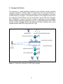
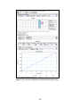
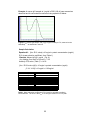
※本プロトコールは参考用の資料になり
ます。商品ご購入の際は必ず商品に添付
されている資料をご参照ください。
HRP-Antibody All-in-One Conjugation V.05.27.10
HRP-Antibody All-in-OneTM Conjugation Kit
User Manual
Catalog No. A-9002-001
Table of Contents
Chapter 1: Introduction ........................................................................................................................ 5
A.
B.
C.
D.
User Manual ...................................................................................................................................... 5
Purpose of Manual ............................................................................................................................ 5
Intended Users .................................................................................................................................. 5
Customer Service and Technical Support .......................................................................................... 5
Chapter 2: Overview of Conjugation .................................................................................................. 6
A. Product Description ........................................................................................................................... 6
B. All-in-One Technology ....................................................................................................................... 6
C. All-in-OneTM Conjugation Process Summary...................................................................................... 9
D. Materials Provided and Storage Conditions .................................................................................... 10
E. Additional Materials Required But Not Provided ............................................................................ 10
Chapter 3: All-in-OneTM Conjugation Protocol ............................................................................... 11
A. IgG Sample Preparation (10 minutes) ............................................................................................. 11
B. Buffer Exchange IgG (3 minutes) ..................................................................................................... 11
C. HyNic Modify IgG (2 hours) ............................................................................................................. 12
D. Buffer Exchange IgG (3 minutes) ..................................................................................................... 13
E. Conjugate Formation (2 hours) ....................................................................................................... 13
F. Buffer Exchange Conjugate (3 minutes) .......................................................................................... 14
G. Q Spin Column Purification (40 minutes) ........................................................................................ 14
H. Buffer Exchange Conjugate (3 minutes) .......................................................................................... 15
Chapter 4: Appendix............................................................................................................................ 17
A.
B.
C.
D.
E.
F.
G.
H.
I.
J.
K.
Polyclonal & Monoclonal IgG-HRP Conjugates: Some Examples .................................................... 17
Direct ELISA Assay Using an IgG-HRP All-in-OneTM Conjugate......................................................... 18
Bradford Protein Assay .................................................................................................................... 19
Using a NanoDropTM to Measure Antibody Concentration ............................................................. 21
HRP Absorption Spectrum (Unmodified Horseradish peroxidase) ................................................. 23
4FB-modified HRP Absorption Spectrum ........................................................................................ 23
Bovine IgG-HRP Conjugate Absorption Spectrum (All-in-OneTM Purified) ...................................... 24
Concentration of Dilute Antibody Solutions.................................................................................... 24
Troubleshooting Guide .................................................................................................................... 26
Component Stability on Storage ..................................................................................................... 27
References ....................................................................................................................................... 28
2
The products offered here are for research use only. Any commercial application will require a license
from Solulink. The Solulink Conjugation System is patented and has multiple patents pending. Please
contact Solulink for information regarding licensing information. Solulink products and methods may be
covered by one or more of the following United States patents Nos. 6,686, 461, 6,800,728, 7,102,024, 7,
173, 125, 7, 462, 689 and other pending patent applications. Information in this manual is subject to
change without notice and does not constitute a commitment on the part of Solulink, Inc. It is supplied on
an “as is” basis without any warranty of any kind, either explicit or implied. Information may be changed
or updated in this manual at any time. This document may not be copied, transferred, reproduced,
disclosed, or duplicated, in whole or in part, without the prior written consent of Solulink, Inc. This
documentation is proprietary information and protected by the copyright laws of the United States and
international treaties. The manufacturer of this documentation is Solulink, Inc.
2009 Solulink - The Conjugation Company, 9853 Pacific Heights Blvd. Suite H, San Diego, California
92121. All trademarks, trade names, service marks or logos referenced herein belong to their respective
companies.
No license is granted or implied to any patents to technologies for which the end user applies our
products. For research purposes only. Not for diagnostic use
3
Safety Information
WARNING – CHEMICAL HAZARD. Some chemicals used can be potentially hazardous,
and can cause injury or illness.
• Read and understand the Material Safety Data Sheets (MSDS) provided with this kit (flash
drive) before you store, handle, or work with any chemicals or hazardous materials.
• Minimize contact with and inhalation of chemicals. Wear appropriate personal protective
equipment when handling chemicals (e.g. safety glasses, gloves, or clothing). For additional
safety guidelines consult the MSDS.
• Check regularly for chemical leaks or spills. If a leak or spill occurs, follow the manufacturer’s
clean-up procedures as recommended in the MSDS.
• Comply with all local, state/provincial, or national laws and regulations related to chemical
storage, handling and disposal.
4
Chapter 1: Introduction
A. User Manual
This manual provides instructions for using the HRP-Antibody All-in-OneTM Conjugation
Kit.
This chapter contains the following sections:
Purpose of Manual
Intended Users
Customer Service and Technical Support
B. Purpose of Manual
Each HRP-Antibody All-in-OneTM Conjugation Kit provides all the necessary reagents
and components to produce two (2) HRP-antibody conjugates.
Use of the kit (for each antibody) results in:
•
The modification of a user-supplied antibody (100 µg) with S-HyNic.
•
The conjugation of a HyNic-modified antibody with 4FB-HRP, resulting in the
formation of an HRP-antibody conjugate.
•
The isolation of highly purified HRP-antibody conjugate using a rapid spin filter
(Q spin filter)
C. Intended Users
The HRP-Antibody All-in-OneTM Kit is designed for users with minimal or no conjugation
experience allowing them to prepare customized, high purity, ready-to-use HRPantibody conjugates in a single day.
D. Customer Service and Technical Support
Additional technical information can be found at:
Telephone
Email
1-888-625-0670 (Toll Free)
[email protected]
Fax
Address
1-858-625-0770
Solulink-The Conjugation Company
9853 Pacific Heights Blvd, Ste H
San Diego, CA 92121
5
Chapter 2: Overview of Conjugation
A. Product Description
Each HRP-Antibody All-in-OneTM Conjugation Kit provides all the necessary
components to generate two (2) highly purified antibody-HRP conjugates. The kit
requires the user to provide 100 µg of starting antibody for each conjugate. The
components of this unique kit feature a pre-activated, high-activity horseradish
peroxidase (>250U/mg) as well as a novel Q spin filter to purify the conjugate in high
yield. Conjugates produced are free of both residual antibody and HRP, thus
providing maximum signal to noise in your assay. Any suitably purified monoclonal
or polyclonal mammalian antibody (regardless of IgG subclass) can be conjugated
and purified within 5 hours (~1 h hands-on).
All-in-OneTM conjugation kits are based on Solulink’s proven HydraLinkTM chemistry.
This chemistry involves the reaction of an aromatic hydrazine with an aromatic
aldehyde to form a stable hydrazone bond. HydraLinKTM conjugation is so efficient
that it converts 100% of the antibody to the conjugate form. This linking efficiency is
made possible because of the recent discovery that small quantities of aniline
catalyze hydrazone bond formation between the two functional groups (1, 2, 3).
Aniline increases both the rate and efficiency of conjugate formation under mild
reaction conditions; leading to quantitative conversion of free antibody to HRP
conjugate.
Complete conversion of antibody to conjugate greatly simplifies downstream
purification. Purification consists of selectively binding the conjugate to a novel Q
spin filter membrane that allows excess HRP to flow through unbound. The spin filter
provides high purity without sacrificing conjugate yield. Conjugates made with All-inOneTM kit are compatible with all downstream applications such as Westerns,
ELISAs, or IHC. Each kit provides sufficient reagents to perform two (2) conjugation
reactions; each yielding between 50-70 µg of high purity HRP-antibody conjugate.
B. All-in-OneTM Technology
1) Conjugation Chemistry
HydraLinKTM chemistry is based on the use of two complementary heterobifunctional
linkers; S-HyNic and Sulfo-S-4FB (Figure 1). S-HyNic (Succinimidly-6-hydrazinonicotinamide) is first used to modify and incorporate protected aromatic hydrazines
(HyNic groups) into the antibody via acylation of lysine residues. In a similar fashion
a second linker, Sulfo-S-4FB (Sulfo-N-succinimidly-4-formylbenzamide) is used by
Solulink to provide a pre-activated high activity HRP called 4FB-HRP (included).
Incubation of HyNic-modified antibody with pre-activated 4FB-HRP in the presence
of aniline catalyst leads to rapid and efficient conversion of the antibody to conjugate
through formation of stable bis-arylhydrazone bonds (Figure 2).
6
Figure 1. Structure of S-HyNic and Sulfo-S-4FB linkers used for conjugating
HRP to antibody.
Figure 2. Aniline catalyzed conjugation of HyNic-modified antibody with preactivated 4FB-HRP.
7
2) Conjugate Purification
The efficiency of aniline-catalyzed hydrazone bond formation greatly simplifies
conjugate purification. Aniline’s ability to increase both the rate and efficiency of
conjugate formation under mild reaction conditions leads to quantitative conversion
of free antibody to conjugate. The complete absence of free antibody at the end of
the catalyzed reaction leaves only two components; excess HRP and conjugate.
After conjugation, a novel Q spin filter is used to selectively bind the conjugate
based on known biophysical properties of IgG (4, 5) while allowing free HRP to flow
through. Purified conjugate can then be eluted from the filter membrane free of both
residual antibody and HRP in high yield (50-70 µg).
IgG-HRP conjugate
+
Bind conjugate to Q Spin Filter
free excess un-conjugated 4FB-modified HRP
spin
Bind conjugate to Q spin filter
free HRP passes through
Q Spin Filter
Wash filter and discard wash
spin
Elute conjugate from filter
spin
100% pure IgG-HRP conjugate
Figure 3. Q spin filter purification of HRP-IgG conjugate.
8
C. All-in-OneTM Conjugation Process Summary
IgG
Buffer Exchange IgG
(3 min)
spin
S-HyNic linker
HyNic-modify IgG
(2 h)
+
HyNic
Excess S-HyNic
spin
Buffer Exchange IgG
(3 min)
4FB
4FB-HRP
Conjugate IgG to HRP
(2 h)
+
Excess 4FB-HRP
spin
Purify Conjugate
(45 min)
spin
Q Spin Column
spin
Purified IgG-HRP conjugate
Figure 4. HRP-Antibody All-in-OneTM conjugation process.
9
D. Materials Provided and Storage Conditions
Components
S-HyNic
4FB-Modified HRP
Buffer A
Buffer B
Buffer C
Spin Column (Red cap)
Spin Column (Yellow cap)
Spin Column (Brown cap)
Spin Column (Blue cap)
Q Spin Filters
Q Collection tubes
DMF
Amount
2 x 100 µg
2 x 50 µl
5 mL
5 mL
0.5 mL
2
2
2
2
2
4
0.5 mL
Diafiltration Spin Filters
(30k)
Collection Tubes
2
16
Flash Drive
1
Storage conditions
Keep refrigerated within
desiccated sealed aluminum
pouch
Keep refrigerated (2-8oC)
Keep refrigerated (2-8oC)
Keep refrigerated (2-8oC)
Keep refrigerated (2-8oC)
Keep refrigerated (2-8oC)
Keep refrigerated (2-8oC)
Keep refrigerated (2-8oC)
Keep refrigerated (2-8oC)
Room temperature or
refrigerated (2-8oC)
Room temperature or
refrigerated (2-8oC)
Room temperature or
refrigerated (2-8oC)
Room temperature or
refrigerated (2-8oC)
Room temperature or
refrigerated (2-8oC)
Room temperature
E. Additional Materials Required But Not Provided
Bradford Protein Assay Reagents (verification of initial IgG concentration)
Conventional UV-VIS or NanoDropTM Spectrophotometer (optional)
Semi-micro quartz cuvette (50-100 µL, 1-cm path length) (For conventional UV-VIS
Spectrophotometer)
Calibrated pipettes (P-2 or P-10, P-100, P-1000) and tips
Variable speed centrifuge (e.g. Eppendorf or MicroMax)
1.5 ml microfuge tubes
10
Chapter 3: All-in-OneTM Conjugation Protocol
IMPORTANT- Before using the kit, remove from refrigerated storage and allow
components to warm up to ambient or room temperature for at least 30 minutes.
A. IgG Sample Preparation (10 minutes)
Antibodies come in two physical forms, solids or liquids. Individual samples can
vary significantly in the amount of packaged IgG (protein mass) and/or
concentration (mg/ml). We highly recommend that IgG concentrations be
confirmed either by Bradford protein assay or A280 whenever possible. The All-inOneTM conjugation protocol requires antibody samples to be free of protein
carriers such as BSA or gelatin before proceeding. A 100 µg mass of antibody is
required at the start of the procedure. Depending on the initial form of your sample
(solid or liquid), proceed as follows:
Antibody is in Solid Form (e.g. lyophilized powder)
Resuspend lyophilized antibody (100 µg free of protein additives gelatin or BSA)
in 25 µl Buffer A to obtain a 4 mg/mL solution. If the antibody sample contains
less than 100 µg per vial (e.g. 50 µg), resuspend the requisite number of vials
equivalent to a 100 µg in 25 µl Buffer A to obtain a 4 mg/mL solution.
Proceed to step B below.
Antibody is in Liquid Form (e.g. PBS or TBS Buffer)
If the antibody sample is in liquid form at 4 mg/ml, simply transfer 25 µl to a
labeled microfuge tube. If the sample is in liquid form at a concentration greater
than 4 mg/ml, transfer a volume equivalent to 100 µg antibody to a labeled
microfuge tube and add Buffer A to obtain 4 mg/ml solution. If a sample is at a
concentration less than 4 mg/ml, concentrate the sample to 25 µL and 4 mg/mL
using a diafiltration spin filter (e.g. Amicon or VivaSpin 500) as described in the
Appendix. A concentration filter is provided in the kit for this purpose (if
necessary).
Proceed to step B below.
B. Buffer Exchange IgG (3 minutes)
1. Prepare a spin column (red cap) by twisting off the bottom closure and
loosening the red cap (do not remove). Place the spin column into a
collection tube (provided).
11
2. Mark the top of the red cap using an indelible pen to identify the sample and
place a vertical mark on the side of each spin column as shown on the next
page.
3. Place the assembly into the centrifuge and orient the vertical mark on the spin
column aiming outward and away from the center of the rotor.
4. Centrifuge at 1,500 x g for 1 minute. Discard the flow through from the
collection tube. The column matrix will appear white in color. Place the
column back into a new, empty collection tube (provided).
5. Remove the red cap; load the antibody sample (25 µL at 4 mg/mL) to the top
of the dry resin bed; loosely recap and place the column back into the
collection tube.
6. Orient the spin column mark outward as before and centrifuge at 1,500 x g
for 2 minutes. Use an appropriate balance tube opposite the assembly.
Important- Rotor speed must be set to 1500 x g (RCF) and not 1500 x rpm
(RPM). The volume recovered should always be approximately the same
volume loaded onto the spin column (e.g. 25 + 5 µL). If the recovered
volume is low, the centrifuge may require recalibration. If recovered volume
is low; re-centrifuge at the appropriate speed in an attempt to recover the
full volume (i.e. 25 µL).
7. Transfer the buffer exchanged IgG solution (25-30 µL) from the bottom of the
collection tube to a new 1.5 mL tube and label appropriately.
C. HyNic Modify IgG (2 hours)
1. Add 20 µl DMF to the vial of S-HyNic reagent. Pipette the solution up and
down to resuspend the reagent pellet. Note- a small but visible pellet can be
seen at the bottom of the vial.
2. Add 1.5 µl dissolved S-HyNic reagent to the antibody solution (25 µl @ ~ 4
mg/mL). Pipette the solution up and down to mix.
3. Incubate the reaction for 2 h at room temperature.
12
D. Buffer Exchange IgG (3 minutes)
1. Five minutes before the end of the HyNic modification reaction (section c step
3), prepare a spin column (yellow cap) by twisting off the bottom closure and
loosening the yellow cap (do not remove). Place the spin column into a
collection tube (provided).
2. Mark the top of the yellow cap using an indelible pen to identify the sample.
Also place a vertical mark on the side of each spin column as shown on the
next page.
Label the lid w/sample ID
Place pen mark on
Side of spin column
Collection tube
3. Place the assembly into the centrifuge and orient the vertical mark on the spin
column aiming outward and away from the center of the rotor. Use an
appropriate balance tube opposite the assembly.
4. Centrifuge at 1,500 x g for 1 minute. Discard the flow through from the
collection tube. The column matrix will appear white in color. Place the
column back into a new, empty collection tube (provided).
5. Open the yellow cap; load the now completed antibody/HyNic modification
reaction (~ 25-35 µL) to the top of the dry resin bed; loosely cap and place the
column back into the collection tube.
6. Orient the spin column mark outward and use an appropriate balance tube
opposite the spin filter. Centrifuge at 1,500 x g for 2 minutes.
7. After centrifugation, transfer the solution (~ 25-35 µL) from the bottom of the
collection tube to a 1.5 mL tube. Label the sample appropriately (e.g. HyNicIgG).
E. Conjugate Formation (2 hours)
1. Briefly spin the dark brown vial containing 4FB-modified HRP (5 seconds @
1000 x g) to collect the contents at the bottom of the tube.
2. Transfer 50 µl 4FB-modified HRP to the tube containing HyNic-modified
antibody (25-35 µL); pipette up and down to mix.
3. Incubate the reaction for 2 h at room temperature to form the conjugate.
13
F. Buffer Exchange Conjugate (3 minutes)
1. After completion of the conjugation reaction, prepare a spin column (brown
cap) by twisting off the bottom closure and loosening the brown cap (do not
remove). Place the spin column into a collection tube (provided).
Remember- equilibrate all kit components to ambient room temperature
before use.
2. Mark the top of the brown cap using an indelible pen to identify the sample.
Also place a vertical mark on the side of each spin column as shown below.
Label lid w/conjugate ID
Place pen mark on
side of spin column
Collection tube
3. Place the assembly into the centrifuge and orient the vertical mark on the spin
column aiming outward and away from the center of the rotor. Use an
appropriate balance tube opposite the spin filter
4. Centrifuge at 1,500 x g for 1 minute. Discard the flow through from the
collection tube. The column matrix will appear white in color. Place the
column back into a new, empty collection tube (provided).
5. Open the brown cap; load ~75-90 µL conjugate solution (section e, step 3) to
the top of the dry resin bed; loosely cap and place the column back into the
collection tube.
6. Orient the spin column mark outward and centrifuge at 1,500 x g for 2
minutes.
7. After centrifugation, add 350 µL Buffer B to the conjugate solution located at
the bottom of the collection tube and pipette up and down to mix. Set aside on
the bench for the time being.
G. Q Spin Column Purification (40 min)
1. Pre-wet a Q Spin Filter (refer to next page) by adding 200 µL Buffer B to the
top of the filter unit and incubate for 2 minutes.
14
filter unit
collection tube
2. Place the filter assembly into the centrifuge and orient the letter Q towards the
center of the rotor; spin at 2,000 x g for 4 minutes using an appropriate
balance tube; discard the flow-through from collection tube and place the filter
back into the empty collection tube.
3. Load the antibody-HRP conjugate (~430-450 µL) from the previous section
(section f, step 7) to the top of the filter unit and allow it to incubate inside the
filter for 2 minutes on the bench top.
4. Place the oriented assembly in the centrifuge and spin at 2,000 x g for 4
minutes using an appropriate balance tube opposite the filter unit; discard the
flow-through from the bottom collection tube and place the filter back into the
empty collection tube. Note- a light brown coloring will appear on the top of
the Q filter membrane (bound conjugate).
5. Add 400 µL Buffer B to the filter unit; orient and balance in the centrifuge and
spin at 2,000 x g for 4 minutes; discard the flow-through from the bottom
collection tube and place the filter back into the empty collection tube.
6. Repeat step 5 two (2) additional times to complete the washing procedure.
7. Remove the top filter unit from the collection tube and place it into a new
collection tube (provided)
8. Add 100 µL Buffer C to the top of the brown-colored filter membrane and
incubate for 5 minutes on the bench top.
9. Place the oriented assembly in the centrifuge and balance, spin at 2,000 x g
for 4 minutes; add an additional 50 µL Buffer C to the top of the filter
membrane and spin the oriented assembly for another 4 minutes at 2,000 x
g. A slightly brown-colored conjugate solution (150 µL) will now be visible at
the bottom of the collection tube. Set the collection tube aside on the bench.
H. Buffer Exchange Conjugate (3 minutes)
1. Prepare a spin column (blue cap) by twisting off the bottom closure and
loosening the blue cap (do not remove). Place the spin column into a
collection tube (provided).
2. Mark the top of the blue cap using an indelible pen to identify the sample.
Place a vertical mark on the side of each spin column as shown on the next
page.
15
3. Place the assembly into the centrifuge and orient the vertical mark on the spin
column aiming outward and away from the center of the rotor. Use an
appropriate balance tube opposite the spin filter
4. Centrifuge at 1,500 x g for 1 minute. Discard the flow through from the
collection tube. The column matrix will appear white in color. Place the
column back into a new, empty collection tube (provided).
5. Remove the blue cap; load 150 µL conjugate from the bottom of the Q spin
filter collection tube (section g, step 9) to the top of the dry resin bed; loosely
recap and place the column back into the collection tube.
6. Orient the spin column mark outward as before and centrifuge at 1,500 x g
for 2 minutes.
7. After centrifugation, transfer the buffer exchanged conjugate (150 µL) from
the bottom of the collection tube to a new 1.5 mL tube and label appropriately.
8. Measure the final protein concentration of the purified conjugate using either
a Bradford or BCA protein assay (see Appendix).
16
Chapter 4: Appendix
A. Polyclonal & Monoclonal IgG-HRP Conjugates: Some Examples
Figure 5. Coomassie-stained (4-12% SDS-PAGE) gels illustrating typical conjugation
results. Note- horseradish peroxidase is a 44 kD highly glycosylated protein that
migrates as a broad band when the protein sample is not heated (70oC) before loading
on an SDS-PAGE gel.
17
B. Direct ELISA Assay Using an IgG-HRP All-in-One Conjugate
Figure 6. Direct ELISA curves generated using an HRP conjugate made with the All-inOne kit. A mouse anti-FITC monoclonal antibody was conjugated to HRP as described
in the manual. Antigen consisting of FITC-labeled BSA (FITC MSR = 2) was coated on
plates in a 2-fold dilution series (100 µl @ 500, 250, 125, 62.5, 31.25, 15.625, 7.8, 3.90,
and 1.95 ng/ml) using standard methods. Immobilized antigen was then detected at 3
different conjugate concentrations (1 µg/ml. 0.5 µg/ml. 0.25 µg/ml) using TMB substrate
(20 minutes @ 450 nm) on a Molecular Devices plate reader.
18
C. Bradford Protein Assay
Solulink highly recommends that whenever IgG is not limiting or its concentration,
source, or quality are unknown that the sample be assayed for initial protein
concentration using a Bradford protein assay prior to conjugation. The starting
quality and quantity of antibody is critical to the success of the procedure. A
reference assay protocol is provided for measuring antibody or conjugate protein
concentrations using Bradford protein reagents (not provided in the kit).
Bradford Microtiter Plate Procedure
Required Materials
Bradford Reagent (Bio-Rad, Hercules, CA, Cat. #500-0006)
96-well microtiter plate (standard flat bottom)
PBS (phosphate buffered saline)
P-200 and P-1000 pipettes
Bovine IgG Antibody Standard: 2 mg/ml (Pierce/ThermoFisher, Cat. # #23212)
Molecular grade water
Assay Protocol
1) Prepare 2 ml of a Bradford working solution by adding 400 µl dye reagent to
1600 µl molecular grade water (1:4 ratio).
2) Prepare the following protein dilution standards and blank as follows:
Add 160 µl 2 mg/ml bovine IgG standard to 240 µl PBS (0.8 mg/ml standard)
Add 150 µl 0.8 mg/ml standard to 50 µl PBS (0.6 mg/ml standard)
Add 75 µl 0.6 mg/ml standard to 25 µl PBS (0.4 mg/ml standard)
Add 50 µl 0.4 mg/ml standard to 50 µl PBS (0.2 mg/ml standard)
Add 50 µl 0.2 mg/ml standard to 50 µl PBS (0.1 mg/ml standard)
Add 50 µl PBS (buffer blank)
3) Pipette 5 µl of each standard (and blank) along with duplicates of antibody
sample into separate microtiter wells.
4) Add 100 µl of previously diluted dye reagent (1:4) to each well and mix
thoroughly. Always replace pipette tips between additions.
5) Incubate at room temperature for 5-10 minutes (but no more than 60 minutes).
6) Measure absorbance at 595 nm on a suitable microtiter plate reader.
7) A typical Bradford microtiter assay result from a commercial plate reader is
illustrated below in Figure 7.
19
Figure 7. Print out from a Bradford microplate-based protein assay.
20
D. Using a NanoDropTM to Measure Antibody Concentration
If an antibody sample is free of protein-based carriers (e.g. BSA, gelatin) or certain
interfering preservatives such as thimerosal, then a simple non-destructive scan of the
IgG sample on a NanoDropTM spectrophotometer can be used to estimate antibody
concentration saving the trouble of conducting a Bradford protein assay to confirm
concentration. To estimate antibody concentration using a NanoDropTM spectrophotometer, proceed as follows.
1. Turn on the NanoDropTM spectrophotometer and click on the NanoDropTM icon to
launch the software.
2. Place a 2 µl drop of molecular grade water on the clean pedestal, click OK.
3. When the main menu appears, select the A280 menu option. Note- do not use the
UV-VIS menu option on the NanoDropTM to read an antibody sample.
4. After the A280 menu appears, click-off the 340 nm normalization option using the
mouse. Note-some instruments do not have this normalization feature in which case
this step can be ignored.
5. In the window labeled Sample Type, select ‘Other protein E1%’ option from the pulldown menu. Enter the appropriate E1% value (Table 1 on the next page)
corresponding to your particular antibody sample type. For example, 14.00 for
mouse IgG.
6. Blank the NanoDropTM spectrophotometer by placing a 2 µl drop of the appropriate
sample buffer (e.g. PBS) and click on the ‘Blank’ icon.
7. Immediately re-click the ‘Measure’ icon to validate the baseline (i.e. flat across the
bandwidth). Clean the pedestal and repeat (if necessary) until a flat baseline is
obtained. Note-sometimes air bubbles can become trapped on the pedestal during
sample loading and cause baseline offsets. If necessary, remove air bubbles and
rescan to insure a proper baseline.
8. Place a 2 µl volume of antibody solution on the clean pedestal and click the
‘Measure’ icon. Wait until the spectrum (220-350 nm) appears in the window. Notefor precious or limited samples the majority of the 2 µl aliquot can be recovered from
the pedestal.
9. Record the antibody concentration directly from the NanoDropTM display window
[mg/ml]. Alternately, calculate the antibody concentration (manually) as illustrated on
the following page.
21
Example: A mouse IgG sample at 1 mg/ml in PBS (100 µl) was scanned as
described and its concentration confirmed using equation #1 below.
Figure 8. A mouse IgG sample 100 µl @ 1 mg/ml in PBS pH 7.2, scanned on the
TM
as described in the text.
NanoDrop
Sample Calculation
Equation #1: [A280 /E1% value] x 10 mg/ml = protein concentration (mg/ml)
E1% (mass extinction coefficient, from Table 1)
Example: Mouse IgG @ 1 mg/ml (Fig. 8)
A280 reading (from scan in Figure 8) = 1.34
Antibody E1% value (Table 1) = 14.00
[A280 / E1% bovine IgG] x 10 mg/ml = protein concentration (mg/ml)
[1.34 / 14.00] x 10 mg/ml = 0.96 mg/ml
Antibody Source
Human IgG
Human IgE
Rabbit IgG
Donkey IgG
Horse IgG
Mouse IgG
Rat IgG
Bovine IgG
Goat IgG
Antibody E1% (1-cm path)
13.60
15.30
13.50
15.00
15.00
14.00
14.00
12.40
13.60
Table 1. Mass extinction coefficients (E1%) used for calculating antibody
concentrations. The E1% is the A280 of a 10 mg/ml solution in a 1-cm path.
22
E. HRP Absorption Spectrum (Unmodified Horseradish peroxidase)
Figure 9. NanoDropTM absorption spectrum of unmodified horseradish peroxidase (220550 nm) @ 0.66 mg/ml (sodium phosphate buffer, pH 6.0, 1 mm path length)
F. 4FB-modified HRP Absorption Spectrum
Figure 10. NanoDropTM absorption spectrum of 4FB-modified horseradish peroxidase
(220-550 nm) @ 0.66 mg/ml (sodium phosphate buffer, pH 6.0, 1 mm path length)
23
G. Bovine IgG-HRP Conjugate Absorption Spectrum (All-in-One Purified)
Figure 11. NanoDropTM absorption spectrum of All-in-One IgG-HRP conjugate (220-550
nm) @ 0.96 mg/ml (sodium phosphate buffer, pH 6.0, 1 mm path length).
H. Concentration of Dilute Antibody Solutions
The HRP-Antibody All-in-One Conjugation protocol requires that initial antibody
protein concentration be at 4-5 mg/ml and 25 µl. Many antibody vendors
package their products at significantly more dilute concentrations (e.g. 0.25 to 1.5
mg/ml). In these instances, IgG samples will need to be concentrated to 4-5
mg/ml and 25 µl before proceeding. The All-in-One kit provides two (2)
diafiltration filters (M.W.C.O. 30 kD) for this purpose (Figure 12). Carefully follow
these instructions to avoid antibody loss or aggregation on the filter’s surface.
Note-Dilute antibody solutions require at least 125 μg of starting antibody (e.g.
500 µl @ 0.25 mg/ml) since diafiltration filters recover ~80% of input antibody. If
antibody samples are not inlimiting supply, we recommend antibody
concentrations be confirmed using a Bradford protein assay before proceeding.
Concentrator body
Filtrate tube
Figure 12. Diafiltration spin filter used for concentrating dilute antibody samples prior to
the start of All-in-One conjugation protocol.
24
Antibody Concentration Protocol
Note- the diafiltration spin filters illustrated is made to contain and process a
maximum volume of 500 μl or less. If a volume greater than 500 μl is to be
concentrated, multiple loadings will be required.
1) Open the lid of a diafiltration spin filter device.
2) Transfer 500 μl (or less) of dilute protein solution (equivalent to 125 μg antibody)
to the center of the filter cup.
3) Close the lid and orient the spin filter in the centrifuge so that the volume
markers face toward the center of the centrifuge rotor. Use an appropriate
balance tube opposite the spin filter.
4) Centrifuge for 2 minutes @ 5,000 x g. Note-never centrifuge for longer periods
of time
5) Open the filter unit and visually inspect the remaining volume. If the volume
remaining in the concentrator body is greater than 25 μl, gently pipette the
solution up and down to mix; taking care not to touch or puncture the filter
surface during this step.
6) Repeat steps 4 and 5 until the volume in the filter cup reaches the 25 μl mark.
Once the final volume reaches 25 μl, do not pipette up and down to avoid sample
loss. Note-if the volume goes lower than 25 μl at this stage, add a small aliquot
of Buffer A to bring the final volume to 25 μl.
7) Carefully transfer the concentrated IgG solution (25 μl) to a new 1.5 ml microfuge
tube and proceed with the rest of the procedure in Chapter III, section B.
25
I. Troubleshooting Guide
Problem
Poor conjugate yield
Possible Cause
-initial antibody concentration and
volume were incorrect or
unknown.
Recommended Action
-whenever possible verify
the original starting
antibody concentration
using a Bradford protein
TM
assay or NanoDrop to
assure efficient
conjugation.
-concentrate or dilute the
antibody sample to be
conjugated into the
required range (4-5
mg/ml and 25 µl)
Poor conjugate yield
Starting antibody concentration
and volume are incorrect or
unknown.
-preservatives can
interfere with the accuracy
of a Bradford protein
assay. Remove all
interfering preservatives
such as thimerosal or
proclin before performing
a Bradford protein assay.
Poor HyNic
modification
-presence of protein carrier (e.g.
BSA or gelatin) is contaminating
the antibody sample.
-remove and purify away
all protein carriers such as
BSA or gelatin using
affinity chromatography or
other methods
Poor HyNic
modification
-improper mixing of HyNic reaction
components
-make sure to properly
mix the antibody- HyNic
reaction mixture
-use a calibrated P-10
pipette to insure accuracy
of small volumes
-presences of amine contaminants
-remove all non-protein
amine contaminants such
as glycine or Tris before
modification
-improper storage of S-HyNic
reagent can lead to hydrolysis of
this NHS ester
-keep and store S-HyNic
sealed in the aluminum
pouch provided that
contains dessicant.
26
-initial antibody concentration was
too low or too high.
measure the initial
antibody concentration
before proceeding
(Bradford or NanoDrop)
-concentrate or dilute the
antibody sample into the
recommend range (4-5
mg/ml and 25 µl) before
proceeding
Low conjugate
and/or antibody
recovery
-low spin column recovery volume
-use a properly calibrated
variable-speed centrifuge
Follow recommended spin
speed/time. Altered spin
speeds can adversely
compromise protein
and/or volume recovery
J. Component Stability on Storage
Component
Stability
Unopened Kit
1 yr
Refrigerated (2-8 C)
1 yr
Keep in sealed
aluminum pouch
o
provided (2-8 C).
S-HyNic
o
24 h after re-suspending
S-HyNic in DMF
HRP-Antibody
Conjugate
All other kit components
Flash Drive
Storage Condition
9 month
Room temperature
o
Refrigerated (2-8 C) in
final conjugate solution.
o
1 yr
Refrigerated (2-8 C)
(50% glycerol)
1 yr
Refrigerated (2-8 C)
1 yr
Room temperature
o
27
K. References
1. Dirksen, A., Hackeng, T., Dawson, P.,(2007). Nucleophilic Catalysis of Oxime
and Hydrazone Reactions by Aniline. ACS Poster
2. Dirksen, A., Hackeng, T., Dawson, P., (2006). Nucleophilic Catalysis of Oxime
Ligations. Angew. Chem. Int. Ed. 45, 7581-7584
3. Dirksen, A., Dirksen, S., Hackeng, T., Dawson, P., (2006). Nucleophilic
Catalysis of Hydrazone Formation and Transimination: Implications for Dynamic
Covalent Chemistry. JIAICIS Communications.
4. Lim,S., Manusu, H.P., Gooley, A.A., Williams, K. L., Rylatt D.B.,(1998).
Purification of monoclonal antibodies from ascitic fluid using preparative
electrophoresis. Journal of Chromatography A. Vol. 827, Issue 2, 11, Pages 329335.
5. Chiodi,F., Sidén, A., Ösby, E., (2005). Isoelectric focusing of monoclonal
immunoglobulin G, A and M followed by detection with the avidin-biotin system.
Electrophoresis, Vol. 6 Issue 3, 124-128.
28