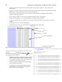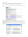
1
An Introduction to
Bioinformatics for
Biological Sciences Students
Department of Microbiology and Immunology, McGill University
Version 2.5 (For the BIOC-300 lab), March 2006
2
AN INTRODUCTION TO BIOINFORMATICS FOR BIOLOGICAL SCIENCES STUDENTS
Contributors
The first edition of the Introduction to Bioinformatics for biological sciences students was written during the summer of
2004 at McGill University for the Bioinformatics Project (BIP) as part of the U2 undergraduate laboratory in Microbiology
and Immunology (MIMM-386).
What you are holding in your hands is the second edition of the manual put together by a new group of students during the
summer of 2005. From the first edition, it contains only the section on biological databases and the main institutes that
develop and maintain them (other parts were included in an “extended” version of the manual instead). The biggest change
from the first edition is that the manual now includes the exercise sheets and tutorial written during the course of the BIP’s
first year of existence, making this volume the comprehensive resource students need to understand the material covered in
the BIP, but also to perform the exercises.
This version has been adapted for the BIOC-300D Laboratory in Biochemistry course lab on bioinformatics.
Main contributors to the abridged version
•
Cédric Sam ([email protected])
•
Oksana Kapoustina
•
Abrar Khan
Contributors to all sections including those not in the abridged version
•
Belinda Befort (PROSITE, Phylip)
•
Scott Bunnell (Editing)
•
Mansoureh Hakimi (Exercises review)
•
Oksana Kapoustina (BLAST, ClustalW)
•
Abrar Khan (Editing)
•
François Pepin (Editing)
•
Cédric Sam (Institutes, Databases, Editing, Exercises, original layout)
•
Sean Wiltshire (Introduction, editing)
Faculty members who contributed to this manual
•
Dr Silvia Vidal ([email protected])
•
Dr Nicholas Acheson
•
Dr Malcolm Baines
Copyright © 2004, 2005 Department of Microbiology and Immunology of McGill University. All rights reserved.
3
Table of Contents
Table of Contents............................................................................................................................................................ 3
Chapter 1: Bioinformatics Institutes.............................................................................................................................6
1.1 NCBI: The National Center for Biotechnology Information (USA)............................................................................... 6
1.1.1 Database resources at the NCBI..............................................................................................................................6
1.1.2 PubMed: The ultimate biomedical literature database............................................................................................6
1.2 EBI: The European Bioinformatics Institute................................................................................................................... 7
1.3 SIB: The Swiss Institute of Bioinformatics..................................................................................................................... 8
1.3.1 How to access SIB’s resources?..............................................................................................................................8
1.4 Bioinformatics in Canada...........................................................................................................................................8
Chapter 2: Molecular Biology Databases..................................................................................................................... 9
2.1 Introduction......................................................................................................................................................................9
2.2 Nucleotide Sequence Databases...................................................................................................................................... 9
2.2.1 The Big Three: GenBank, DDBJ and EMBL....................................................................................................... 10
2.2.2 Entrez: NCBI’s multi-purpose search engine....................................................................................................... 10
Refine your search....................................................................................................................................................10
NCBI UniGene website............................................................................................................................................10
2.2.3 NCBI’s UniGene...................................................................................................................................................10
dbEST and UniGene.................................................................................................................................................11
What does UniGene contain exactly?...................................................................................................................... 11
How do you search UniGene?..................................................................................................................................11
NCBI UniGene website............................................................................................................................................12
2.3 Protein Sequence Databases.......................................................................................................................................... 12
2.3.1 What can you find in a curated protein database?................................................................................................ 12
2.3.2 Swiss-Prot and TrEMBL.......................................................................................................................................12
TrEMBL: Translation of EMBL nucleotide sequence database.............................................................................. 12
Searching Swiss-Prot/TrEMBL................................................................................................................................13
2.3.3 PIR-PSD: The Protein Sequence Database...........................................................................................................13
2.3.4 UniProt.................................................................................................................................................................. 13
Searching UniProt ................................................................................................................................................... 13
Reading a UniProt entry........................................................................................................................................... 14
Uniprot website........................................................................................................................................................ 14
2.4 Protein Families and Domains Databases......................................................................................................................14
2.4.1 PROSITE ............................................................................................................................................................. 15
What does the Prosite database contain?..................................................................................................................15
4
AN INTRODUCTION TO BIOINFORMATICS FOR BIOLOGICAL SCIENCES STUDENTS
Search PROSITE using ScanProsite........................................................................................................................ 15
Operation and interpretation of ScanProsite............................................................................................................ 15
2.4.2 Pfam: Protein families database of alignments and HMMs..................................................................................16
What’s in Pfam?....................................................................................................................................................... 16
What can I do with Pfam?........................................................................................................................................ 16
2.5 3-D Structure Databases................................................................................................................................................ 17
2.5.1 PDB: The Protein Data Bank................................................................................................................................ 17
Contents ...................................................................................................................................................................18
Searching PDB......................................................................................................................................................... 18
An example: Myoglobin...........................................................................................................................................19
Query Tutorial.......................................................................................................................................................... 20
PDB Structure Explorer............................................................................................................................................20
2.5.2 Viewing Structures with RasMol..........................................................................................................................20
Customizing Structures ........................................................................................................................................... 21
Using “select” to change the display options for specific residues..........................................................................21
2.6 Other databases..............................................................................................................................................................22
2.7 References......................................................................................................................................................................22
Chapter 3: Tutorials..................................................................................................................................................... 23
Tutorial: How to use BLAST to search for homologous sequences? (and using NCBI ORF finder).................................23
Tutorial: How to use ClustalW to perform multiple sequence alignments and build phylogenetic trees?......................... 31
Tutorial: How to use PDB and Rasmol to find and manipulate three-dimensional Structures........................................... 37
Tutorial: How to use InterPro to find conserved protein domains?.....................................................................................43
Glossary..........................................................................................................................................................................46
Appendix: How to write the report?........................................................................................................................... 52
TABLE OF CONTENTS
5
6
Chapter 1: Bioinformatics
Institutes
This section will be an overview of the major actors in the field of bioinformatics, what are the services they offer and what
sort of databases they each manage. These research institutes were all established in different countries, but their reach,
their funding sources, their staff are now well worldwide.
1.1 NCBI: The National Center for Biotechnology Information
(USA)
The NCBI is a unit of the National Library of Medicine (NLM), which is in turn a branch of the National Institutes of
Health (NIH). The NCBI is located in Bethesda, MD, in the outskirts of Washington DC.
1.1.1
Database resources at the NCBI
Here’s an overview of a few of the databases hosted by NCBI and the services which come with them1.
•
Database Retrieval Tools - Entrez is an integrated retrieval system for the databases hosted by NCBI. Taxonomy
indexes over 150 000 organisms that are represented by at least one nucleotide or protein sequence. LocusLink
provides a single query interface to curated sequence and descriptive information about genetic loci.
•
BLAST family of sequence-similarity search programs.
•
Resource for gene-level sequences - UniGene is a system which partitions GenBank sequences into nonredundant set of gene-oriented clusters. There are many other specialized databases for single nucleotide
polymorphisms (dbSNP), and for information on Major Histocompatibility Complex (dbMHC).
•
Resources for genome-scale analysis - Entrez Genomes provides access to genomic data and includes genomes
spanning from microbes to multicellular organisms.
•
Eukaryotic Genomic Resources - Map Viewer displays genome assemblies using sets of aligned chromosomal
maps.
•
Structural databases - The NBCI MMDB is built by processing entries from the Protein Data Bank.
1.1.2
PubMed: The ultimate biomedical literature database
MEDLINE is the NLM’s premier bibliographic database covering the fields of medicine, nursing, dentistry, veterinary
medicine, the health care system, and the preclinical sciences. MEDLINE contains bibliographic citations and author
abstracts from more than 4,800 biomedical journals published in the United States and 70 other countries. The database
contains over 12 million citations dating back to the mid-1960. Coverage is worldwide, but most records are from Englishlanguage sources or have English abstracts.
PubMed is NCBI’s biomedical literature database giving access to citations compiled in databases such as MEDLINE. To
the average user, Pubmed just equals Medline, although a website describes the difference between both concepts:
(http://www.nlm.nih.gov/pubs/factsheets/dif med pub.html).
What you need to know is that PubMed is a biomedical literature giving access to the MEDLINE database, but also to
certain non-medical article featured in MEDLINE journals. What you read in a textbook today has almost always been
1
Source: Database resources of the National Center for Biotechnology Information, Nucleic Acids Res.2004 Jan 1; 32 Database issue: D35-40
7
published and debated through peer-reviewed journals. Reading “review” articles in prominent journals like Science or
Nature is a good way to start familiarizing yourself with peer-reviewed journals.
NCBI’S PUBMED - HTTP://WWW.NCBI.NIH.GOV/PUBMED
1.2 EBI: The European Bioinformatics Institute
“The European Bioinformatics Institute (EBI) is a non-profit academic organization that forms part of the European
Molecular Biology Laboratory (EMBL).” (EBI website, http://www.ebi.ac.uk/) The EBI is located in Cambridgeshire,
United Kingdom, and was established in 1992. It is the European equivalent of the NCBI. In 2004, EBI was funded
primarily by the EMBL (45%) and the European Union (25%), but also by the National Institutes of Health (NIH) in the
USA (accounting for about 10%). Many applications are available from EBI through a web interface. Here are some
examples2:
•
Homology & Similarity - the BLAST or Fasta programs can be used to look for sequence similarity. (Note: The
BLAST provided by EBI is different from the one provided by NCBI (it’s “WU-BLAST”, by Washington U in
St.Louis, rather than “NCBI-BLAST”).
•
Protein Functional Analysis - InterProScan can be used to search for motifs in your protein sequence.
•
Sequence Analysis - ClustalW a sequence alignment tool.
•
Structural Analysis - MSDfold or DALI can be used to query your protein structure and compare it to those in the
Protein Data Bank (PDB).
•
Tools Miscellaneous - Expression Profiler a set of tools for clustering, analysis and visualization of gene
expression and other genomic data.
As well as applications, the following are databases maintained by EBI3:
•
EMBL Nucleotide Database - Europe’s primary collection of nucleotide sequences is maintained in collaboration
with Genbank (USA) and DDBJ (Japan). (Note: These are the three partners of The International Nucleotide
Sequence Database Collaboration (INSD). See Science, Brunak et al 298 (5597):1333)
•
UniProt Knowledgebase - a complete annotated protein sequence database. It is a central repository of protein
sequence and function created in 2002 by joining the information contained in Swiss- Prot/TrEMBL (SwitzerlandEurope), and PIR (USA). See Curr Opin Chem Biol 2004 Feb 8(1):76-80, a recent article on Uniprot and Protein
sequence databases at large.
•
Macromolecular Structure Database - European Project for the management and distribution of data on
macromolecular structures.
•
ArrayExpress - for gene expression data.
2
(Source: EBI Services - http://www.ebi.ac.uk/services/)
3
(Source: EBI Databases - http://www.ebi.ac.uk/databases/)
8
•
Ensembl - Providing up to date completed metazoic genomes and the best possible automatic annotation.
THE EUROPEAN BIOINFORMATICS INSTITUTE WEBSITE - HTTP://WWW.EBI.AC.UK/
1.3 SIB: The Swiss Institute of Bioinformatics
“The SIB is an academic not-for-profit foundation established on March 30, 1998 whose mission is to promote research, the
development of databanks and computer technologies, teaching and service activities in the field of bioinformatics, in
Switzerland with international collaborations.” (SIB website, http://www.isb-sib.ch/)
The SIB maintains a number of important databases such as the Swiss-Prot/TrEMBL protein databases, the PROSITE
protein families and domains database and the SWISS-2DPAGE database of 2D-gels, plus many other specialized
databases.
The SIB is also active in developing software tools like Melanie for the analysis of 2-D gels, Swiss- PdbViewer for the
visualization of 3-D structures (such as those found in the Protein Data Bank, or PDB, database), and SWISS-MODEL, a
fully-automated server which takes protein sequences and tries to model their 3-D structure from known 3-D structures
found in the PDB.
1.3.1
How to access SIB’s resources?
EXPASY: THE SWISS PROTEOMICS SERVER - HTTP://CA.EXPASY.ORG/ (CANADIAN MIRROR)
ExPASy (Expert Protein Analysis System, http://ca.expasy.org/) is the SIB’s proteomics web
server. ExPASy is the website to use to access to all of the aforementioned SIB databases and
analytical tools (and Swiss-Jokes http://www.expasy.org/cgi-bin/sw-jokes.pl).
1.4
Bioinformatics in Canada
The website of the Canadian Bioinformatics Resource in Ottawa hosts well-known
bioinformatics applications, such as BLAST, ClustalW and a web version of the popular
phylogenetics program Phylip. (http://cbr-rbc.nrc-cnrc.gc.ca/)
9
Chapter 2: Molecular Biology
Databases
Databases are large collections of data arranged for ease of search and retrieval. Common databases such as GenBank, PDB
or Swiss-Prot exist as (extremely) large files which can be downloaded from public sites for various data manipulations on
local private computers, or more practically, consulted on-line by molecular biologists at large using search tools such as
BLAST.
2.1 Introduction
This section will cover nucleotide sequence, protein structure and protein sequence databases. Some of the main databases
are found below4:
•
Biomedical literature: PubMed.
•
Species-specific: SGD, FlyBase, WormBase, MGI.
•
Nucleotide sequences: GenBank, EMBL, DDBJ.
•
Genome sequences: Entrez Genome, TIGR databases.
•
Protein sequences: GenPept, Swiss-Prot/TrEMBL, PIR.
•
Macromolecular 3-D Structures: Protein Data Bank, MMDB.
•
Protein and peptide mass spectroscopy: PROWL.
•
Post-translational modifications: RESID.
•
Biochemical and biophysical information: ENZYME, BIND.
•
Biochemical pathways: PathDB, KEGG, WIT.
•
Microarray chips data: ArrayExpress, SMD.
•
2D-PAGE: SWISS-2DPAGE.
•
Protein families and domains: PROSITE, Pfam, InterPro, ProDom.
2.2 Nucleotide Sequence Databases
Nucleotide sequences (DNA and RNA) are essential pieces of information. Researchers might use protein- coding
nucleotide sequences to produce large quantities of protein for various experiments in the wet lab.
4
This listing is vaguely based on the one found in “Developing bioinformatics computer skills” by Cynthia Gibas and Per
Jambeck, O’Reilly & Associates, 2001.
10
2.2.1
The Big Three: GenBank, DDBJ and EMBL
NCBI’s GenBank (USA), EBI’s EMBL Nucleotide Sequence Database (Europe), and the DNA Databank of Japan (DDBJ,
Japan) are the three biggest nucleotide sequence databases in the world. Their main sources for DNA and RNA sequences
are direct submissions from individual researchers, genome sequencing projects and patent applications.
The NCBI hosts the most well-known database, GenBank. As a result of the International Nucleotide Sequence Database
(INSD) Collaboration between the NCBI, EMBL and DDBJ, new submissions are shared between databases, leading them
to have similar content (although the annotations can differ). This collaboration between the three institutes has existed for
16 years5.
2.2.2
Entrez: NCBI’s multi-purpose search engine
Entrez can be used to search any of the NCBI-hosted databases. Pubmed is one of NCBI’s databases; it is the scientific
publications database.
The NCBI website is not easy to navigate and takes a lot of fooling-around before one can safely sail from place to place6.
You can use Entrez directly from NCBI’s homepage http://www.ncbi.nih.gov/, but you will be missing out on many of the
search options. If you want to search PubMed or another database, just click on the upper bar link, with Entrez being the
cross-database search (useful when you want all the information about a specific gene).
Refine your search
Some parameters can be used to refine your search. In general, you might want to start by limiting your searches. So these
options are only accessible through each specific Entrez flavor. Using the “Limits” link at the bottom of the Entrez search
bar/box, you specify many parameters, such as the fields you want to limit your search to. There are also limits specific to
the type of Entrez you are using: for example, with Entrez Nucleotide (GenBank), you can decide the type of nucleotide
(genomic DNA/RNA, mRNA or rRNA), date of modification, the subset of GenBank it belongs to, etc. In Entrez PubMed,
you can conveniently specify a range for the date of publication or choose the publication type, among other things.
NCBI UniGene website
To search Genbank, use Entrez: http://www.ncbi.nih.gov/Entrez/ (case-sensitive).
2.2.3
NCBI’s UniGene
“Each UniGene cluster contains sequences that represent a unique gene, as well as related information such as the tissue
types in which the gene has been expressed and map location.” (NCBI website)
UniGene is not a database per se, it is rather a system for “automatically partitioning GenBank sequences, including
expressed sequence tags (ESTs), into a non-redundant set of gene-oriented clusters”. (Wheeler DL, et al. Database
Resources of the National Center for Biotechnology. Nucl Acids Res 31:28-33;2003)
The importance of UniGene is its role in organizing the numerous sequences contained in public databases that can relate to
a single gene. Means of organizing the information have been overwhelmed by the deluge of sequences coming from the
various genome projects, and UniGene is an effort to group nucleotide sequences (ESTs and mRNA) of selected organisms
by genes they are related to.
5
See: Nucleotide Sequence Database Policies, Science 298 (5597): 1333 15 Nov 2002
6
The Entrez Help Document is a useful resource (http://web.ncbi.nih.gov/entrez/query/static/help/helpdoc.html)
11
BY DEFAULT, THE RESULTS OF AN ENTREZ SEARCH IN GENBANK ARE FORMATTED FOR THE “SEQUENCE” VIEW.
A PULL-DOWN MENU ALLOWS YOU TO SELECT THE VIEW FITTING YOUR NEEDS (SUCH AS FASTA, ONE OF THE
POPULAR FORMATS ACCEPTED BY SEQUENCE ANALYSIS PROGRAMS).
dbEST and UniGene
Complementary DNA (cDNA) is single-stranded DNA synthesized from a mature mRNA template. Now, what are
“Expressed Sequence Tags” (ESTs)? They are short sequences generated by sequencing the ends of these cDNA molecules.
ESTs are important gene mapping and discovery tools because they can be used as primers to amplify genomic DNA
spanning a region presumably bounded on one side by the EST. The EST database (dbEST) is one of the many divisions of
GenBank, the NCBI nucleotide database. As of September 2004, there were some 5.7 million Homo sapiens ESTs in
dbEST.
By design, ESTs in dbEST may be redundant, as several different ESTs might be derived from mRNA expressed by the
same gene. This is where UniGene comes into play.
What does UniGene contain exactly?
UniGene regroups ESTs, mRNAs, high-throughput cDNAs (HTC), etc., representing a unique gene into clusters. UniGene
is an automated system, and has so far reduced 4.6 million Homo sapiens sequences to some 107,014 gene clusters. Clusters
are never stable, they can be merged together at any point based on certain criteria as new sequences are added to
GenBank/UniGene.
Every cluster has its own webpage through UniGene’s web interface. From that page, related information about the cluster
can be found: tissue types in which the gene is expressed, protein similarities with clusters in model organisms (say if you
wanted to express a human gene in mice using the murine counterpart), LocusLink report for the gene and its location in the
genome.
How do you search UniGene?
Each species has its own summary page with all kinds of nice statistics about the number and size of the clusters as
sequences get added to the system by the thousand every week.
Typically, you might know the name of the gene, or could be looking for the cluster to which your nucleotide sequence
belongs to. UniGene is the database, and Entrez is NCBI’s all-purpose search engine, so Entrez UniGene is naturally the
way to go.
12
NCBI UniGene website
http://www.ncbi.nih.gov/UniGene
2.3 Protein Sequence Databases
Protein databases are the natural extension of nucleotide sequences. They come in two varieties: “Sequence Repositories”
and “Universal Curated Databases”. Sequence repositories are generally just places where protein sequences are compiled
with minimal attention given to provide non-redundant entries. GenBank is a well-known example of sequence repository.
In contrast, universal curated databases are manually organized and looked after by experts.
Among protein databases, NCBI’s GenPept is an example of a sequence repository. It contains the translation of nucleotide
sequences contained in the GenBank-EMBL-DDBJ triumvirat mentioned earlier. Curated databases contain information
validated by expert biologists and thus considered highly reliable. Swiss-Prot/TrEMBL and PIR are examples of such
databases, and we will look more closely at their history and modes of functioning before talking about UniProt, an effort
by Apweiler, Bairoch and Wu’s groups to establish networks for sharing protein information around the world.
2.3.1
What can you find in a curated protein database?
Information on each protein is very specific and presumably highly reliable. Anything essential such as the accession
number, the source organism and (sometimes very many) references can be found. Cross- references with other databases
can be useful for researchers who might be interested to learn more about a given structural domain contained in the protein,
or the related nucleotide sequences if one wishes to express the protein for various assays.
2.3.2
Swiss-Prot and TrEMBL
“Swiss-Prot is a curated biological database of protein sequences created in 1986 by Amos Bairoch during his PhD and
developed by the Swiss Institute of Bioinformatics and the European Bioinformatics Institute. It strives to provide a high
level of annotation (such as the description of the function of a protein, its domain structure, post-translational
modifications, variants, etc.), a minimal level of redundancy and high level of integration with other databases. As of July
5, 2004, Swiss-Prot (release 44.0) contains 153,871 entries.” (Wikipedia)
THE SWISS-PROT LOGO.
Each entry of Swiss-Prot (http://ca.expasy.org/sprot/) is carefully inspected by specialists from around the world to ensure a
high quality of the information contained. This is a long process, and more and more sequences are added to the database
every day. That’s where TrEMBL comes to the rescue.
TrEMBL: Translation of EMBL nucleotide sequence database
TrEMBL is a computer-annotated supplement to Swiss-Prot introduced in 1996 as a solution to preserve the high editorial
standards of Swiss-Prot while making new sequences available to the public.
TrEMBL contains translations of all coding regions in the DDBJ-EMBL-GenBank nucleotide databases, and protein
sequences extracted from the literature or submitted to UniProt, which are not yet integrated into Swiss-Prot. TrEMBL
allows these sequences to made publicly available quickly without diluting the high quality annotation found in Swiss-Prot.
13
Searching Swiss-Prot/TrEMBL
In an effort to create a single source of protein information, the UniProt consortium was established. Searching and using
Swiss-Prot/TrEMBL is similar to searching and using the UniProt databases, so this section will actually be covered below.
2.3.3
PIR-PSD: The Protein Sequence Database
“The Protein Information Resource (PIR), located at Georgetown University Medical Center, is an integrated public
bioinformatics resource that supports genomic and proteomic research and scientific studies.” (PIR website,
http://pir.georgetown.edu/) The PIR maintains the Protein Sequence Database (PSD), an annotated protein database similar
to Swiss-Prot. The PSD grew out of the Atlas of Protein Sequence and Structure (1965-1978) edited by the late Margaret
Dayhoff7.
2.3.4
UniProt
Nucleotide sequence databases were united under the International Nucleotide Sequence Database (INSD) Collaboration,
but curated protein databases didn’t have their equivalent body until 2002, when the UniProt consortium was established
between the developers of the main existing annotated protein databases: the EBI/SIB (Swiss-Prot & TrEMBL) and the PIR
(Protein Sequence Database (PSD)).
UniProt is a very recent addition that aims to replicate the efforts of UniGene in the amino acid sequence world. The first
version (1.0) of UniProt was officially launched 15-Dec-2003, and its second version (2.0) on 5-Jul-2004. Both of these
were in fact the most current versions of Swiss-Prot/TrEMBL and PSD merged together.
Databases making up UniProt are:
•
The UniProt Archive (UniParc) is the most comprehensive publicly accessible non-redundant protein sequence
database available. It includes sequences from databases hosted by the founding members of UniProt but also
sequences derived from other public databases such as PDB, RefSeq or EMBL. As its name implies, UniParc is an
archive, so every time a change is made to an entry on the native database, UniParc takes note of it, updates the
sequence and keeps the old version.
•
Initially, the UniProt Knowledgebase (UniProt) consists of the merging of the Swiss-Prot, TrEMBL and PSD
entries, but will later be derived from the UniParc database8. UniProt will retain the organization of the SwissProt/TrEMBL duo (Swiss-Prot as a manually-curated database and TrEMBL as a computer-annotated database)
and integrate data from PIR-PSD that’s not already in Swiss- Prot/TrEMBL.
•
The UniProt Non-redundant Reference (UniRef) is, as its name implies, a collection of non- redundant protein
sequences. UniRef sequences come from the UniProt knowledgebase, and the non- redundancy is generated
automatically. Sequences are compared with each other and if there is sequence homology, they are merged
together and added as a single entry in UniRef.
Searching UniProt
The search interface is slightly different among the three UniProt associates, even if the same tools are essentially offered.
Because all computers connecting from North America are redirected to the PIR’s UniProt site, we will only consider that
version of the search interface. (PIR’s UniProt website: http://www.pir.uniprot.org/)
The two main search tools are:
1.
Text Search, which allows you to search in one field in particular or all of them at once for plain query strings.
One of UniProt’s layers/databases (as discussed in the previous section) must be selected. Query boxes can be
added as you go by clicking the add input box or + box button, with the corresponding boolean operator (and, or,
not) which are used to concatenate the query terms.
2.
BLAST, which is a sequence-alignment program to search a protein sequence against a database, UniProt in our
case.
7
Dr Dayhoff (1925-1983) was considered a pioneer of bioinformatics. She developed a number of algorithms for alignment and comparison, as well as
protein and DNA databases. A footnote (!) in her biography would probably be the single-letter code for amino acids she came up with...
8
The difference between database and knowledgebase is subtle. The web defines knowledgebase as “ A collection of in- formation used to answer
questions”, while a database is “ A collection of data arranged for ease and speed of search and retrieval”
14
But that’s not it... “There are various tools and analyses available from the individual UniProt consortium member web sites
and other sites that complement the UniProt Databases. These are categorized as Similarity Search, Multiple Sequence
Alignment, Batch Retrieval, Proteomics, and Bibliography. There is also a section for Comprehensive Tools/Links Lists.”
(http://www.uniprot.org/search/tools.shtml on the UniProt website)
Reading a UniProt entry
A UniProt entry is just text organized in a consistent format. Every entry contains information about the following items:
1.
Entry Information: Entry name, Accession Nb, etc.
2.
Name and origin of the protein: The protein’s full name, a description, the species.
3.
References: Articles referring to this protein.
4.
Comments: Combination of various fields concerning that protein, like a description of the protein’s function, etc.
5.
Database Cross-References: Links to other databases concerning the protein of interest (such as domains it
contains).
6.
Features: A description of the domains, disulfide bonds, transmembrane regions, etc., with begin/end position and
length.
7.
Sequence: The peptide sequence in plain text.
The default view from UniProtPIR is of course the PIR view. Probably because UniProt is still in its infant stages, the EBI
format (SRS) and the SIB format (Niceprot) are also offered as alternatives. All views show the same information, with
fields ordered slightly differently.
Uniprot website
http://www.uniprot.org/
2.4 Protein Families and Domains Databases
Before talking about Protein families and domains databases, it is important to outline some of the concepts of molecular
evolution (itself a major field of study in bioinformatics and biomathematics). A protein family is a group of evolutionarily
related proteins (Wikipedia).
Evolution is an expensive process, in the sense that if an enzyme doesn’t “work”, you die. Most of the mutations will
appear as neutral (there’s a nucleotide change, but it’s either in non-coding regions, or it doesn’t change the amino acid the
codon ultimately coded for) or as having deleterious effect (the aa sequence is changed, modifying the structure and
function of the protein, and you die!), and a few mutations will slightly change the structure of a protein, in the long run
conferring a selective advantage to the organism carrying it.
Within a protein, a structural domain (“domain”) is an element of overall structure that is self-stabilizing and often folds
independently of the rest of the protein chain. Many domains are not unique to the protein products of one gene or one gene
family but instead appear in a variety of proteins. Domains often are named and singled out because they figure
prominently in the biological function of the protein they belong to; for example, the “calcium-binding” domain of
calmodulin. Because they are self-stabilizing, domains can be “swapped” by genetic engineering between one protein and
another to make chimeras. A domain may be composed of one, more than one or not any structural motifs. (Wikipedia)
But such change is slow. If evolution depended only on point mutations, we wouldn’t be here today. Instead, we must see
proteins as collections of domains. Protein domains themselves are made of sequences of the simplest secondary structures,
α-helices, β-sheets and turns (segments between helices and sheets). Therefore, in an oversimplified conclusion, the
swapping, deletion, or duplication of these building blocks (entire genes, domains, secondary structures) are at the origin of
most significant evolutionary changes.
Proteins are all somehow evolutionarily related and the information obtained from protein families and domains databases is
crucial to understand the relationships between proteins, to infer function for newly discovered proteins and the biological
importance of certain protein domains.
15
2.4.1
PROSITE
Prosite is both a database and a collection of tools. As a database, it serves to collect the amino acid sequence patterns for
different peptide motifs. The collection of motifs is drawn from analysis of the amino acid sequences in the SWISSPROT/Tremble database. The main tool of interest to the user is the peptide scan function of ScanProsite, which detects the
presence motifs from the database in any amino acid sequence of interest. Other tools available but not covered in this
manual include the motif scan function of ScanProsite, tools which scan against other motif databases and tools which allow
the user to scan various databases in search of as yet unnoted motifs and create profile for them.
Prosite was written by L. Falquet, M. Pagni, P. Bucher, N. Hulo, C.J. Sigrist, K. Hoffmann and A. Bairoch, was produced
by a collaboration between the Swiss Institute of Bioinformatics(SIB) and the European Bioinformatics Institute (EBI) and
is hosted on ExPASy (Expert Protein Analysis System) the proteomics server of SIB. It is available in Canada via the mirror
site at http://ca.expasy.org/prosite/.
THE PROSITE LOGO.
What does the Prosite database contain?
The Prosite database consists of only two files: the data file PROSITE.DAT and the documentation file PROSITE.DOC.
Both are text files and both contain exactly one entry for each motif which has been identified by Prosite.
The format of each data file entry depends on whether it represents a pattern or profile described motif. While both give a
data file identification name, data file accession number and pointer to the motif’s documentation entry, a motif pattern
entry give a one-line pattern description and a motif profile entry will give a multiple-line weight matrix. A pattern
description defines the exact amino acid sequence expected for the motif whereas a profile weight matrix defines gives
score values for all the different amino acids for each site.
The entries of the documentation file all conform to a single format and each contain the documentation entry accession
number, the corresponding data entry accession number and identification name and any important documentation
information regarding the entry in free-format text (ex. Biochemical, taxonomic, anatomical and source information).
Search PROSITE using ScanProsite
The aim of ScanProsite is to identify the occurrences of any motifs from the Prosite database in the sequence indicated by
the user. To do this, the tool scans through the entire amino acid sequence once with each motif entry in the
PROSITE.DAT file. The scanning process consists of progressively aligning the pattern or profile with different positions in
the sequence.
The first alignment matches the first position of the pattern or profile to the first position of the sequence and compares all
the now aligned sites. If the pattern finds a match or the profile score is high enough (both situations are called hits), the
positions are marked as being the pattern or profile’s motif. The pattern or profile’s alignment head then moves forward one
site in the sequence to begin comparison all over again. Thus, identified specific motifs may overlap. As this scanning
process is done with all the different patterns and profiles, different identified motifs may also overlap. Because too many or
too extensive overlaps are likely meaningless, once all possible motifs have been identified, Prosite implements an
algorithm to select among them.
Operation and interpretation of ScanProsite
The Quick Scan tool on the main page of Prosite performs exactly the same function as ScanProsite with all but one default
option set. The input can be a sequence (in raw, FASTA or Swiss-Prot format), an accession number for a protein sequence
in the Swiss-Prot/TrEMBL database or an ID for a protein sequence in the Protein Data Bank (PDB).
16
There is one option whose setting must be considered by the user: Exclude patters (and profiles) with a high probability of
occurrence.
2.4.2
Pfam: Protein families database of alignments and HMMs
Pfam is a large collection of multiple sequence alignments and hidden Markov models9 (HMMs) covering many common
protein domains and families.
While there is only one Pfam database in circulation, there are many websites from which it is accessible from (same db,
different interfaces). These sites aren’t mirrors of each other, but the services offered on them are equivalent. There are
Pfam sites in Sweden, South Korea and France, but the main ones are Sanger Institute’s in the UK10 and Washington
University’s in St.Louis, MI11.
ALIGNMENTS, PHYLOGENETIC TREES, STRUCTURES AND OTHER RELEVANT INFORMATION CAN BE DOWNLOADED
FROM A PFAM ENTRY PAGE.
What’s in Pfam?
Pfam is divided in two sets of protein families
9
•
Pfam-A families are based on curated multiple alignments. A certain number of proteins (ranging from around 10
to a few thousands) are chosen to form the “seed” group representing a protein family. An example of protein
family can be the “Class I Histocompatibility antigen, domains alpha 1 and 2” (Code: MHC I), which regroups
MHC-I-like proteins based on HMMs .
•
Pfam-B is based on an automated clustering of the proteins in UniProt not already in Pfam-A from a database
called ProDom.
A hidden Markov model is essentially a statistical model, which has found interesting applications in describing protein families, as well as in
computerized speech recognition.
10 http://www.sanger.ac.uk/Software/Pfam/
11 http://pfam.wustl.edu/
17
HOW TO VISUALIZE A MARKOV MODEL. . . (X: STATES OF THE MARKOV MODEL, A: TRANSITION
PROBABILITIES, B: OUTPUT PROBABILITIES, Y: OBSERVABLE OUTPUTS.) (PICTURE: WIKIPEDIA)
The data in a Pfam entry will include the following:
•
A seed alignment which is a hand edited multiple alignments representing the family.
•
Hidden Markov Models (HMM) derived from the seed alignment which can be used to find new members of the
domain and also take a set of sequences to realign them to the model. One HMM is in ls mode (global) the other is
an fs mode (local) model.
•
A full alignment which is an automatic alignment of all the examples of the domain using the two HMMs to find
and then align the sequences.
•
Annotation which contains a brief description of the domain links to other databases and some Pfam specific data.
To record how the family was constructed.
What can I do with Pfam?
With full alignments and hidden Markov models, one has a lot of raw information representing a family of protein. Keeping
up with the MHC-I family of proteins, some viruses, like HCMV, encode MHC-I-like proteins in order to evade killing
from Natural Killer cells. Using the data contained in the MHC I Pfam entry, one could then build programs to scan viral
protein databases for novel candidates in viral host resistance. This is a first step in an experiment that will necessarily
include a wet lab component.
2.5 3-D Structure Databases
From the protein sequence, the ultimate goal would be to decipher the function based on the sequence alone. While
sequence comparisons are somewhat useful in this manner, knowing the three-dimensional structure can get us a step closer
to this goal. This is done, in part, by elucidating the interaction between macromolecules and by comparing the spatial
arrangement of the polypeptide chain. As well, 3-D structures have been of prime importance in the rational development of
new drugs (versus the good old trial and error approach). PDB is the most comprehensive structural database, and we will
now go more in depth about it.
2.5.1
PDB: The Protein Data Bank
The Protein Data Bank (PDB, http://www.rcsb.org/pdb/) is the single worldwide repository of (despite its name) protein,
nucleic acid and other biologically-relevant 3-D structures. “These data, typically obtained by X-ray crystallography or
NMR spectroscopy, are submitted by biologists and biochemists from around the world, are released into the public domain,
and can be accessed for free. The database is the central repository for biological structural data.” (Wikipedia)
PDB is hosted and managed by a three research centers based in the United States that are also part of the Research
Collaboratory for Structural Bioinformatics (RCSB) consortium. For reference, they are Rutgers University, the San Diego
Supercomputer Center (SDSC), and the Center for Advanced Research in Biotechnology (near Washington DC)
18
AS OF MARCH 2006, THE PDB IS HOLDING A TOTAL OF 35579 3-D STRUCTURES (27204 IN SEPT 2004),
AMONG WHICH 32519 ARE PROTEINS, PEPTIDES OR VIRUSES, 1448 ARE PROTEINS/NUCLEIC ACIDS COMPLEXES,
1510 ARE NUCLEIC ACIDS ONLY, AND 102 ARE OTHER COMPOUNDS.
Determining the 3-dimensional structure of macromolecules, in particular of proteins, is a daunting task involving X-ray
crystallography or NMR spectroscopy. The success of these experimental techniques is difficult to predict, and structure
determination is often likened to an art. X-ray crystallography, for instance, requires the growth of a protein crystal up to 1
mm of size from a highly purified protein source12.
Contents
What information is contained in each entry of PDB? “A variety of information associated with each structure is available,
including sequence details, atomic coordinates, crystallization conditions, 3-D structure neighbors computed using various
methods, derived geometric data, structure factors, 3-D images, and a variety of links to other resources.” (PDB website)
Two file formats are available to represent the structural data contained in a PDB entry (and other information such as name
of molecule, references, etc˙). They are namely the “PDB” and the “macromolecular Crystallographic Information File” (or
“mmCIF”) formats, which consist essentially of plain text specifying spatial coordinates of the atoms and bonds of the
molecule in question. You could read all of this code if you opened the files from a text editor like Notepad, but that’s not
generally what you’d want. Instead, visualization programs are used to convert the text information into molecules you can
twist and turn in space.
Searching PDB
This section will now cover methods for searching the PDB database for information that’s relevant to us.
1.
The simplest search tool is SearchLite, which is available directly on PDB’s homepage (as the text input box right
in the middle of the webpage). You may enter a query as a PDB ID (a unique 4-character alphanumerical string,
scientific papers sometimes use these), the authors of the structure or the full text search, which is basically any
text that’s found associated with an entry.
12http://www.whatislife.com/reader/techniques/techniques.html
19
2.
3.
Other search tools are linked from the homepage:
•
QuickSearch: Searches the entries like SearchLite, but also all of the support pages making up PDB.
•
SearchFields: Searches against specific fields of information - for example, deposition date or author.
•
Status Search: Searches on the status of an entry, on hold or released.
Interactive Search: Among structures obtained through one of the types of search previously mentioned, you can
choose a subset of structures to perform additional searches. Structures to search within can be selected through a
pull-down menu on a query results page, or manually by checking the box corresponding to a target entry.
THE PROTEIN DATA BANK HOMEPAGE (HTTP://WWW.PDB.ORG/)
2.5.2
Viewing Structures with RasMol and derivatives
RasMol is considered the grandfather of many molecular visualization tools out there. Its first version was released by
Roger Sayle at the University of Massachusetts in 1992. Molecular visualization tools back then had to be run on graphics
workstations, but RasMol, being an extremely well optimized program, could run on then moderately powerful computers.
It is still being used nowadays, although technology has since evolved and several convenient web-based tools have been
developed. One piece of software adopted by the PDB is Jmol, which does not need to be installed and can be run from the
web on any computer equipped with a browser and Java. Jmol borrows its scripting language from RasMol, and users can
interact with the program using a command-line interface, which allows the user to perform several tasks that would
otherwise be too complex in a point-and-click fashion (such as selecting a range of amino acids and to "highlight" them).
Another nice program to view and customize structures is DeepView (aka Swiss Pdb-Viewer) developed by the Swiss
Bioinformatics Institute and GlaxoSmithKline. Another fairly popular downloadable program is PyMol. Both of these
programs are used in a production environment and have advanced view and customization features you will not find in
RasMol or web-based applications.
20
2.6 Other databases
There’s bound to be a database suited to your needs. For instance, the McGill Center for Bioinformatics hosts a database
called HERA, which compiles all human proteins known to reside in the endoplasmic reticulum.
Amos Bairoch, the founder of the Swiss-Prot database, lists many databases of biological relevance, which takes more than
a half of his already extensive links page! (http://www.expasy.org/alinks.html)
2.7 References
•
General references
1.
Introduction to Molecular Biology Databases (by Rolf Apweiler, EBI’s Swiss-Prot coordinator)
http://www.ebi.ac.uk/swissprot/Publications/mbd1.html
2.
The NCBI Handbook
http://www.ncbi.nlm.nih.gov/books/bv.fcgi?rid=handbook
•
3-D structure databases
1.
A reference article used when citing PDB: H.M. Berman, J. Westbrook, Z. Feng, G.
Gilliland, T.N. Bhat, H. Weissig, I.N. Shindyalov, P.E. Bourne: The Protein Data Bank.
Nucleic Acids Research, 28 pp. 235-242 (2000)
2.
More publications from PDB are available on the PDB Info webpage
http://www.rcsb.org/pdb/info.html. It’s information you can skim through during your spare time.
3.
Examples of mmCIF, the file format used in PDB:
http://ndbserver.rutgers.edu/mmcif/examples/
•
Protein sequence databases
1.
UniProt User Manual
http://ca.expasy.org/sprot/userman.html
2.
Protein Sequence Databases (by Apweiler, Bairoch and Wu): A short overview of the
existing protein sequence databases, and what differ between them (Curr Opin Chem Biol
Feb 2004 8(1):76-80)
21
Chapter 3: Tutorials
Tutorial: How to use BLAST to search for homologous
sequences? (and using NCBI ORF finder)
By Oksana Kapoustina <[email protected]> and Abrar Khan <[email protected]> (layout: Cedric Sam)
Version 2.0, August 2005.
BLAST (Basic Local Alignment Search Tool) is a bioinformatics tool that is used to compare an unknown sequence (from
now on we will call this sequence a query sequence) to millions of known sequences in a database. Therefore the choice of,
the completeness and the integrity of the database are essential to a BLAST search. BLAST hosted by NCBI works by
comparing a query sequence to all the sequences in the NCBI databases. It does so by looking for “regions of similarity”
between the query sequence and sequences contained in the database.
Part 1: Using the web-based version of BLAST
BLASTN
BLASTX
BLASTP
BLAST can be found on the NCBI
website:
http://www.ncbi.nih.gov/blast/
When you enter the BLAST
website you will be given a choice
of different BLAST programs. For
our purposes we will be using
blastn, blastp and blastx.
•
blastn is used to compare
an unknown nucleotide
sequence to the NCBI
nucleotide database
•
blastp compares a protein
sequence to a protein
database
•
and blastx takes a
nucleotide sequence,
converts it into its protein
complement and compares
it to the protein database
Note: Just remember that you have to compare nucleotides with nucleotides and proteins with proteins, unless you are using
blastx.
22
AN INTRODUCTION TO BIOINFORMATICS FOR BIOLOGICAL SCIENCES STUDENTS
Part 2: The BLAST form
Once you have chosen the appropriate BLAST program you will see the BLAST input window. You can enter the
query sequence into the search window. The sequence can be in plain text format. FASTA (.fa) format will also be
accepted.
Insert sequence
Here we inserted a query sequence into the search window. In this case we are using blastn and the sequence inserted
is a nucleotide sequence.
Choose a database
Choosing a database:
It is now time to choose the database that BLAST will use to search matches for the query sequence. As it was already
mentioned there is a number of databases that BLAST can use. All of them contain sequences that have already been
identified by the researchers. If you click on “choose database” and scroll down you will notice that some databases are
specific for an organism and some can only be used for nucleotides or proteins.
•
The database fit for our purposes is the nr database.
•
For the complete list and description of BLAST databases you can refer to or the manual.
•
After entering the sequence and selecting a database click on the BLAST button
CHAPTER 3: TUTORIALS
23
Click BLAST
Now you should see the following screen. It will let you know the estimated search time and the ID of your request. To
continue click the “Format!” button. This will produce a window where the results of your query will be displayed after the
BLAST program will have processed it.
Click Format! To continue
Part 3: The BLAST results page
The wait may be quite long in the case of long query sequences, or during peak hours. Once the search is over you will see
the BLAST Results window. If you scroll down you will see a picture representation of your search, which will look
something like this:
•
Each of the lines on the picture represents a match between a database sequence and a query sequence.
24
AN INTRODUCTION TO BIOINFORMATICS FOR BIOLOGICAL SCIENCES STUDENTS
•
There can me numerous matches for one query sequence. Only the top few matches are shown in the picture
representation.
•
The picture is colour-coded (you can see the colour map on top of the picture) Red lines represent matches with the
highest scores (> 200), green lines are for the lowest scores and so on.
•
If you click on any line in the picture you will be taken to a page that shows the alignment of the matching
sequence with the query sequence.
•
If you scroll down further you will see a list of all the matching sequences in the database
•
On the left is the gi number – it is a unique identifier for a sequence within a database.
•
Clicking on the gi identifier will summon a new page with a complete description of the sequence provided by
GenBank
•
On the right you can see the scores and E-values
•
Clicking on the score takes you to the alignment of the database match with the query
gi
Score
E-value
Name of the sequence
If you keep moving down the
page you will notice that most of it is taken up by the alignments that look something like the next picture below.
This line lets you know the score and the E-value of this specific
alignment
This line shows the number of residues in the query sequence and
in the alignment that are identical
•
Alignments represent two sequences, the query sequence
and the matching sequence, lined up against each other.
This will help you determine how many mutations there
are and where exactly they are located.
•
The query sequence is usually on top and the database
match is usually on the bottom.
•
The numbers on each side of the sequence represent
residue numbers. (eg: the first line of the alignment
shows residues from 1 through 60).
CHAPTER 3: TUTORIALS
25
Part 3: Interpreting the BLAST results page
Scores: Scores in BLAST represent the extent of similarity between the query sequence and a database sequence. They are
based on the percent identity/conservation observed when the sequences are optimally aligned against each other. Naturally,
the higher is the score the more similar are the sequences.
E-Values: E-values, also called the Expect values, are the measures of the “background noise” in an alignment. They
represent the number of matches with high scores that can occur in a searched database purely by chance. Eg., E-value of 1
that means that there could be at least one sequence in a database that has a high alignment score (i.e. it will be considered a
matching sequence for the query) but it is not really a match for the query sequence (it has a high score purely by chance).
Therefore, your goal is to find E-values that are closest to “0”. An E-value of “0” means that the match is one of a kind and,
therefore, it is significant. If you are wondering why some unrelated sequences can have high alignment scores with the
query sequence refer to the BLAST section of the manual that you were given but largely, this question is beyond the scope
of our course.
Hint: For the purposes of this exercise we will concentrate on the scores rather than the E-values. i.e. As long as
your result has a very high score it does not have to have a perfect E-value.
Choosing the best matching sequence
•
Once the alignment is complete and you have examined the results you can choose the sequence that matches the
query sequence the best. Remember, when choosing the optimal matching sequence you want it to have the lowest
E-value and the highest alignment score~
•
In the previous example the first sequence in the list of would be a perfect match for the query since it has the
highest score and an E-value of “0”.
Score
gi|56792951|gb|AY842936.1| Influenza A virus (A/tiger/Thail... 2642
E-value
0.0
•
From the name of the match you can infer that the query sequence represents an Influenza A virus gene or a part of
it. This gene most likely belongs to the A/tiger/Thailand/CU-T3/2004(H5N1) strain and it codes for the
neuraminidase gene.
•
(You can obtain all of this information by clicking on the score beside the gene name and examining the header of
the alignment.) You can get more information about the Influenza A gene from GenBank by clicking on the gi
number beside it.
Part 4: Using Blastp to search the protein databases
As stated previously, the BLAST search pages allow you to select from several different programs (blastn, blastp, blastx).
The blastp database takes an amino acid input sequence and compares it with millions of protein sequences within the Blast
database. It then provides you with a list of the closest matches found.
Protein-protein means you are in the right database!
Depending on what you are looking for, you can modify
the database to search within
The following is a list of some important databases used in
blastp searches:
nr (default):All non-redundant GenBank CDS
translations+PDB+SwissProt+PIR+PRF
month:All new or revised GenBank CDS
translation+PDB+SwissProt+PIR released in the last 30
days.
swissprot: The last major release of the SWISS-PROT
protein sequence database.
pat:
Protein sequences derived from the Patent division of GenBank.
pdb:
Sequences derived from the 3-dimensional structure Protein Data Bank.
26
AN INTRODUCTION TO BIOINFORMATICS FOR BIOLOGICAL SCIENCES STUDENTS
Blast Options
Limited to Entrez query: BLAST searches can
be limited to the results of an Entrez query
against the database chosen. This can be used to
limit searches to subsets of the BLAST
databases.
Filtering: Mask off segments of the query
sequence that have low compositional
complexity.
Expect: The statistical significance threshold for
reporting matches against database sequences;
the default value is 10, meaning that 10 matches
are expected to be found merely by chance
Word size: BLAST is a heuristic that works by
finding word-matches between the query and
database sequences. One may think of this
process as finding "hot-spots" that BLAST can
then use to initiate extensions that might lead to
full-blown alignments.
Matrix: A key element in evaluating the quality of a pairwise sequence alignment is the "substitution matrix", which
assigns a score for aligning any possible pair of residues. The matrix used in a BLAST search can be changed depending on
the type of sequences you are searching.
Part 5: Analyzing Conserved Domains using Blastp
If you are lucky enough to have a sequence that is highly annotated, you may be able to determine the protein function
of specific open reading frames through the use of conserved domains using the blastp database.
Conserved domains are a region in a protein sequence that are retained in the 3-D structure of a protein and confer a
special function for the protein (i.e. zinc finger domain, Ribonuclease domain)
Conserved Domains
Clicking on the colored conserved domains above will open a more detailed outlook of the various domains and their
positions within your ORF.
CHAPTER 3: TUTORIALS
27
The domain relatives button looks
for similarity of domain
architecture between different
taxonomical groups.
Clicking on the conserved domains in
the graphical view or the tabular view
will direct you to the Pfam website
and provide you with more
information about the structure and
nature of that domain.
The domain relatives page (above) is
useful in analyzing homology of the
domains between evolutionary
species. It also shows domains in
close proximity within other species,
which may be useful in defining its
function.
Part 6: Analyzing Taxonomy reports
An interesting feature of the Blastn output is the Taxonomy reports, which can provide you with valuable information of the
taxonomic relationships among the records returned from a BLAST search. The taxonomy report link can be found just
above the Blast Hits on the Results page.
Clicking on the Taxonomy Reports link on the BLAST results page will generate taxonomy reports in three formats: a
Lineage Report, an Organism Report, and a Taxonomy Report.
•
The Lineage Report gives a simplified view of the relationships between the organisms generating database hits to
the query sequence by showing how closely these organisms are related to a “focus organism”, according to the
taxonomy database. This focus organism is the organism giving the strongest BLAST hit and this will often be the
source organism of the query sequence.
•
In the Organism Report, the BLAST results are grouped into blocks by species. Within each species block, the
records are sorted by BLAST score. The order of species blocks themselves is based on the BLAST score of the
best hit within the block.
•
The Taxonomy Report summarizes the relationships among all of the organisms found in the BLAST results.
Using this report, it is easy to see how many records are found within broad taxonomic groups such as the
mammalia, or the archaea.
28
AN INTRODUCTION TO BIOINFORMATICS FOR BIOLOGICAL SCIENCES STUDENTS
Part 7: Using NCBI’s Orf Finder
NCBI ORF finder website: http://ncbi.nih.gov/gorf/
“The ORF Finder (Open Reading Frame Finder) is a graphical analysis tool which finds all open reading frames of a
selectable minimum size in a user's sequence or in a sequence already in the database. This tool identifies all open reading
frames using the standard or alternative genetic codes. The deduced amino acid sequence can be saved in various formats
and searched against the sequence database using the WWW BLAST server.” (NCBI website)
Enter the accession # of the
sequence you would like to search
within, or you can paste the fasta
(.fa) format of nucleotides in the box
below.
To limit your search to within a specific
genetic code, you may select it from the below
toolbar.
ORF Finder Results
The Orf Finder results page lists the largest open
reading frames, along with a graphical view of
their relative positions.
The graphical view shows all three reading
frames both in the positive and the negative
directions.
Clicking on an ORF will highlight it in the list
and present you with its sequence.
You can also examine alternative initiation
codons, as opposed to the deafult ATG codon
that ORF Finder uses.
CHAPTER 3: TUTORIALS
29
Tutorial: How to use ClustalW to perform multiple sequence
alignments and build phylogenetic trees?
By Cedric Sam <[email protected]> Version 2.0, September 2005.
Part 1: Using the web-based version of ClustalW
For this tutorial, we will be showing you how to use the web interface for ClustalW hosted by the European Bioinformatics
Institute. If you can't remember the website, Google will: just search for “clustalw” and you will get it as your first hit.
If you don't want to look, ClustalW can be found here: http://www.ebi.ac.uk/clustalw/
(ClustalW is a program. Many interfaces exist for it, and we show only show you the web version. You will also find
ClustalW bundled with DS Gene, and another ClustalW called “ClustalX” which can be downloaded and run on your home
computer!)
Part 2: The ClustalW form and importing data to it
First, take your time and look around. The ClustalW form has many options to be toggled. For the purpose of our tutorial
and upcoming exercise, we will keep all the default settings, minus trivial things such as the name we want to give to
identify our query and an e-mail to send results to.
For now, don't touch the output and phylogenetic tree sections. By default, ClustalW will output a “multiple sequence
alignment” (or MSA), which takes several sequences, amino acid or nucleotide, and gives you the best alignment it can find
between all the sequences. As part of the "alignment" process, and depending on the sequences given, gaps are inserted and
"similar" letters aligned together. ClustalW is just one of many alignment programs. It has its strength and its weaknesses,
but the details are beyond the scope of this course.
30
AN INTRODUCTION TO BIOINFORMATICS FOR BIOLOGICAL SCIENCES STUDENTS
What sort of data does ClustalW take?
Many formats are supported by ClustalW, but we will use the format called "FASTA" (the name of another
alignment program), a fairly standard and simple format to use. The FASTA format looks like this:
Each sequence is given as a block of text with a description header on a single line starting with a "Greater Than" symbol
(">"). The first entry of the example given is called "SARS_coronavirus_spike” and the sequence goes like
"MFIFLL...VKLHYT". The second ">" symbol indicates the start of the second sequence (you don't need to put a space
between sequences and the next description line like we did here).
Enter sequences... ->
or Upload a file ->
[Note: FASTA files are written as plain text. Plain text, as opposed to formatted text, consists only of characters without
information pertaining to font, size, etc. Formatted text files, like Word documents, are encoded and can only be opened
using specific programs, like MS Word, and cannot be interpreted by programs such as ClustalW. You must therefore use a
plain text editor, like Windows' Notepad, to write your own FASTA files and save them with any Windows extension (.txt
here). Remember this nuance, and always use plain text for anything that doesn't require formatting (such as sequences).]
For such short sequences, we might be waiting for a few minutes to get our results. For longer queries, it is
possible that the query takes up to 20-30 minutes (especially if you send at peak periods - daytime for North
American time zones). From the ClustalW form, it's possible to change the "Results" field (at the top) from the
default "interactive" to "e-mail", where a link to your results is sent to you when your query has been processed.
CHAPTER 3: TUTORIALS
31
Part 3: Interpreting the Multiple Sequence Alignment (MSA)
After processing, you get your first set of results: the MSA. Along with other data, it is displayed in your browser window
as follows:
We're not showing the whole page here, but be aware of the output you get.
The "Output file" shows what the program outputs as it runs. ClustalW first does a pairwise alignment between each
sequence inputted, and then puts them together for the multiple alignment. This file contains important data about the %
identity between each sequence.
The "Alignment file" is the MSA itself. Symbols below each column of the MSA roughly indicates the level of identity for
the aligned nucleotide/amino acid. No symbols are seen in the image because there is not enough identity in this section of
the alignment.
32
AN INTRODUCTION TO BIOINFORMATICS FOR BIOLOGICAL SCIENCES STUDENTS
The guide tree is constructed by ClustalW to infer a MSA. It is based on pairwise alignments, and is not a valid substitute of
a "true phylogenetic tree" (itself built from a MSA).
Part 4: Building the phylogenetic tree
Now save your multiple alignment, because
we need it for the next step.
Depending on the system you work on, you
may open you .aln directly from your browser
window. Copy its contents to the Clipboard
(or Notepad, if you're afraid to lose it).
Otherwise, you should save you .aln file.
Right-click on the link at the top of the page.
Alternatively, you may also copy the
alignment as seen on the results webpage.
ClustalW is smart and will interpret it (but
only if you didn't copy any junk before and
after the alignment!).
Now return to your original ClustalW form
(http://www.ebi.ac.uk/clustalw/) and paste
your multiple sequence alignment, as ugly as
it might be. (Or choose to upload the .aln file
you saved - it's always a good idea to save
every file you use in a safe place. Like a good
experiment in a real lab requires you to keep
track of anything you do in a lab book.)
The only modification you have to make is at the level of "tree type" in the Phylogenetic Tree section. This will tell
ClustalW that we don't want the default MSA, but rather a phylogenetic tree, as the output. "Phylip" is one of the existing
tree formats, which we'll show you briefly on the next page.
Press run, and after the usual wait
screen, you will get the following
results page:
Again, the "output file" is a semimisnomer: it is what the program
ClustalW outputs while it runs.
Here, nothing really "useful"
comes out of it, but the length of
the sequences and the name of the
input format. The ".ph" file is what
really interests us.
Every ClustalW results page comes
with a java applet displaying a
simple representation of the tree
(the ph file). So, what is actually
the tree? How do you represent a
tree, if not as something visual?
CHAPTER 3: TUTORIALS
Here's the tree data -->
Not that you need to
understand the format of the
.ph file, but it insightful to
know that so little is used to
define the appearance of the
tree.
At the bottom is a
representation of the tree by
the java applet built into the
ClustalW web version
webpage.
33
34
AN INTRODUCTION TO BIOINFORMATICS FOR BIOLOGICAL SCIENCES STUDENTS
Part 5: Using TreeView to view tree files (.ph)
The next step is to view .ph files in a program somewhat more flexible than the ClustalW webpage's java applet.
The program we use is called TreeView (while the Phylip suite contains a tree viewing utility with more viewing
options, TreeView is much easier to manipulate).
TreeView has a Windows version that can be downloaded from this website:
http://taxonomy.zoology.gla.ac.uk/rod/treeview.html
Clicking one of the buttons on the top will allow you to change the "view" of the tree. Here, the same tree as
before is now view as an unrooted tree, more appropriate with this example
(different species of coronaviruses with no specified evolutionary ancestry).
CHAPTER 3: TUTORIALS
35
Tutorial: How to use PDB and Jmol to find and manipulate
three-dimensional Structures
By Cedric Sam <[email protected]>, Version 2.5 (March 2006)
Part 1: Protein Data Bank (PDB) to find structures
PDB homepage: http://www.rcsb.org/pdb (or search "pdb beta" on Google)
[As of September 2005, this tutorial shows the use of the beta site of PDB, found at http://pdbbeta.rcsb.org/pdb/]
PDB is a repository for 3-D
structures of biological
relevance. Although PDB
means Protein Data Bank, it
is also a database where you
can find structures of nucleic
acids and other
macromolecules (although
proteins are by far the most
well-represented category).
Just to illustrate the use of
PDB and molecular
visualization tools, we will
use a major
histocompatibility complex
class 1 (MHC-I) molecule
from mice throughout this
tutorial.
Step 1 [Getting started]:
Open a browser window and
google “PDB”. Then, click
on the first link (the site's
address is
http://www.rcsb.org/pdb, but
it's easier to find through
Google).
Step 2 [Searching by PDB ID]: Now that you are on the PDB website, use the main search form at the top of the page to
find the struture you want to study. If you know the PDB ID (a unique four-character ID for all structures found in PDB),
you may input it in the main search form. Otherwise, you may search the PDB database by keywords and browse the results
for a suitable structure.
36
AN INTRODUCTION TO BIOINFORMATICS FOR BIOLOGICAL SCIENCES STUDENTS
Step 3 [The Structure Summary Page]: Searching for "1MHC" will lead you to the structure's webpage.
Various information is given on the "Structure Summary Page" of each structure in PDB:
•
Title: A description of the structure.
•
Primary citation: Reference published when this structure was submitted.
•
Molecular Description: A summary of the structure's chains (a single structure can be made of several polypeptide chains).
•
Source: The organism from which the protein originally comes from, how it was amplified for crystallization, etc.
•
SCOP Classification: A manual classification of similar structures into hierarchized categories.
Step 4 [The Structure Explorer bar]: At the top of each PDB entry page, you will also find the "Structure
Explorer" bar, which you will use to find more information about a structure, as well to download the structure
for viewing in RasMol, an external program which allows you to manipulate a structure, and make cosmetical
changes to it.
CHAPTER 3: TUTORIALS
37
Step 5 [viewing the structure's
sequence]: Before we go download the
structure, we will look over some of the
features of the Structure Explorer. The
first one is called "Sequence Details",
which shows the amino acid sequence of
each chain of the structure file. From the
top menu, select Sequence Details.
The sequence of the structure can be
downloaded in "Fasta" format from the
button below the secondary structure
overview.
All sorts of data concerning the protein
sequence can be found on this page.
Occasionally, the structure will have a
link going to the corresponding SwissProt page, which would contain curated
data on the protein.
Step 6 [viewing the structure]:
If you are using an external molecular visualization tool, you may choose to
download the PDB file from the "Download Files" sub-menu, or under one of the.
However, our preferred mode of viewing would be with one of the web-based
viewers, using Jmol for instance, under "Images and Visualization" on the rightside menu.
38
AN INTRODUCTION TO BIOINFORMATICS FOR BIOLOGICAL SCIENCES STUDENTS
Part 2: Using Jmol to visualize PDB files
Step 1 [getting the structure]:
From the Structure Explorer page, you have clicked
on one of the links leading to Jmol. The applet will
load and display the structure in its most basic view,
in ribbons without annotation. Each single chain is
coloured differently, and non-protein compounds
are shown in sticks and balls.
Pressing the middle wheel button allows you to
zoom in and out the molecule.
Pressing CTRL and dragging with the right button
allows to move the molecule around (translation)
within the window.
Double-click on an atom (easier to perform when
the molecules are in spacefill mode) will display a
meter that will show the distance between it and any
other atom you are pointing to (and to subsequent
atoms as well).
Single-click on an atom and the status bar of your
browser (view>status bar) or the console (if opened)
will display details on what you have just clicked.
The right-click menu allows you to perform many
manipulations on the appearance of the molecule.
The context menu can also be access by singleclicking the Jmol logo.
Step 2 [manipulating the image]:
By default, all of the molecule (or sometimes, just
the protein chains) are selected. The application
keeps in memory what molecules you have
selected, and performs the rendering commands
on these only.
By using the context menu, you may select a
whole group of molecules under the "Select" submenu. However, for greater flexibility (for
selecting a range of amino acids for instance), you
must use the Jmol Console instead.
CHAPTER 3: TUTORIALS
39
Step 3 [using the console]:
In our example '1MHC', we know from the Sequence Details of the
structure that it made up of four polypeptide chains. By using the
Console, we can select a particular chain, and then perform various
aesthetic changes on the selection. The syntax of the scripting
commands is fully described on the Jmol documentation website:
http://jmol.sourceforge.net/docs/. (The scripting documentation also
shows interactive example of some commands)
For instance, if we wanted to select all of chain A, and show the
electromagnetic contours of the molecules, we would have to
execute "select *a" and "spacefill".
We can also decide to select by amino acid, by using the
'select' command by appending a number range to the chain
letter. For instance, to select amino acids 300 to 400 in
chain B, we would call the command "select 300-400b". If
successful (if the range delimiters exist - verify with the
sequence details page), the console will display that it has
just selected a certain number of molecules.
[The chain name is optional, and if omitted, it will select
molecules on all chains. This might not be important
because many structures have one single polypeptide chain,
or many subunits of the same polypeptidic chain.]
To put emphasis on these selected molecules, the user can
use various view customization commands, such as 'color
<colorname>' and 'spacefill'.
To select co-crystallized compounds (peptides, single
nucleic acids, chemicals, solvent, etc), it is more convenient
to use the context menu, because their selection name may
not be standard. If these chemicals are listed, it will be
under "Chemical Component" on the main page.
40
AN INTRODUCTION TO BIOINFORMATICS FOR BIOLOGICAL SCIENCES STUDENTS
Tutorial: How to use InterPro to find conserved protein
domains?
By Cedric Sam <[email protected]> and Abrar Khan <[email protected]>
Version 2, August 2005.
InterPro is a database of protein families, domains and functional sites in which identifiable features found in
known proteins can be applied to unknown protein sequences.
Part 1: Using InterProScan to search InterPro
Go to http://www.ebi.ac.uk/InterProScan/ (case-sensitive)
•
Enter an e-mail address if
you want the results sent to
your inbox.
•
InterPro integrates data
from various Protein Family
database, the most notorious
of which are ProSite (a
product of the Swiss
Bioinformatics Institute)
and Pfam (originally
developed by the Sanger
Institute in the UK). It's OK
to choose the default
options(*).
•
This is where you paste
your sequence. You would
typically use a protein
sequence, but the system
will take a nucleotide
sequence, or even multiple
protein sequences. You may
also use a file containing all
sequences already.
(*) HMMPfam looks in Pfam; ScanRegExp looks in Prosite; TMHMM predicts transmembrane
domain; and SignalPHMM predicts the presence of signal peptides.
Part 2: Gathering the results
After InterProScan has looked through the database using the programs you selected, you will get a set of results, as shown
below.
CHAPTER 3: TUTORIALS
41
Button for table view
First “block” that
contains
a set of hits for the
same motif from
different databases
First set of hits, which
found a “TNFR
…”domain in the query
This picture shows the default "graphical" view of the results. Each block represents a set of hits from several
programs/databases for one documented protein domain/family.
In this example, the first hit is for TNFR cysteine-rich domains, which are said in the literature to be repeated
four times in members of the TNFR superfamily of receptors, which we used here in our example. Boxes show
the relative location of each conserved domain (so, we only see three repeated domains, but this is probably
because this is a truncated version of the protein don’t appear). If you are using Internet Explorer, you may hover
on each rectangle to obtain numerical values for the start-end amino acids of the hit, as well as an "E-value"
determining the goodness of the hit (lower is better). If you use a different browser, you may need to click the
Table View (see figure below) button to see these details. The hits in Table View are sorted by the InterPro
accession number, which has the form of IPRXXXXXX with X being a digit.
Motif position
42
AN INTRODUCTION TO BIOINFORMATICS FOR BIOLOGICAL SCIENCES STUDENTS
The ID next to each row represents the families found using each different program. Each link leads you to a
description of the domain found. To simplify things, we can limit ourselves to the InterPro description (ID
starting with "IPR...") since all domains listed in one block are equivalent. Numbers to the right represent the
location of the domain within the sequence and the letters beside the numbers signify the status of the hit T for
True, F for False positive or (?) for unknown. For hits with Negative (N) and partial (P) status the positions are
undefined and cannot be shown in graphical view.
Part 3: Gathering information from the PFAM database
Pfam is a database of protein domain families. Pfam contains curated multiple sequence alignments for each
family, as well as profile hidden Markov models (profile HMMs) for finding these domains in new sequences.
Pfam contains functional annotation, literature references and database links for each family.
Pfam is a member of the InterPro consortium and has, like the other member databases, contributed annotation
and families to the InterPro project. InterPro aims to provide an integrated view of the diverse protein family
databases and one of its strengths is that a comprehensive set of annotations has been created through the
merging of information from each member.
GO Terms
Pfam alignments
Species Distribution
Tree
GO Terms: This link is a browser of the Gene Ontology at the EBI. It is a site that describes gene products in
terms of their associated biological processes, cellular components and molecular functions in a speciesindependent manner.
Pfam Alignment: This link leads to the output alignment file that Pfam uses to determine the domains within a
query sequence
Species Distribution Tree: This link leads to a level-based visualization of the phylogenetic tree, and allows
the user to view alignments and& domain organisation by species.
CHAPTER 3: TUTORIALS
43
Glossary
(Selected from the 2can glossary on the EBI website: http://www.ebi.ac.uk/2can/glossary/)
A
function. Clustering expression data enables the researcher to
discern patterns of co-regulation in groups of genes.
Accession number: An identifier supplied by the curators of
Complexity (of gene sequence): The term "low complexity
the major biological databases upon submission of a novel
entry that uniquely identifies that sequence (or other) entry. sequence" may be thought of as synonymous with regions of
locally biased amino acid composition. In these regions, the
Algorithm: A series of steps defining a procedure or formula sequence composition deviates from the random model that
for solving a problem, which can be coded into a
underlies the calculation of the statistical significance (Pprogramming language and executed. Bioinformatics
value) of an alignment. Such alignments among low
algorithms typically are used to process, store, analyze,
complexity sequences are statistically but not biologically
visualize and make predictions from biological data.
significant, i.e., one cannot infer homology (common
Analogy: Reasoning by which the function of a novel gene ancestry) or functional similarity.
or protein sequence may be deduced from comparisons with
other gene or protein sequences of known function.
Identifying analogous or homologous genes via similarity
searching and alignment is one of the chief uses of
Bioinformatics.
Conformation: The precise three-dimensional arrangement
of atoms and bonds in a molecule describing its geometry
and hence its molecular function.
Bioinformatics:
1.The field of endeavor that relates to the collection,
organization and analysis of large amounts of biological data
using networks of computers and databases (usually with
reference to the genome project and DNA sequence
information).
2. Computational biology, sometimes, is used
interchangeably with the term
Deletion: A chromosomal alteration in which a portion of
the chromosome or the underlying DNA is lost.
Consensus sequence: A single sequence delineated from an
alignment of multiple constituent sequences that represents a
Annotation: A combination of comments, notations,
"best fit" for all those sequences. A "voting" or other
references, and citations, either in free format or utilizing a
selection procedure is used to determine which residue
controlled vocabulary, that together describe all the
(nucleotide or amino acid) is placed at a given position in the
experimental and inferred information about a gene or
event that not all of the constituent sequences have the
protein. Annotations can also be applied to the description of identical residue at that position.
other biological systems. Batch, automated annotation of
D
bulk biological sequence is one of the key uses of
Bioinformatics tools.
Database: Any file system by which data gets stored
following a logical process.
B
C
Cluster: The grouping of similar objects in a
multidimensional space. Clustering is used for constructing
new features which are abstractions of the existing features
of those objects. The quality of the clustering depends
crucially on the distance metric in the space. In
bioinformatics, clustering is performed on sequences, highthroughput expression and other experimental data. Clusters
of partial or complete gene sequences can be used to identify
the complete (contiguous) sequence and to better identify its
Domain (protein): A region of special biological interest
within a single protein sequence. However, a domain may
also be defined as a region within the three-dimensional
structure of a protein that may encompass regions of several
distinct protein sequences that accomplishes a specific
function. A domain class is a group of domains that share a
common set of well-defined properties or characteristics.
E
F
FASTA format: A sequence in FASTA format begins with a
single-line description, followed by lines of sequence data.
The description line is distinguished from the sequence data
by a greater-than (">") symbol in the first column. It is
recommended that all lines of text be shorter than 80
characters in length. An example sequence in FASTA format
is:
44
AN INTRODUCTION TO BIOINFORMATICS FOR BIOLOGICAL SCIENCES STUDENTS
>gi|532319|pir|TVFV2E|TVFV2E envelope protein
ELRLRYCAPAGFALLKCNDADYDGFKTNCSNVSVVHCTNLMNTTVTTGLLLNGSYSENRT
QIWQKHRTSNDSALILLNKHYNLTVTCKRPGNKTVLPVTIMAGLVFHSQKYNLRLRQAWC
HFPSNWKGAWKEVKEEIVNLPKERYRGTNDPKRIFFQRQWGDPETANLWFNCHGEFFYCK
MDWFLNYLNNLTVDADHNECKNTSGTKSGNKRAPGPCVQRTYVACHIRSVIIWLETISKK
TYAPPREGHLECTSTVTGMTVELNYIPKNRTNVTLSPQIESIWAAELDRYKLVEITPIGF
APTEVRRYTGGHERQKRVPFVXXXXXXXXXXXXXXXXXXXXXXVQSQHLLAGILQQQKNL
LAAVEAQQQMLKLTIWGVK
A FASTA file can also contain multiple sequences:
>VECTOR32
Synthetic vector sequence #32
ATGAGCGGCGGCCCCATGGGCGGCAGGCCCGGCGGCAGGGGCGCCCCCGCCGTGCAGCAG
AACATCCCCAGCACCCTGCTGCAGGACCACGAGAACCAGAGGCTGTTCGAGATGCTGGGC
>VECTOR33
Synthetic vector sequence #33
ACGAGCGGCGGTCCCATGGGCGCCAGGCCCGGCGGCAGGGGCGCTGCCGCCGTGCAGCAC
ATCATCCCCAGCACCCTGCAGCAGGACCACGAGTACCAGAGGCTGTTCGAGATGCTGGGC
>VECTOR34
Synthetic vector sequence #34
GTGAGCGGCGGCTACTTGGGCGGCAGGCCCGGCGGCAGGGGCGCCCACGCCGTGCAGCAG
Sequences are expected to be represented in the standard
IUB/IUPAC amino acid and nucleic acid codes with these
exceptions: lower-case letters are accepted and are mapped
into upper-case; a single hyphen or dash can be used to
represent a gap of indeterminate length; and in amino acid
sequences, U and * are acceptable letters (see below). Invalid
characters (digits, blanks) are automatically removed.
H
G
Homeodomain: A 60 amino-acid protein domain coded for
by the homeobox region of a homeotic gene.
Heterodimer: Protein composed of 2 different chains or
subunits.
Homeobox: A highly conserved region in a homeotic gene
composed of 180 bases (60 amino acids) that specifies a
protein domain (the homeodomain) that serves as a master
Frameshift: A deletion, substitution, or duplication of one or genetic regulatory element in cell differentiation during
more bases that causes the reading-frame of a structural gene development in species as diverse as worms, fruitflies, and
humans.
to shift from the normal series of triplets.
Gaps (affine gaps): A gap is defined as any maximal,
consecutive run of spaces in a single string of a given
alignment. Gaps help create alignments that better conform
to underlying biological models and more closely fit patterns
that one expects to find in meaningful alignment. The idea is
to take in account the number of continuous gaps and not
only the number of spaces when calculating an alignment.
Affine gaps contain a component for gap insertion and a
component for gap extension, where the extension penalty is
usually much lower than the insertion penalty. This mimics
biological reality as multiple gaps would imply multiple
mutations, but a single mutation can lead to a long gap quite
easily.
Gap penalties: The penalty applied to a similarity score for
the introduction of an insertion or deletion gap, the extension
of a gap, or both. Gap penalties are usually subtracted from a
cumulative score being determined for the comparison of
two or more sequences via an optimization algorithm that
attempts to maximize that score.
Homology: (strict) Two or more biological species, systems
or molecules that share a common evolutionary ancestor.
(general) Two or more gene or protein sequences that share a
significant degree of similarity, typically measured by the
amount of identity (in the case of DNA), or conservative
replacements (in the case of protein), that they register along
their lengths. Sequence "homology" searches are typically
performed with a query DNA or protein sequence to identify
known genes or gene products that share significant
similarity and hence might inform on the ancestry, heritage
and possible function of the query gene.
I
in silico (biology): (Lit. computer mediated). The use of
computers to simulate, process, or analyse a biological
experiment.
Iteration: A series of steps in an algorithm whereby the
processing of data is performed repetitively until the result
exceeds a particular threshold. Iteration is often used in
Gene: Classically, a unit of inheritance. In practice, a gene is multiple sequence alignments whereby each set of pairwise
a segment of DNA on a chromosome that encodes a protein alignments are compared with every other, starting with the
and all the regulatory sequences (promoter) required to
most similar pairs and progressing to the least similar, until
control expression of that protein.
there are no longer any sequence-pairs remaining to be
aligned.
Gene families: Subsets of genes containing homologous
sequences which usually correlate with a common function.
J
CHAPTER 3: TUTORIALS
Junk DNA: Term used to describe the excess DNA that is
present in the genome beyond that required to encode
proteins. A misleading term since these regions are likely to
be involved in gene regulation, and other as yet unidentified
functions.
K
L
45
a start codon, and end with a termination codon. No
termination codons may be present internally. The
identification of an ORF is the first indication that a segment
of DNA may be part of a functional gene.
Operator: A segment of DNA that interacts with the
products of regulatory genes and facilitates the transcription
of one or more structural genes.
Operon: A unit of transcription consisting of one or more
Library: A large collection of compounds, peptides, cDNAs structural genes, an operator, and a promoter.
or genes which may be screened in order to isolate cognate
Ortholog: Orthologs are genes in different species that
molecules.
evolved from a common ancestral gene by speciation.
Normally, orthologs retain the same function in the course of
M
evolution. Identification of orthologs is critical for reliable
prediction of gene function in newly sequenced genomes.
Map unit: A measure of genetic distance between two
linked genes that corresponds to a recombination frequency (See also Paralogs.)
of 1%.
Motif: A conserved element of a protein sequence alignment
that usually correlates with a particular function. Motifs are
generated from a local multiple protein sequence alignment
corresponding to a region whose function or structure is
known. It is sufficient that it is conserved, and is hence likely
to be predictive of any subsequent occurrence of such a
structural/functional region in any other novel protein
sequence.
P
PAM matrix: PAM (percent accepted mutation) and
BLOSUM (blocks substitution matrix) are matricies that
define scores for each of the 210 possible amino acid
substitutions. The scores are based on empirical substitution
frequencies observed in alignments of database sequences
and in general reflect similar physiochemical properties (e.g.
a substitution of leucine for isoleucine, two amino acids of
similar hydrophobicity and size, will score higher than a
substitution of leucine for glutamine)
Multigene family: A set of genes derived by duplication of
an ancestral gene, followed by independent mutational
Paralog: Paralogs are genes related by duplication within a
events resulting in a series of independent genes either
clustered together on a chromosome or dispersed throughout genome. Orthologs retain the same function in the course of
evolution, whereas paralogs evolve new functions, even if
the genome.
these are related to the original one.
Multiple (sequence) alignment: A Multiple Alignment of k
Parameters: Parameters are user-selectable values, typically
sequences is a rectangular array, consisting of characters
experimentally determined, that govern the boundaries of an
taken from the alphabet A, that satisfies the following
algorithm or program. For instance, selection of the
conditions: There are exactly k rows; ignoring the gap
character, row number i is exactly the sequence sI; and each appropriate input parameters governs the success of a search
column contains at least one character different from "-". In algorithm. Some of the most common search parameters in
practice multiple sequence alignments include a cost/weight bioinformatics tools include the stringency of an alignment
search tool, and the weights (penalties) provided for
function, that defines the penalty for the insertion of gaps
mismatches and gaps.
(the "-" character) and weights identities and conservative
substitutions accordingly. Multiple alignment algorithms
Protein families: Sets of proteins that share a common
attempt to create the optimal alignment defined as the one
evolutionary origin reflected by their relatedness in function
with the lowest cost/weight score.
which is usually reflected by similarities in sequence, or in
primary, secondary or tertiary structure. Subsets of proteins
Mutation: An inheritable alteration to the genome that
with related structure and function.
includes genetic (point or single base) changes, or larger
scale alterations such as chromosomal deletions or
Q
rearrangements.
Query (sequence): A DNA, RNA of protein sequence used
N
to search a sequence database in order to identify close or
Naked DNA: Pure, isolated DNA devoid of any proteins that remote family members (homologs) of known function, or
sequences with similar active sites or regions (analogs), from
may bind to it.
whom the function of the query may be deduced.
O
R
Open reading frame (ORF): Any stretch of DNA that
potentially encodes a protein. Open reading frames start with Reading frame: A sequence of codons beginning with an
intiation codon and ending with a termination codon,
46
AN INTRODUCTION TO BIOINFORMATICS FOR BIOLOGICAL SCIENCES STUDENTS
typically of at least 150 bases (50 amino acids) coding for a
polypeptide or protein chain (see ORF and URF).
Repeats (repeat sequences): Repeat sequences and
approximate repeats occur throughout the DNA of higher
organisms (mammals). For example, the Alu sequences of
length about 300 characters, appear hundreds of thousands of
times in Human DNA with about 87% homology to a
consensus Alu string. Some short substrings such as TATAboxes, poly-A and (TG)* also appear more often than by
chance. Repeat sequences may also occur within genes, as
mutations or alterations to those genes. Repetitive sequences,
especially mobile elements, have many applications in
genetic research. DNA transposons and retroposons are
routinely used for insertional mutagenesis, gene mapping,
gene tagging, and gene transfer in several model systems.
transfer of structure/function information to a potentially
homologous protein is straightforward when the sequence
similarity is high and extended in length, but the assessment
of the structural significance of sequence similarity can be
difficult when sequence similarity is weak or restricted to a
short region.
Structure prediction: Algorithms that predict the
secondary, tertiary and sometimes even quarternary structure
of proteins from their sequences. Determining protein
structure from sequence has been dubbed "the second half of
the Genetic Code" since it is the folded tertiary structure of a
protein that governs how it functions as a gene product. As
yet most structure prediction methods are only partially
successful, and typically work best for certain well-defined
classes of proteins.
Repetitive elements: Repetitive elements provide important
clues about chromosome dynamics, evolutionary forces, and
mechanisms for exchange of genetic information between
organisms The most ubiquitous class of repetitive elements
in the DNA sequence in primate genomes is the Alu family
of interspersed repeats which have arisen in the last 65
million years of evolution Alu repeats belong to a class of
sequences defined as short interspersed elements (SINEs).
Approximately 500,000 Alu SINEs exist within the human
genome, representing about 5% of the genome by mass.
Substitution matrix: A model of protein evolution at the
sequence level resulting in the development of a set of
widely used substitution matrices. These are frequently
called Dayhoff, MDM (Mutation Data Matrix), BLOSUM or
PAM (Percent Accepted Mutation) matrices. They are
derived from global alignments of closely related sequences.
Matrices for greater evolutionary distances are extrapolated
from those for lesser ones.
S
U
Selectivity: Selectivity of bioinformatics similarity search
algorithms is defined as the significance threshold for
reporting database sequence matches. As an example, for
BLAST searches, the parameter E is interpreted as the upper
bound on the expected frequency of chance occurrence of a
match within the context of the entire database search. E
may be thought of as the number of matches one expects to
observe by chance alone during the database search.
Unidentified reading frame (URF): An open reading frame
encoding a protein of undefined function.
the detection of significant extended sequence similarity to a
protein of known structure, or of a sequence pattern
characteristic of a protein family. Statistical methods are less
successful but more general and are based on the derivation
of structural preference values for single residues, pairs of
residues, short oligopeptides or short sequence patterns. The
X
T
V
Variation (genetic): Variation in genetic sequences and the
detection of DNA sequence variants genome-wide allow
studies relating the distribution of sequence variation to a
population history. This in turn allows one to determine the
density of SNPS or other markers needed for gene mapping
Sensitivity: Sensitivity of bioinformatics similarity search
algorithms centers around two areas: First, how well can the studies. Quantitation of these variations together with
method detect biologically meaningful relationships between analytical tools for studying sequence variation also relate
genetic variations to phenotype.
two related sequences in the presence of mutations and
sequencing errors; Secondly how does the heuristic nature of
the algorithm affect the probability that a matching sequence W
will not be detected. At the user's discretion, the speed of
Weight matrix: The density of binding sites in a gene or
most similarity search programs can be sacrificed in
sequence can be used to derive a ratio of density for each
exchange for greater sensitivity - with an emphasis on
element in a pattern of interest. The combined individual
detecting lower scoring matches.
density ratios of all elements are then collectively used to
build a scoring profile known as a weight matrix. This profile
Similarity (homology) search: Given a newly sequenced
can be used to test the prediction of the identification of the
gene, there are two main approaches to the prediction of
selected pattern and the ability of the algorithm to
structure and function from the amino acid sequence.
Homology methods are the most powerful and are based on discriminate them from non-pattern sequences.
Y
Z