1
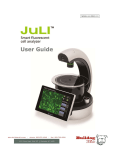
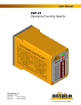
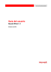
User Manual
Contents
Product Components
Product Overview
Description
Installation Arthur™
4
5
6-7
8
Operation
9-36
Settings
9-16
Set date & time
Set counting settings
9
10
Align cameras
11-12
Calibrate
13-15
Update firmware
16
Recommend actions
17
Selecting assay
18
Naming the sample
19
Loading sample
22
Measuring background (optional)
23
24
Review captured image
25
Layers
26
Gating
Rename
32
Delete
32
Review
33
Annotate
33
Export data
34-35
Cell analysis report
Cleaning & Maintenance
Warranty
Safety precautions
Product specifications
Ordering information
Technical support
36
37
38
39-41
42
43
44-45
27-28
29-30
Viability
29
Apoptosis
29
Cell cycling
30
Green fluorescence
30
2014 NanoEntek Inc.
31
24-28
Capture
Result
31-36
Select data file
20-21
Focusing
Running sample
Data
Arthur™, Image based cytometer
3
Product components
Arthur™ is shipped with the following components.
Please check all components listed below when an instrument was delivered. If any item is
missing or damaged, contact your local distributor or e-mail [email protected].
Image based cytometer
1 EA
Power cord (4 pcs/set, for U.S./Canada/
Taiwan/Japan, Europe or UK)
1 SET
USB drive
4 GB
1 EA
Calibration beads
(Green Cali.Beads, Red Cali. Beads and
Alignment Beads)
1 KIT
User manual
Quick manual
1 EA
1 EA
4
Cell analysis slide
(Cat. No. AC0050)
1 Box (50 slides/box)
Product overview
The Arthur™, Image based cytometer, is a 3-channel (bright field, green fluorescence, red fluorescence) desktop image
analyzer that allows users to perform assays for cells in suspension, including GFP and RFP expression, apoptosis,
cell viability (live, dead, and total cells), cell cycle, and cell counting assays using state-of-the-art optics. It is compatible
with a wide variety of eukaryotic cells.
Depending on complexity of the assay and number of fields captured, it takes 10 seconds to 2 minutes that Arthur™
counts for a typical assay with only 25 μL of sample volume. In addition to the bright field channel, the Arthur™ features
two fluorescent channels (green and red), enabling it to simultaneously count green or red fluorescent stains, as well as
cells expressing GFP and RFP.
The Arthur™ offers an intuitive user interface, and provides the option to save data and generate a report, which can
then be transferred to a PC using the USB drive supplied with the instrument or available separately.
The Arthur™ is supplied with disposable Arthur™ Cell analysis slide, which are also available separately.
Features
Is designed for compact desktop design for fast, easy-to-use, and accurate 3-channel assay.
Uses disposable Arthur™ Cell analysis slide that eliminate washing steps and cross
contamination between samples.
Provides comprehensive test results with graphic data and the cell analysis report allowing
the export of data as .csv (comma separated value), .jpg, .fcs, and .pdf files for archiving
and sample comparisons.
2014 NanoEntek Inc.
Arthur™, Image based cytometer
5
Description
Front View
Power button
The power button is used to turn the instrument on and off.
The blue status light indicates that the instrument is on.
LCD touch screen
The LCD touch screen display, located in the front of the
instrument, provides buttons for all the functions available
and shows data from the cell count.
Slide port
The slide port is used to insert the Arthur™ Cell analysis
slide containing the sample into the counter for analysis.
USB port
The USB port allows you to transfer and save the cell count
data and image to your computer for record keeping and
printing purposes. The USB drive supplied with the
instrument or any other standard USB drive is inserted into
the USB port for data transfer.
Focus knob
The focus knob is used to adjust the image quality to obtain
better contrast between the cells and the background, as
well as between cells that have taken up Arthur™ reagents
and unstained cells.
Power button
USB port
Slide port
Focus knob
LCD touch screen
6
Description
Rear and Side view
Power inlet
The power inlet connects the instrument to an electrical
outlet through the supplied power cord and
the appropriate plug, based on the electrical outlet
configuration in your country.
On/Off switch
The on/off switch is the main power switch that is not required to use for day-by-day operation of the Arthur™.
On/Off switch
Focus knob
Power inlet
2014 NanoEntek Inc.
Arthur™, Image based cytometer
7
Installation
Installing Arthur™
1. After unpacking the instrument, place the instrument on a flat, level, dry surface.
2. Plug one end of the supplied power cord into the Arthur™. To the other end attach the
appropriate plug adaptor, based on the electrical outlet configuration in your country.
3. Plug the power cord into the electrical outlet. Be sure to use only the power cord
supplied with your instrument. Powering the instrument with an unapproved power cord
may damage the instrument.
4. Turn on the main power switch (i.e., On/Off switch) located above the power inlet port
(page7).
5. When you are ready to use, turn on the Arthur™ by pressing the Power button on the
front of the instrument.
6. When the Arthur™ is turned on, the instrument starts up and displays the Home screen.
From the Home screen, you can proceed immediately to Arthur™ Assays functions
including Quick count, Cell Health, Cell Cycle, or Green/Red, and you can use the navigation bars to access saved data (Data screen) or adjust instrument settings (Settings screen).
IMPORTANT
When installing or moving Arthur™, it is recommended to perform auto-alignment
(page 11).
8
Operation
Settings
1. Press the Settings menu on the navigation bar to see the Settings screen.
The Settings screen allows you to:
Calibrate and align the instrument to ensure optimal performance (page 13).
Update to install new firmware version as it become available (page 16).
Set up date and time (see below).
Adjust counting settings (sensitivity and circularity) (page 10).
Settings
Set date & time
1. The date and time is preset. Press the Settings menu to reset date and time based on
your environment.
2. Choose the date and time by moving up and down the roller wheel.
After completing date/time setting, the updated date/time will be displayed
on the top right corner of the screen.
2014 NanoEntek Inc.
Arthur™, Image based cytometer
9
Operation
Settings
Set counting settings
1. Press the Settings menu to adjust counting setting. The counting parameter of Arthur™
is preset in order to perform accurate cell counting for the majority of cultured cell types
without changing default counting parameter. The global parameters for counting
described below:
Sensitivity refers to the contrast of the objects from the background. Increasing the
sensitivity makes the instrument more sensitive to objects in the bright field, while
decreasing the sensitivity makes the instrument less sensitive, which is useful if there is
a significant amount of debris in the sample.
Circularity is used to determine which objects to include in the measurement based on
their roundness. Increasing the circularity value requires objects to be rounder for
inclusion in the measurement and may be useful if you need to distinguish perfectly
round cells from more oddly-shaped cells. Decreasing the circularity allows objects to
be less round for inclusion in the measurement and may be useful if the cell type is not
particularly circular or perhaps oddly shaped.
2. Change sensitivity and circularity by moving slider. If necessary to return to default
values, press the Default button.
In addition to sensitivity and circularity, you can also adjust the parameters for the cell
size and green and/or red-fluorescence, which are specific to the assay in use (page 27).
The proper alignment of the two cameras inside the Arthur™ is required to obtain the
most accurate data for rescent image with the bright field image.
10
Operation
Settings
Align cameras
1. Load 25 µL of Arthur™ Alignment Beads on the Arthur™ Cell analysis slide (page 20).
Make sure to mix the bead thoroughly before pipeting. Load the bead gently to spread
it evenly in the cell analysis slide.
2. Press the Align cameras to start the alignment procedure.
3. Insert the Arthur™ cell analysis slide containing the Arthur™ Alignment beads into
the instrument.
4. Adjust the focus using the Focus knob.
4-1. Touch the screen to magnify the image.
5. Press the Run button to start the auto-alignment.
2014 NanoEntek Inc.
Arthur™, Image based cytometer 11
Operation
Settings
Align cameras
6. Press the OK button to confirm the correct alignment of the cameras.
Press the Cancel button to cancel the alignment.
You can also manually adjust the cameras by positioning the green circles over the
corresponding Alignment beads using the directional keys on this screen, but this is
not recommended.
IMPORTANT
When installing or moving Arthur™, it is recommended to perform auto-alignment.
12
Operation
Settings
Calibrate
Calibrating the green and red fluorescent channels of the Arthur™ is to set the dynamic
range of the instrument. The calibration procedure is most effective when performed
after alignment sequence.
1. Press the Calibrate Green/Red button on the Setting menu. The Calibration dialog
will appear.
2. Load 25 µL of Arthur™ Green calibration beads on the Arthur™ Cell analysis slide.
Make sure that the beads are mixed thoroughly before pipetting. (page 20).
Make sure to mix the bead thoroughly before pipeting. Load the bead gently to spread
it evenly in the cell analysis slide.
3. Insert the slide with calibration beads into the slide port of instrument and press the
Insert sample button.
4. Adjust the focus using the Focus knob.
When correctly focused, the Calibration beads will appear round rather than oblong.
Correct focus
2014 NanoEntek Inc.
Incorrect focus
Arthur™, Image based cytometer 13
Operation
Settings
5. Press the Run button to start the auto calibration of the green channel.
Calibrate
6. When the green channel calibration is complete, the cell analysis slide will be ejected
automatically and the red channel calibration dialog will appear.
7. Load 25 µL of Arthur™ Red Calibration Beads on the Arthur™ Cell analysis slide
(page 20). Make sure that the beads are mixed thoroughly before pipetting.
8. Insert the slide with calibration beads into the slide port for instrument and press the
Insert sample button.
9. Adjust the focus using the Focus knob.
10. Press the Run button to start the auto calibration of the red channel.
14
Operation
Settings
Calibrate
11. Press the OK button to return to the Settings menu. Arthur™ does not require
recalibration each time the instrument is turned on.
IMPORTANT
Insert the slide with calibration beads just at the time of use, to prevent drying of
solution in slide.
Allow the beads to settle in the slide before pressing run. There should be no
movement of beads on the image.
The entire calibration can take up to 20 minutes to complete.
2014 NanoEntek Inc.
Arthur™, Image based cytometer 15
Operation
Settings
Update firmware
1. Periodically, Nanoentek adds functionality and other improvements to Arthur™ user
interface. We recommend keeping your Arthur™ update with the latest firmware using the
Update buttons.
If you have any questions about the software updates, contact [email protected].
2. Visit our website at www.nanoentek.com to download update software which will be
supplied as a .zip file to your desktop.
3. Unzip the Arthur Update.zip file and save the software in a USB drive.
One folder / one “.ini” , two “.exe”, six “.dll”
IMPORTANT
The files must reside individually on the USB drive. Do not transfer as a folder or
put the files in a folder.
4. Connect the Arthur™ USB drive to main body of the Arthur™.
5. Check if the version of software and firmware are the latest one.
Press the Update firmware button. The update process takes a few seconds.
6. The Update dialog will appear. Press the Update button to start updating.
7. After updating, press the OK button to restart the Arthur™ automatically.
16
Operation
Recommend
actions
To obtain the best results, follow these recommendations:
Wear gloves during sample handling.
Do not touch the optical surfaces of the Arthur™ Cell analysis slides. Hold the
slides by the edges.
Use the Arthur™ at room temperature only (5 – 40 °C).
The Arthur™ contains delicate optics. Place the instrument on a flat, dry surface that is
free from excessive vibration. Do not spill liquids on the surface of the instrument or
introduce liquids into its interior.
The Arthur™ can accurately count cells 5 μm to 60 μm in diameter.
5
The recommended sample concentration range for the Arthur™ is 1 × 10 to
1 × 10⁷ cells/mL; however, the sample concentration does not need to be exact to
perform an assay.
For accurate viability count results, ensure the counting area is covered with cell
suspension and count cells immediately after staining per the assay protocol.
Re-calibrate the Arthur™ after updating the firmware or when using assay reagents not
formulated for the instrument (page 13).
The internal working memory of the Arthur™ is 145 Gigabytes, sufficient for storing
comprehensive data from 500 sample runs. However, we recommend that you save your
data to the USB drive after completing each experiment. Using the USB drive, you may
then transfer the data to your PC as described in “Export data” (page 34).
After using Arthur™ Cell analysis slides, appropriately dispose of them as biohazardous
waste. Do not reuse the Arthur™ Cell analysis slides.
Turn off the Arthur™ at the end of the day.
2014 NanoEntek Inc.
Arthur™, Image based cytometer 17
Operation
Selecting assay
1. To select a Arthur™ assay, press the appropriate assay button (Quick count, Cell Health,
Cell Cycle, or Green/Red) on the Home screen. For example, press the Cell Health button
for the viability and apoptosis assay options.
2. Next, Apoptosis assay is selected from the options, giving you the option to name the
sample now or later.
18
Operation
Naming the
sample
1. Press the Name now button to name the sample before performing the assay.
The alpha-numeric keyboard appears.
2. Type the name of the sample series using up to 40 alpha-numeric characters, and then
press the Save button. Each sample run in the series will be appended with a number to
reflect the order in which it was run.
(Press the Close button to return to the previous screen without naming the sample.)
If pressing the Name later button, the name for each sample series by date and time
will be automatically assigned. Go to the Data menu to rename, if necessary (page 32).
2014 NanoEntek Inc.
Arthur™, Image based cytometer 19
Operation
Loading sample
1. Load 25 μL of your sample (per slide chamber) on the Arthur™ Cell analysis slide using
pipette as described below.
Make sure to mix the sample thoroughly before pipeting. Load the sample gently to
spread it evenly in the cell analysis slide.
IMPORTANT
Follow the guidelines below to load your samples on the Arthur™ Cell analysis slide.
Do not touch the optical surfaces of the Arthur™ Cell analysis slides. Hold the slides by
the edges.
Use 25 μL of sample volume per slide chamber. Do not overfill or underfill the
slide chambers.
Pipette the sample at an angle of approximately 80° into the half moon-shaped
sample loading area. The sample is loaded into the chamber through capillary action.
Take care to avoid forming bubbles in the sample.
The Arthur™ counts the cells in the chamber opposite the inserting direction.
For example, to count the sample in Chamber A, you need to insert the Arthur™ Cell
analysis slide into the instrument with the Chamber B-side first until it stops.
For inserting the Arthur™ Cell analysis slide into the instrument.
The Arthur™ Cell analysis slides are specifically designed for use with the Arthur™
exclusively. Use of other slides result in inaccurate cell counts and can damage
the Arthur™.
20
Operation
Loading sample
2. The Arthur™ counts the cells in the chamber opposite inserting direction.
Inserting direction
Sample loaded
IMPORTANT
To count the sample in Chamber A, insert the Arthur™ Cell analysis slide into the
slide port of the instrument with the Chamber B-side first until it stops.
3. Press the Press to insert new sample button, the slide will automatically be pulled into
the instrument.
If you are performing a background measurement, the button for inserting the sample
will read Press to insert unstained control (page 23).
2014 NanoEntek Inc.
Arthur™, Image based cytometer 21
Operation
Focusing
1. Adjust the focus using the Focus knob on the right side of the instrument.
2. Press the Zoom button and select either 4× or 16× magnification enhancement to
review the image.
Correctly focused images have uniformly dark-colored cells surrounded by bright
halos (see example on the left, below).
Cells may be undercounted when the transition between the background
and the edges of the cells are fuzzy and the cells have undefined boundaries
(see example in the middle, below).
Cells maybe overcounted when they have bright centers and dark
perimeters (see example on the right, below).
[Image is correctly focused]
* Cell type : Hep3B
22
[Incorrect –undercounted cells]
[Incorrect –overcounted cells]
Operation
Measuring
background
(optional)
1. Press the Background tab to see screen opens.
The Background feature is available for Arthur™ Viability, Apoptosis, Green + Red,
and Cell Cycle assays except for the Quick count assay, which does not use
fluorescent reagents.
“No Background” will be displayed on the dropdown menu, if there are the saved
background measurements for the assay on the instrument. The fluorescence threshold,
which you can change manually after the assay, will be set up when choosing not to run
a background control.
The latest background will be displayed on the dropdown menu, it there are the saved
background measurements on the instrument.
2. Select the File name from the drop-down menu to use a previous background
measurement. If you want to change a previously obtained background measurement,
select the other file name from the drop-down menu.
3. To perform a new background measurement, load the slide on the unstained cell
control, insert the slide into the instrument, and press the Press to insert new unstained
cell control button.
4. Press the Press to run unstained cell control button after Arthur™ cell analysis slide is
completely pulled into the instrument.
2014 NanoEntek Inc.
Arthur™, Image based cytometer 23
Operation
Running sample
Capture
1. Select the number of images from the drop-down menu to specify the number of fields
of view to capture.
2. Press the Press to run sample button to capture the selected number of images.
3. After capturing the specified number of fields of view, the Arthur™ cell analysis slide will
be ejected automatically. The analysis result data including average cell size, the counted
number of cells, total cell concentration, assay-specific data, other specific data and
histogram plots will be displayed in the data window located in the sample tab.
4. If necessary to run the next sample, insert the new slide, and repeat the procedure.
24
Operation
Running sample
Review captured
image
1. Press the thumbnail of the image displayed in the Zoom tab to select a captured field of
view for review.
2. Move the Green square in the slider bar to zoom in and out the image.
3. Press the image to display the navigation tool for reviewing a different section of an
image viewed at 4x or 16x magnification.
4. Select the appropriate area on navigation tool to display the desired section of
the image.
5. Press the image outside the navigation tool to hide the tool and review the image.
The navigation tool is only available for reviewing the images at 4× and
16× magnification.
2014 NanoEntek Inc.
Arthur™, Image based cytometer 25
Operation
Running sample
Layers
1. Press the thumbnail of the image and the Layers tab to review images captured
through the bright field, the green fluorescence, and the red fluorescence.
The number of layers that are available for viewing and the labels for those layers
depend on the Arthur™ Assay performed. In the example above, the buttons for the
fluorescent channels are labeled Green and Red, because the Arthur™ Green + Red Assay
is selected. However, if the Arthur™ Apoptosis Assay was selected, these same buttons
would be labeled Annexin V (for the green channel) and PI (for the red channel).
2. Press the Bright field/Green/Red buttons as necessary. When multiple layers are turned
on, they appear overlaid on the image.
3. Press the Circle button to identify the cells counted through a particular channel.
Arthur™ circles the cells that were analyzed as follows:
Blue: cells counted in bright field channel
Green: cells counted in green fluorescence channel
Red: cells counted in red fluorescence channel
Yellow: cells counted in both green and red channels
Black: cells excluded from the counting results
26
Operation
Running sample
Gating
The procedure for setting gates and thresholds is identical for all Arthur™ Assays,
but the buttons on the touch-screen are labeled according to the specific assay selected.
For example, the histogram thumbnails that are labeled Cell size, Green, and Red for the
Arthur™ Green + Red Assay (see image below) are labeled Cell size, Annexin V, and PI for
the Arthur™ Apoptosis Assay. The thin blue lines on the histograms represent the
set thresholds.
1. Press the cell size thumbnail to set the gating parameters for cell size.
2. The “Cell size vs. # of cells” histogram will appear. Move the two green square buttons on
the slider bar to set the minimum cell size and maximum cell size.
3. Press the Apply button to confirm and return to the previous screen.
2014 NanoEntek Inc.
Arthur™, Image based cytometer 27
Operation
Running sample
4. Press the Green histogram thumbnail to set the threshold for the Green fluorescence.
Gating
5. Press the Sample fluorescence button or the Control fluorescence button on the
bottom left.
6. Press on the minimum or maximum RFU value (relative fluorescence unit) to set the
x-axis scale and type in a new value using the popup (alpha-numeric) key pad.
7. Move the arrow button on the slider bar to set the threshold. Press the RFU threshold
button and type in the threshold value using the (alpha-numeric) popup key pad.
8. Press the Apply button to confirm. Or press the Close button to return to the previous
screen without setting.
9. Press the Red histogram thumbnail to set the threshold for Red fluorescence.
IMPORTANT
When using the Arthur™ Cell Cycle Assay, the Propidium Iodide (PI) histogram will have
four individual thresholds, which are used for partitioning the cell population into the
stages of the cell cycle. Each threshold can be set as described above and the color of
each partition will correspond to the appropriate data set in the data table.
Biological molecules found within cells fluoresce upon excitation and result in
background fluorescence. Because the Arthur™ is a highly sensitive instrument, it detects
this background fluorescence and displays it as a peak closest to the 0 RFU value.
To eliminate the background fluorescence from your calculations, adjust the threshold
to exclude this peak.
28
Operation
Result
Viability
Result
Apoptosis
2014 NanoEntek Inc.
Arthur™, Image based cytometer 29
Operation
Result
Cell cycling
Result
Green fluorescence
30
Operation
Data
1. Press the Data menu to see the stored data files in a list on the right side of the screen.
Select data file
2. Move up and down the Green square button on the scrollbar to see all data files.
3. Select the data file to review by touching the corresponding line on the file list.
Select Multiple button to select more than one file at a time. The Select Multiple button
will be disabled when pressing it again.
Select All button to select all files in the list.
The internal working memory of the Arthur™ is 145 Gigabytes, sufficient for storing comprehensive data from 500 sample runs. However, we recommend that you save your data
to the USB drive after completing each experiment. Using the USB drive, you may then
transfer the data to your PC as described in “Export Data” (page 34).
2014 NanoEntek Inc.
Arthur™, Image based cytometer 31
Operation
Data
Rename
1. Select the Data file to rename.
2. Press the Rename button to rename the selected files. The alpha-numeric keyboard
will appear.
3. Enter the file name using the keyboard, and then press the Save button. Press the
Close button to return to the previous screen without renaming.
Data
Delete
1. Select the Data file(s) to be deleted.
Select Multiple button to select more than one file at a time. The Select Multiple button
will be disabled when pressing it again.
Select All button to select all files in the list.
2. Press the Delete button to delete the selected file(s).
32
Operation
Data
Review
Data
Annotate
1. Select the File name from the Data menu to review the saved data.
2. Press Layers, Zoom, or Analysis tabs to display the corresponding screens.
1. Select the File name to annotate, and press the Notes box above the data window.
The alpha-numeric keyboard will appear.
2. Enter your comments using the keyboard, and then press the Save button.
Your comments in the Notes box is be attached to the file and shown on all Data tabs
(i.e., Layers, Zoom, and Analysis). Press the Close button to return to the previous screen
without your comment.
2014 NanoEntek Inc.
Arthur™, Image based cytometer 33
Operation
Data
Export data
Data table contains comprehensive information about the count with size and
fluorescence intensity of each individual particle/cell in the sample and provided in
.csv file and .fcs file format.
Images exports a single image in each channel collected for each field of view. Images
are exported as .jpg files.
Report contains only the aggregate results and calculations from the run such as the
total cell count and concentration, counts and concentrations of fluorescent cells and
their relative abundance in the sample, thumbnails of the bright field image of each field
of view captured, as well as the relevant histograms and instrument settings
in a .pdf file (page 36).
34
Operation
Data
Export data
1. Connect the Arthur™ USB drive (or any other USB drive) into the Arthur™ .
2. Select the Data file to export. Press the Select all to highlight all files.
3. Press the Data table (.csv and .fcs) button, Image (.jpg) button, or Report (.pdf)
button to export the data in the designated formats. Press the All formats button
to export data in all these formats.
4. Transfer the files on the USB drive to your PC. You may open the exported files using
the appropriate programs.
2014 NanoEntek Inc.
Arthur™, Image based cytometer 35
Operation
Data
Cell analysis report
36
The image below shows an example of a data analysis report exported as a .pdf.
Cleaning & Maintenance
Clean the surface of the Arthur™ with a damp cloth. To clean the LCD screen, turn off
Arthur, disconnect the power cable, and clean the LCD screen with a soft cloth lightly
moistened with LCD cleansing detergent. Cleaning the screen with excessive force can
damage the LCD screen. Wipe the screen dry immediately. Do not reuse the cell
counting slides.
Arthur™ does not need regular maintenance. To troubleshoot problems with Arthur™,
contact Technical Support (page 44).
IMPORTANT
Never disassemble or service Arthur™ by yourself.
Unauthorized repairs may damage Arthur™ or alter its functionality, which will void your
warranty. Contact [email protected] or your local Arthur™ distributor to arrange
for service.
Always wipe surfaces with ethanol-soaked paper towels.
Do not directly spray ethanol anywhere on Arthur™.
Avoid exposing Arthur™ to UV light
UV light may degrade components, including plastic. Damage from UV exposure is not
covered under the manufacturer’s warranty.
2014 NanoEntek Inc.
Arthur™, Image based cytometer 37
Warranty
Nanoentek warrants the Arthur will be free from defects in material and workmanship for
a period of one (1) year from date of purchase.
If any defects occur in Arthur during this warranty period, NanoEnTek will repair or replace
the defective parts at its discretion without charge.
The following defects, however, are specifically excluded:
1. Defects caused by improper operation.
2. Repair or modification done by anyone other than Nanoentek or an authorized agent.
3. Damage caused by substituting alternative parts.
4. Use of fittings or spare parts supplied by anyone other than Nanoentek.
5. Damage caused by accident or misuse.
6. Damage caused by disaster.
7. Corrosion caused by improper solvent or sample.
For your protection, Arthur™ units being returned must be insured against possible
damage or loss. Nanoentek cannot be responsible for damage incurred during shipment
of a defective instrument. It is recommend that you save the original packing material in
which the instrument was shipped. This warranty is limited to the replacement of
defective products.
For any inquiry or request for repair service, contact [email protected]
or your local distributor.
38
Safety precautions
Review and follow the safety instructions below :
If water or other material enters the instrument, adaptor, or power inlet, disconnect the
power cord and contact a service person. For operating environment, refer to Product
Specifications (page 42).
Do not touch the main plug or power cord with wet hands.
Always ensure that the power supply input voltage matches the voltage available
at your location.
This instrument is air-cooled and its surfaces may become hot during operation.
During installing, leave a space of more than 10 cm (4 inches) around the instrument and
do not place any objects between the instrument and the walls.
Do not install the instrument on a slant or a place prone to vibrations, which induces the
risk of instrument malfunction or damage of the instrument.
Never insert any objects into the air vents of the instrument as this could result in
electrical shock, personal injury, and equipment damage.
Plug the power cord firmly into the wall outlet and AC adapter.
To avoid potential shock hazard, make sure that the power cord is properly grounded.
Be sure to position the equipment such that it is easy to disconnect.
Turn off the instrument before unplugging the power cord and/or moving
the instrument.
If the instrument is dropped or broken, disconnect the power cord and contact a
service person. Disassembly of case will void warranty.
Use only authorized accessories (adaptor, power cord, and USB drive).
Warning
Class A equipment is intended for use in an industrial environment. In the documentation for
the user, a statement shall be included drawing attention to the fact that there may be potential
difficulties in ensuring electromagnetic compatibility in other environments, due to conducted
as well as radiated disturbances.
2014 NanoEntek Inc.
Arthur™, Image based cytometer 39
Consignes de securite
Examinez et suivez les consignes de securite ci-dessous :
N’installez pas l’instrument dans un endroit humide comme une serre ou un
incubateur pour eviter un risque de choc electrique. Si de l’eau ou tout autre
materiau penetre dans l’instrument, l’adaptateur, ou l’entrees d’alimentation, debranchez
le cordon d’alimentation et contactez un technicien de service. Pour l’environnement
d’exploitation, reportez-vous aux specifications du produit.
Ne touchez pas la fiche ou le cordon d’alimentation principale avec les mains mouillees.
Assurez-vous toujours que la tension d’entree d’alimentation correspond a la tension
disponible dans votre endroit.
Cet instrument est refroidi a l’air de sorte que ses surfaces peuvent devenir chaudes
pendant le fonctionnement.
Lors de l’installation de l’instrument, laisser un espace de plus de 10 cm (4 pouces) autour
de cet instrument et ne placez aucun objet entre l’appareil et le mur.
N’installez pas l’instrument sur une pente ou un endroit soumis a des vibrations, ce qui
induit le risque de dysfonctionnement ou d’endommagement de l’instrument.
N’inserez jamais aucun objet dans les orifices d’aeration de l’instrument, car cela
pourrait entrainer un choc electrique, des blessures chez les utilisateurs et des
dommages d’equipement.
Branchez le cordon d’alimentation fermement dans la prise murale et l’adaptateur
secteur aussi.
Pour eviter un risque potentiel de commotion electrique, assurez-vous que le cordon
d’alimentation est correctement mis a la terre.
Assurez-vous de positionner l’instrument de telle sorte qu’il soit facile de debrancher
l’instrument.
Eteignez l’instrument avant de debrancher le cordon d’alimentation et / ou de deplacer
l’instrument.
Si l’instrument est casse ou qu’il soit tombe, debranchez le cordon d’alimentation et
contactez un technicien de service. Ne demontez pas l’instrument et la garantie sera
annulee en cas de demontage.
Utilisez uniquement les accessoires autorises (l’adaptateur, le cordon d’alimentation,
et le lecteur USB).
Warning
Le produit de classe A est conçu pour l’utilisation dans un environnement industriel.
Dans la documentation de l’utilisateur, la déclaration doit être incluse pour attirer l’attention
sur le fait qu’il peut y avoir des difficultés potentielles pour assurer la compatibilité
électromagnétique dans d’autres environnements, en raison des perturbations rayonnées et
conduites par l’électricité.
40
Safety precautions
Review and follow the safety instruction below:
Symbol
Meaning
Caution & Warning
Protective earth (Ground)
This instrument and consumables conforms to the EC Declaration of Conformity.
FCC Compliance
This equipment has been tested and found to comply with the limits for a Class A digital
device, pursuant to Part 15 of the FCC Rules.
These limits are designed to provide reasonable protection against harmful interference
when the equipment is operated in a commercial environment.
This equipment generates, uses, and can radiate radio frequency energy and, if not
installed and used in accordance with the instruction manual, may cause harmful
interference to radio communications.
Operation of this equipment in a residential area is likely to cause harmful interference in
which case the user will be required to correct the interference at his own expense.
Disposal of your old appliance1. When this crossed-out wheeled bin symbol is attached
to a product it means the product is covered by the European Directive 2012/19/EU.2. All
electrical and electronic products should be disposed of separately from the municipal
waste stream via designated collection facilities appointed by the government or the local
authorities.3. The correct disposal of your old appliance will help prevent potential
negative consequences for the environment and human health.4. For more detailed
information about disposal of your old appliance, please contact your city office, waste
disposal service or visit our web-site, www.nanoentek.com.
This product conforms to UL 61010-1, CAN/CSA C22.2 No.61010-1 “Safety Requirements for
Electrical Equipment for Measurement, Control, and Laboratory Use, Part l: General Requirements.” Instruments bearing the TUV symbol are certified by TUV Product Services to be in
conformance with the applicable safety standard for the US and Canada.
2014 NanoEntek Inc.
Arthur™, Image based cytometer 41
Product specifications
Environmental conditions
Operating power
100 – 240 VAC, 2.5 A, 120 W
Frequency
50 / 60 Hz
Installation site
Indoor use only
Operating temperature
5 - 40 °C
Maximum Relative Humidity
20 - 95 %
Instrument
Counting time
10 sec ~ 2 min.
Cell measurement range
5
7
1 × 10 - 1 × 10 cells/mL
Cell size range
3 channels (bright field, green fluorescence, red fluorescence)
Optics
5 - 40 °C
Excitation
Green channel LED: 458 ± 20 nm
Red channel LED: 530 ± 20 nm
Filters
Green channel: 466/40 EX, 495 LP Di, 525/50 EM
Red channel: 543/22 EX, 580 LP Di, 585 LP EM
Camera
1.3 Mega pixels, 4× objective,
4× or 16× digital zoom
USB drive
4 GB
Dimension
290 mm (W) × 440 mm (D) × 290 mm (H)
Weight
8.7 kg
Cell analysis slide
Material
Polymethy methacrylate (PMMA)
Dimension
110 mm (W) × 24 mm (D) × 1.9 mm (H)
Chamber (sample) volume
25 μL
42
Ordering information
Cat No.
Description
AT1000
Arthur™ Image based cytometer
AC0050
Arthur™ Cell analysis slide (50 slides)
AC0500
Arthur™ Cell analysis slide (500 slides)
AB0100
Arthur™ Calibration beads (100 μL)
2014 NanoEntek Inc.
Arthur™, Image based cytometer 43
Technical support
Visit the our Website at www.nanoentek.com for :
Technical resources, including manuals, FAQs, etc.
Technical support contact information
Additional product information and special offers.
For more information or technical assistance, please call or email.
NanoEnTek Inc. (HQ)
5, Digital-ro 26-gil, Guro-gu, Seoul, 152-740, Korea
Tel :+82-2-6220-7940
Fax:+82-2-6220-7721
NanoEnTek USA Inc.
5627 Stoneridge Drive Suite 304, Pleasanton, CA 94588, USA
Tel :+1-925-225-0108, +1-888-988-0108(Toll free)
Fax:+1-925-225-0109
Email
[email protected]
Website
www.nanoentek.com
44
All the materials in this user guide are protected by Korean
and international copyright laws. They cannot be reproduced, translated,
published or distributed without the permission of the copyright owner.
Arthur™ User manual
Website : www.nanoentek.com
E-mail : [email protected]
Manufactured by : NanoEnTek Inc.
12F, 5, Digital-ro 26-gil, Guro-gu, Seoul, 152-740, Korea
Website : www.nanoentek.com
E-mail : [email protected]
The information in this manual is described as accurately as possible.
Firmware and software changes and updates may change without prior
consent or notification.
Copyright
2014 by NanoEnTek Inc. All rights reserved. Published in Korea.
Documentation : NESMU-ATT-001E
Revision history : V.0.0 Date : SEP 2014
2014 NanoEntek Inc.
Arthur™, Image based cytometer 45
NESMU-ATT-001E (V.0.0)
NanoEnTek Inc. (HQ)
5, Digital-ro 26-gil, Guro-gu, Seoul, 152-740, Korea
Tel : +82-2-6220-7940
Fax: +82-2-6220-7721
NanoEnTek USA Inc.
5627 Stoneridge Drive Suite 304, Pleasanton, CA 94588, USA
Tel : +1-925-225-0108, +1-888-988-0108(Toll free)
Fax: +1-925-225-0109
Email
[email protected]
Website
www.nanoentek.com