1
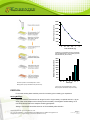
EPIGENTEK Complete Solutions for Epigenetics MethylFlash™ Urine 5-Methylcytosine (5-mC) Quantification Kit (Fluorometric) Base Catalog # P-1040 PLEASE READ THIS ENTIRE USER GUIDE BEFORE USE Uses: The MethylFlash™ Urine 5-Methylcytosine (5-mC) Quantification Kit (Fluorometric) is suitable for detecting total urinary 5-methylcytosine levels resulting from whole body turnover or degradation of methylated DNA/RNA using urine from humans and animals. The urine samples can be in fresh or frozen form. Starting Material and Input Amount of DNA: The volume of urine for each assay can be between 0.5 and 5 µl. For optimal quantification, the input urine volume should be 1 µl. Internal Control: Both a negative control and a positive 5-mC standard are provided in this kit. A standard curve can be performed (range: 0.05 to 1 ng). Because 5-mC levels in urine can vary from individual to individual, and from normal and diseased states, it is advised to run replicate samples to ensure that the signal generated has been validated. This kit will allow the user to quantify an absolute amount of 5-mC and determine the relative turnover states of methylated DNA/RNA of two different urine samples. Precautions: To avoid cross-contamination, carefully pipette the sample or solution into the strip wells. Use aerosol-barrier pipette tips and always change pipette tips between liquid transfers. Wear gloves throughout the entire procedure. In case of contact between gloves and sample, change gloves immediately. 110 Bi County Blvd. Ste. 122, Farmingdale, NY 11735 Tel: 1-877-374-4368 ■ Fax: 1-718-484-3956 ■ E-mail: [email protected] ■ Web: www.epigentek.com © Epigentek Group Inc. All rights reserved. Products are for research use only. Page 1 Printed 2014-10-13 P-1040 EPIGENTEK Complete Solutions for Epigenetics KIT CONTENTS Component 48 Assays Cat. #P-1040-48 96 Assays Cat. #P-1040-96 Storage Upon Receipt WB (10X Wash Buffer) 14 ml 28 ml 4°C BS (Binding Solution) 5 ml 10 ml RT MS (5-mC Standard, 4 µg/ml)* 10 µl 20 µl –20°C ME3 (Negative Control, 50 µg/ml)* 10 µl 20 µl –20°C ME4 (5-mC DNA solution, 320X)* 15 µl 30 µl –20°C ME5 (Capture Antibody, 1000X)* 4 µl 8 µl 4°C ME6 (Detection Antibody, 2000X)* 4 µl 8 µl –20°C ES (Enhancer Solution)* 4 µl 8 µl –20°C FD (Fluoro-Developer)* 10 µl 20 µl –20°C FE (Fluoro-Enhancer)* 10 µl 20 µl 4°C DB (Dilution Buffer) 4 ml 8 ml RT Plate 1 (Assay Plate) 1 (6 strips) 1 (12 strips) 4°C Plate 2 (Sample Preparation Plate) 1 (6 strips) 1 (12 strips) RT User Guide 1 1 RT * Spin the solution down to the bottom prior to use. SHIPPING & STORAGE The kit is shipped in two parts: the first part at ambient room temperature and the second part on frozen ice packs at 4°C. Upon receipt: (1) Store MS, ME3, ME4, ME6, ES, and FD at –20°C away from light; (2) Store WB, ME5, FE, and Plate 1 at 4°C away from light; (3) Store remaining components (BS, DB, and Plate 2) at room temperature away from light. All components of the kit are stable for 6 months from the date of shipment, when stored properly. Note: Check if WB (10X Wash Buffer) contains salt precipitates before use. If so, briefly warm at room temperature or 37°C and shake the buffer until the salts are re-dissolved. MATERIALS REQUIRED BUT NOT SUPPLIED Adjustable pipette Aerosol resistant pipette tips Microplate reader capable of reading fluorescence at 530ex/590em nm. 1.5 ml microcentrifuge tubes Incubator for 37°C incubation 110 Bi County Blvd. Ste. 122, Farmingdale, NY 11735 Tel: 1-877-374-4368 ■ Fax: 1-718-484-3956 ■ E-mail: [email protected] ■ Web: www.epigentek.com © Epigentek Group Inc. All rights reserved. Products are for research use only. Page 2 Printed 2014-10-13 P-1040 EPIGENTEK Complete Solutions for Epigenetics Plate seal or Parafilm M Distilled water 1X TE buffer pH 7.5 to 8.0 Urine sample GENERAL PRODUCT INFORMATION Quality Control: Each lot of the MethylFlash™ Urine 5-Methylcytosine (5-mC) Quantification Kit (Fluorometric) is tested against predetermined specifications to ensure consistent product quality. Epigentek guarantees the performance of all products in the manner described in our product instructions. Product Warranty: If this product does not meet your expectations, simply contact our technical support unit or your regional distributor. We also encourage you to contact us if you have any suggestions about product performance or new applications and techniques. Safety: Suitable lab coat, disposable gloves, and proper eye protection are required when working with this product. Product Updates: Epigentek reserves the right to change or modify any product to enhance its performance and design. The information in this User Guide is subject to change at any time without notice. Thus, only use the User Guide that was supplied with the kit when using that kit. Usage Limitation: The MethylFlash™ Urine 5-Methylcytosine (5-mC) Quantification Kit (Fluorometric) is for research use only and is not intended for diagnostic or therapeutic applications. Intellectual Property: The MethylFlash™ Urine 5-Methylcytosine (5-mC) Quantification Kit (Fluorometric) and methods of use contain proprietary technologies by Epigentek. A BRIEF OVERVIEW Nucleobase 5-methylcytosine (5-mC), a modified form of cytosine converted by cytosine methyltransferases is widespread both in DNA and different cellular RNAs. The biological importance of DNA 5-mC-methylation as a major epigenetic modification in phenotype and gene expression has been recognized widely. Recent data strongly suggests that RNA 5-mC methylation is also involved in the regulation of various biological processes including tRNA stability/recognition and mRNA translation. Urinary excretion of 5-mC including both 5-methyl-2-deoxycytidine and 5-methylcytidine is an indication of the whole body turnover or degradation of methylated DNA and RNA. The urinary 5-mC level can be changed with a change of the bodies’ turnover of methylated DNA/RNA or alteration of cellular DNA/RNA methylation status. A number of studies have indicated that 5-mC excreted in urine has the potential to act as a cancer biomarker, with an increased level arising from disease onset and progression. For example, an elevated level of urinary 5-mC was observed in lung cancer, breast cancer, and leukemia patients with active disease states. It was also shown that urinary 5-mC excretion is effected by Alzheimer’s disease or by radiation treatment. It has been suggested that urinary 5-mC might be applicable as a biological marker for detecting certain types of cancer and monitoring cancer progression after treatment with radiation or demethylation reagents. 110 Bi County Blvd. Ste. 122, Farmingdale, NY 11735 Tel: 1-877-374-4368 ■ Fax: 1-718-484-3956 ■ E-mail: [email protected] ■ Web: www.epigentek.com © Epigentek Group Inc. All rights reserved. Products are for research use only. Page 3 Printed 2014-10-13 P-1040 EPIGENTEK Complete Solutions for Epigenetics Chromatography-based techniques such as HPLC and TLC mass spectrometry are used for detecting 5-mC in urine. These methods are accurate but are time consuming, less sensitive, and have low throughput with high costs. To address this problem, Epigentek offers the MethylFlash™ Urine 5Methylcytosine (5-mC) Quantification Kit to quantify 5-mC or body turnover status of methylated DNA/RNA using urine samples. The kit has the following advantages and features: Innovative fluorometric assay with easy-to-follow steps for convenience and speed. The entire procedure can be finished within 4 hours. 96 strip well microplate format makes the assay flexible: manual or high throughput analysis Innovative kit composition enables background signals to be extremely low, which eliminates the need for plate blocking and allows the assay to be simple, accurate, reliable, and consistent. The level of 5-mC measured in human urine samples using this kit is comparable to that detected by HPLC method. A novel assay principle allows high sensitivity to be achieved. The detection limit can be as low as 0.01 ng/assay well or 0.3 nM of 5-mC. Low input range of urine for each assay with a volume of 0.5 to 5 µl and an optimal volume of 1 µl. Optimized antibody and enhancer solutions allow high specificity to 5-mC, with no crossreactivity to unmethylated cytosine. Negative control and positive standard are included, which are suitable for quantification of 5mC in free form and 5-mC contained in methylated DNA/RNA fragments from different urine samples. PRINCIPLE & PROCEDURE In this ELISA-like inhibitory competitive immunoassay assay, urine samples and 5-mC standard are first incubated with a 5-mC antibody solution and then transferred to the wells coated with methylated DNA (5-mC). The well is washed to remove any unbound reagents after incubation and then a detection antibody is added to generate a signal that can be measured fluorometrically by reading the fluorescent intensity in a microplate spectrophotometer. Because 5-mC in the urine sample inhibits the binding of 5-mC antibody to 5-mC coated on the well, higher concentrations of 5-mC in the urine sample lead to a reduced binding of the antibody to the 5-mC on the strip-well. Therefore the signal or fluorescent intensity measured from the well will be inversely proportional to the amount of 5-mC in the urine sample and the amount of 5-mC in the urine sample can be quantified by a comparison with a predetermined 5-mC standard. 110 Bi County Blvd. Ste. 122, Farmingdale, NY 11735 Tel: 1-877-374-4368 ■ Fax: 1-718-484-3956 ■ E-mail: [email protected] ■ Web: www.epigentek.com © Epigentek Group Inc. All rights reserved. Products are for research use only. Page 4 Printed 2014-10-13 P-1040 EPIGENTEK Complete Solutions for Epigenetics 14000 Relative Fluorescence Unit 12000 10000 8000 6000 4000 2000 0 0 0.1 0.2 0.4 1 2 5-mC standard (ng) 5-mC standard was added into the assay wells at different concentrations and then measured with the MethylFlash™ Urine 5-Methylcytosine Quantification Kit (Fluorometric). Relative Fluorescence Unit 18000 16000 14000 12000 10000 8000 6000 4000 2000 0 20 Schematic procedure of the MethylFlash™ Urine 5Methylcytosine (5-mC) Quantification Kit (Fluorometric) 10 5 2 1 0.5 0 Urine (ul) 5-mC level is quantified from different volumes of human urine using the MethylFlash™ Urine 5Methylcytosine Quantification Kit (Fluorometric). PROTOCOL For the best results, please read the protocol in its entirety prior to starting your experiment. Starting Materials Input Urine Volume: Urine amount can range from 0.5 to 5 µl per assay. An optimal amount is 1 µl per assay. Clear urine samples can be directly used for the assay. Centrifugation at 2500-3000 g for 10 min should be required for the samples containing precipitates. Storage: Urine sample should be stored at -20°C immediately after collection. 110 Bi County Blvd. Ste. 122, Farmingdale, NY 11735 Tel: 1-877-374-4368 ■ Fax: 1-718-484-3956 ■ E-mail: [email protected] ■ Web: www.epigentek.com © Epigentek Group Inc. All rights reserved. Products are for research use only. Page 5 Printed 2014-10-13 P-1040 EPIGENTEK Complete Solutions for Epigenetics 1. Buffer and Solution Preparation a. Prepare Diluted WB 1X Wash Buffer: 48-Assay Kit: Add 13 ml of WB (10X Wash Buffer) to 117 ml of distilled water and adjust pH to 7.2-7.5. 96-Assay Kit: Add 26 ml of WB (10X Wash Buffer) to 234 ml of distilled water and adjust pH to 7.2-7.5. This Diluted WB 1X Wash Buffer can now be stored at 4°C for up to six months. b. Prepare Diluted ME4 1X 5-mC DNA solution: Dilute 1 µl of 320X ME4 with 320 µl of BS Binding Solution to make Diluted ME4. About 80 µl of Diluted ME4 will be required for each assay well. c. Prepare Diluted ME5 Capture Antibody Solution: Dilute ME5 Capture Antibody with Diluted WB 1X Wash Buffer at a ratio of 1:1000 (ex: add 1 µl of ME5 to 1000 µl of Diluted WB 1X Wash Buffer). About 50 µl of Diluted ME5 will be required for each assay well. d. Prepare Diluted ME6 Detection Antibody Solution: Dilute ME6 Detection Antibody with Diluted WB 1X Wash Buffer at a ratio of 1:2000 (ex: add 1 µl of ME6 to 2000 µl of Diluted WB). About 50 µl of Diluted ME6 will be required for each assay well. e. Prepare Diluted ES Enhancer Solution: Dilute ES Enhancer Solution with Diluted WB 1X Wash Buffer at a ratio of 1:5000 (ex: add 1 µl of ES to 5000 µl of Diluted WB). About 50 µl of Diluted ES will be required for each assay well. f. Prepare Diluted MS 5-mC Standard Solution: Suggested Standard Curve Preparation: First, dilute MS to 2 ng/µl (1 µl of MS + 1 µl of 1X TE). Then, further prepare five different concentrations with the 2 ng/µl diluted MS and 1X TE into 0.05, 0.1, 0.2, 0.4, and 1 ng/µl according to the following dilution chart: 1X TE Resulting MS Concentration 1.0 µl 39 µl 0.05 ng/µl 1.0 µl 19.0 µl 0.1 ng/µl 3 1.0 µl 9.0 µl 0.2 ng/µl 4 1.0 µl 4.0 µl 0.4 ng/µl 5 1.5 µl 1.5 µl 1.0 ng/µl Tube MS (2 ng/µl) 1 2 Note: Keep each diluted solution, except Diluted WB 1X Wash Buffer, on ice until use. Any remaining diluted solutions other than Diluted WB should be discarded if not used within the same day. g. Prepare Fluorescence Development Solution: Add 1 µl of FD Fluoro Developer and 1 µl of FE Fluoro Enhancer to every 500 µl of DB Dilution Buffer. 110 Bi County Blvd. Ste. 122, Farmingdale, NY 11735 Tel: 1-877-374-4368 ■ Fax: 1-718-484-3956 ■ E-mail: [email protected] ■ Web: www.epigentek.com © Epigentek Group Inc. All rights reserved. Products are for research use only. Page 6 Printed 2014-10-13 P-1040 EPIGENTEK Complete Solutions for Epigenetics 2. 5-mC Binding a. Predetermine the number of assay strip wells from Plate 1 to be required for your experiment. Carefully remove un-needed strip wells from the plate frame and place them back in the bag (seal the bag tightly and store at 4°C). b. Blank Wells: Add 80 µl of BS to each blank well. c. Negative Control Wells: Add 80 µl of Diluted ME4 to each negative control well. d. Standard Wells: Add 80 µl of Diluted ME4 to each standard well. e. Sample Wells: Add 80 µl of Diluted ME4 to each sample well. f. Cover Plate 1 with plate seal or Parafilm M and incubate at 37°C for 90 min. g. Meanwhile, predetermine the number of wells required in the Plate 2 (Sample Preparation Plate). Cover remaining unused wells with plate seal and set up sample preparation in the subsequent steps (Steps 2h to 2k). h. Blank Wells: Add 50 µl of Diluted ME5 and 50 µl of Diluted WB to each blank well. i. Negative Control Wells: Add 50 µl of Diluted ME5, 49 µl of Diluted WB, and 1 µl of ME3 to each negative control well. j. Standard Wells: Add 50 µl of Diluted ME5, 49 µl of Diluted WB, and 1 µl of Diluted MS at the different concentrations from 0.05 to 1 ng/µl (as shown in the chart in Step 1f) to each standard well. k. Sample Wells: Add 50 µl of Diluted ME5, 49 µl of Diluted WB, and 1 µl of urine sample to each sample well. Note: (1) Plate 1 and Plate 2 set up for the wells are depicted in Table 1 and Table 2 in the “Suggested Strip Well Setup” section of this user guide. (2) As the volume of the control and sample DNA is very small (1 µl), to ensure that the control and sample DNA are completely added into the wells, the pipette tip should be put in the solution contained in the well and aspirated in/out 1-2 times. l. Mix solution by gently tilting from side to side or shaking Plate 2 several times. Cover Plate 2 with plate seal or Parafilm M and incubate at 37°C for 60-90 min. m. Remove the solution from each well of Plate 1 after its incubation period. Wash each well three times with 150 µl of the Diluted WB 1X Wash Buffer each time. This can be done by simply pipetting Diluted WB in and out of the wells. n. Carefully transfer the solution from each well of Plate 2 to the corresponding well of Plate 1 (ex: from strip wells 1A through 1H of Plate 2 to strip wells 1A through 1H of Plate 1. Also see the “Suggested Strip Well Setup” of this user guide). Plate 2 will no longer be used from this point forward. o. Cover Plate 1 with a plate seal or Parafilm M and incubate at 37°C for 60 min. p. Remove the solution from each well. q. Wash each well three times with 150 µl of the Diluted WB each time. 110 Bi County Blvd. Ste. 122, Farmingdale, NY 11735 Tel: 1-877-374-4368 ■ Fax: 1-718-484-3956 ■ E-mail: [email protected] ■ Web: www.epigentek.com © Epigentek Group Inc. All rights reserved. Products are for research use only. Page 7 Printed 2014-10-13 P-1040 EPIGENTEK Complete Solutions for Epigenetics 3. 5-mC Capture a. Add 50 µl of the Diluted ME6 to each well, then cover and incubate at room temperature for 30 min. b. Remove the Diluted ME6 solution from each well. c. Wash each well four times with 150 µl of the Diluted WB each time. d. Add 50 µl of the Diluted ES to each well, then cover and incubate at room temperature for 30 min. e. Remove the Diluted ES solution from each well. f. Wash each well five times with 150 µl of the Diluted WB each time. 4. Signal Detection a. Add 50 µl of Fluorescence Development Solution to each well and incubate at room temperature. Closely monitor for a color change, for about 2 to 4 min away from direct or strong lighting. Fluorescence Development Solution will turn pink in the presence of sufficient methylated products. b. Read the relative fluorescence units (RFU) on a fluorescence microplate reader within 2 to 10 min at 530ex/590em nm. Note: If the strip-well plate frame does not fit in the microplate reader, transfer the solution to a standard 96-well microplate. 5. 5-mC Calculation 5-mC Quantification: To quantify the amount of 5-mC, first generate a standard curve and plot the RFU values against the amount of MS at each concentration point. Next, determine the slope (RFU/ng) of the standard curve using linear regression (Microsoft Excel’s linear regression functions are suitable for such calculation) and the most linear part (include at least 4 concentration points) of the standard curve for optimal slope calculation. Now calculate the amount and concentration of 5-mC in urine sample using the following formulas: 5-mC (ng/ml) = (Sample RFU – Blank RFU)- (ME3 RFU – Blank RFU) x 1000 Slope x Urine Volume* * Volume of urine added at Step 2k, sample well. Example calculation: Average RFU of blank is 800 Average RFU of ME3 is 12800 Average RFU of sample is 8800 Slope is -6000 RFU/ng Urine volume is 1 µl 5-mC (ng/ml) = 8000 – 12000 x 1000 = 667 ng/ml -6000 x 1 110 Bi County Blvd. Ste. 122, Farmingdale, NY 11735 Tel: 1-877-374-4368 ■ Fax: 1-718-484-3956 ■ E-mail: [email protected] ■ Web: www.epigentek.com © Epigentek Group Inc. All rights reserved. Products are for research use only. Page 8 Printed 2014-10-13 P-1040 EPIGENTEK Complete Solutions for Epigenetics Suggested Strip Well Setup Table 1. Plate 2 Setup. The suggested sample preparation plate (U-shaped bottomed wells) setup in a 48assay format depicted below (for a 96-assay format, Strips 7 to 12 can be configured as Sample Wells). The controls and samples can be measured in duplicate. Well # A B C D E F G H Strip 1 Diluted WB ME3 ME4 0.05 ng ME4 0.1 ng ME4 0.2 ng ME4 0.4 ng ME4 1.0 ng Sample Strip 2 Diluted WB ME3 ME4 0.05 ng ME4 0.1 ng ME4 0.2 ng ME4 0.4 ng ME4 1.0 ng Sample Strip 3 Sample Sample Sample Sample Sample Sample Sample Sample Strip 4 Sample Sample Sample Sample Sample Sample Sample Sample Strip 5 Sample Sample Sample Sample Sample Sample Sample Sample Strip 6 Sample Sample Sample Sample Sample Sample Sample Sample Table 2. Plate 1 Setup After Transfer. The suggested assay strip-well plate (flat bottomed wells) setup for standard curve preparation in a 48-assay format depicted below (for a 96-assay format, Strips 7 to 12 can be configured as Sample Wells). The controls and samples can be measured in duplicate. Standard 1: ME4 0.05 ng; Standard 2: ME4 0.1 ng; Standard 3: ME4 0.2 ng; Standard 4: ME4 0.4 ng, and Standard 5: ME4 1 ng. Well # A B C D E F G H Strip 1 Blank NC Standard 1 Standard 2 Standard 3 Standard 4 Standard 5 Sample Strip 2 Blank NC Standard 1 Standard 2 Standard 3 Standard 4 Standard 5 Sample Strip 3 Sample Sample Sample Sample Sample Sample Sample Sample Strip 4 Sample Sample Sample Sample Sample Sample Sample Sample Strip 5 Sample Sample Sample Sample Sample Sample Sample Sample Strip 6 Sample Sample Sample Sample Sample Sample Sample Sample TROUBLESHOOTING Problem Possible Cause Suggestion No signal or weak signal in the negative control wells, the standard wells, and the sample wells Reagents are added incorrectly. Check if reagents are added in the proper order and if any steps in the protocol may have been omitted by mistake. Antibody was not correctly added into the designated wells of Plate 2 (Sample Preparation Plate). Ensure the antibody at a proper dilution is added into the designated wells of the Plate 2 (Sample Preparation Plate). The bottom of the well is not completely covered by the BS Binding Solution. Ensure the solution coats the bottom of the well by gently tilting from side to side or by shaking the plate several times. Incubation time and temperature are incorrect. Ensure the incubation time and temperatures described in the protocol are followed correctly. 110 Bi County Blvd. Ste. 122, Farmingdale, NY 11735 Tel: 1-877-374-4368 ■ Fax: 1-718-484-3956 ■ E-mail: [email protected] ■ Web: www.epigentek.com © Epigentek Group Inc. All rights reserved. Products are for research use only. Page 9 Printed 2014-10-13 P-1040 EPIGENTEK Complete Solutions for Epigenetics Insufficient 5-mC DNA solution is added into the assay wells. Ensure that a sufficient amount of 5-mc DNA solution is added into the wells. Incorrect absorbance reading. Check if appropriate wavelength (530ex/590em nm) is used. Kit was not stored or handled properly. Ensure all components of the kit were stored at the appropriate temperature and the cap is tightly capped after each opening or use. No signal or weak signal in only the sample wells Too much urine sample is used. Ensure the volume of urine samples added into the wells is within the recommended range. Optimal volume is 1 µl. No signal or weak signal in only the standard wells The MS solution is not properly diluted. Properly dilute the MS solution to the different concentrations according to the Dilution Chart in Step 1f. High background present in the blank wells Insufficient washing of wells. Check if washing recommendations at each step is performed according to the protocol. Contaminated by 5-mC DNA solution. Ensure the well is not contaminated from adding 5-mC DNA solution accidentally or from using contaminated tips. RFU values in the negative control wells, and some of the standard and sample wells are out of the fluorescent plate reader’s range Over development of fluorescence. Decrease the development time in Step 5a. Also, RFU can be re-measured after diluting the solution after fluorescence in the wells with PBS at a 1:5 or 1:10 ratio. The RFU ratio remains unchanged in the designated assay wells. Large variation between replicate wells Fluorescence reaction is not evenly added due to inconsistency in pipetting time. Ensure FD Fluoro-Developer solution is added at the same time between replicates or otherwise maintain the consistent timing in between each addition of solutions. Fluorescence reaction is not evenly added due to an inconsistent order of adding solutions. Ensure all solutions, particularly FD FluoroDeveloper solution, are added in the same order each time as all other solutions. The solutions are not evenly added due to inconsistency in pipetting volume. Ensure the solution in each pipette tip is equal in the multi-channel pipette. Equilibrate the pipette tip in any solutions before adding them. Ensure the solutions, especially those with small volume (ex: 1 ul) are completely added into the wells. Solutions or antibodies not actually added into the wells. Do not allow pipette tip to touch the outer edges or inner sides of the wells to prevent solutions from sticking to the surface. Did not sufficiently shake the solutions in the wells evenly after adding sample or positive control at Step 2. Gently shake the plate frame across a flat surface so that the solutions in the wells are better distributed. Do not stir. 110 Bi County Blvd. Ste. 122, Farmingdale, NY 11735 Tel: 1-877-374-4368 ■ Fax: 1-718-484-3956 ■ E-mail: [email protected] ■ Web: www.epigentek.com © Epigentek Group Inc. All rights reserved. Products are for research use only. Page 10 Printed 2014-10-13 P-1040 EPIGENTEK Complete Solutions for Epigenetics Capture Antibody vial appears to be empty or insufficient in volume Did not use the same pipette device throughout the experiment. Use the same multi-channel pipette device throughout the entire experiment, as different pipette devices may have slight variations in performance. Buffer evaporated due to the very small volumes, resulting in a higher concentrated antibody. Add 1X PBS buffer into the Capture Antibody vial until you restore the correct, intended volume according to the Kit Contents described in this User Guide. Mix and centrifuge prior to use. RELATED PRODUCTS P-1034 P-1035 P-1036 P-1037 P-1038 P-1039 MethylFlash™ Methylated DNA Quantification Kit (Colorimetric) MethylFlash™ Methylated DNA Quantification Kit (Fluorometric) MethylFlash™ Hydroxymethylated DNA Quantification Kit (Colorimetric) MethylFlash™ Hydroxymethylated DNA Quantification Kit (Fluorometric) EpiQuik™ Hydroxymethylated DNA Immunoprecipitation (hMeDIP) Kit MethylFlash™ Urine 5-Methylcytosine Quantification Kit (Colorimetric) 110 Bi County Blvd. Ste. 122, Farmingdale, NY 11735 Tel: 1-877-374-4368 ■ Fax: 1-718-484-3956 ■ E-mail: [email protected] ■ Web: www.epigentek.com © Epigentek Group Inc. All rights reserved. Products are for research use only. Page 11 Printed 2014-10-13 P-1040