1
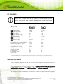
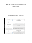
EPIGENTEK Complete Solutions for Epigenetics EpiQuik™ Tissue Methyl-CpG Binding Domain Protein 2 ChIP Kit Base Catalog # P-2018 PLEASE READ THIS ENTIRE USER GUIDE BEFORE USE The EpiQuik™ Tissue Methyl-CpG Binding Domain Protein 2 ChIP Kit is suitable for combining the specificity of immunoprecipitation with qualitative and quantitative PCR, MS-PCR, DNA sequencing, and southern blot, as well as DNA microarray. 110 Bi County Blvd. Ste. 122, Farmingdale, NY 11735 Tel: 1-877-374-4368 ■ Fax: 1-718-484-3956 ■ E-mail: [email protected] ■ Web: www.epigentek.com © Epigentek Group Inc. All rights reserved. Products are for research use only. Page 1 Printed 2015-08-05 P-2018 EPIGENTEK Complete Solutions for Epigenetics KIT CONTENTS Important Information: The amount of components supplied in this kit is designed for reaction count, not sample count, such as negative IgG controls and input DNA. Thus, experiments with samples to be paired with both IgG and input may require additional columns or components to be purchased separately. Please calculate the necessary volumes based on the below kit contents and protocol prior to starting the experiment. Components 24 reactions P-2018-24 48 reactions P-2018-48 CP1 (Wash Buffer) CP2 (Antibody Buffer) CP3 (Lysis Buffer) CP4 (ChIP Dilution Buffer) CP5 (DNA Release Buffer) CP6 (Reverse Buffer) CP7 (Binding Buffer) CP8 (Elution Buffer) Homogenizing Buffer Protease Inhibitor Cocktail (100X)* Normal Mouse IgG (1 mg/ml)* Proteinase K (10 mg/ml)* Anti-MBD2 (0.5 mg/ml)* 8-Well Assay Strips (with Frame) 8-Well Strip Caps F-Spin Column F-Collection Tube User Guide 28 ml 15 ml 4 ml 4 ml 2 ml 2 ml 5 ml 0.6 ml 5 ml 25 µl 10 µl 25 µl 50 µl 3 3 30 30 1 2 x 28 ml 30 ml 6 ml 6 ml 2 x 2 ml 2 x 2 ml 8 ml 1.2 ml 10 ml 40 µl 20 µl 50 µl 100 µl 6 6 50 50 1 * For maximum recovery of the products, centrifuge the original vial after thawing prior to opening the cap. SHIPPING & STORAGE The kit is shipped in two parts: the first part at ambient room temperature and the second part on frozen ice packs at 4°C. Upon receipt, store the following components at 4°C: Protease Inhibitor Cocktail, Normal Mouse IgG, Anti-MBD2, Proteinase K and 8-Well Assay Strips. Store all other components at room temperature. All components of the kit are stable for 6 months from the date of shipment, when stored properly. 110 Bi County Blvd. Ste. 122, Farmingdale, NY 11735 Tel: 1-877-374-4368 ■ Fax: 1-718-484-3956 ■ E-mail: [email protected] ■ Web: www.epigentek.com © Epigentek Group Inc. All rights reserved. Products are for research use only. Page 2 Printed 2015-08-05 P-2018 EPIGENTEK Complete Solutions for Epigenetics MATERIALS REQUIRED BUT NOT SUPPLIED Variable temperature waterbath Vortex mixer Desktop centrifuge (up to 14,000 rpm) Sonicator Orbital shaker Pipettes and pipette tips 1.5 ml microcentrifuge tubes 15 ml conical tube 37% formaldehyde Glycine solution TE buffer (pH 8.0) Ethanol (96-100%) GENERAL PRODUCT INFORMATION Product Updates: Epigentek reserves the right to change or modify any product to enhance its performance and design. Usage Limitation: The EpiQuik™ ChIP kit is for research use only and is not intended for diagnostic or therapeutic application. Intellectual Property: The EpiQuik™ ChIP kits and methods of use contain proprietary technologies by Epigentek. EpiQuik™ is a trademark of Epigentek Group Inc. A BRIEF OVERVIEW MBD2 (methyl-CpG-binding domain protein 2) is a member of the MBD protein family. MBD2 selectively binds to methylated DNA and suppresses transcription from a methylated target gene through recruiting transcriptional repressor complexes, which contain Mi-2/NuRD or HDACs. MBD2 has also been shown to catalyze demethylation by directly removing methyl groups from 5methylcytosine residues in DNA. MBD2 is also demonstrated to be associated with tumorigenesis. For example, deficiency of MBD2 suppresses intestinal tumor formation, indicating that MBD2 is necessary not only for tumor development, but also for tumor growth. The in vivo binding of MBD2 to the methylated CpG region of the gene promoters may be affected by MBD2 mutation and by biochemical or pharmacological intervention. Chromatin Immunoprecipitation (ChIP) is a powerful technique for studying protein-DNA interaction in vivo. ChIP also offers an advantageous tool that allows identification of silenced genes associated with MBD2. Requirement for using ChIP to analyze the binding of MBD2 to the methylated CpGs in the promoter in solid tissues and tumors is rapidly increased. There are several methods used for Chromatin Immunoprecipitation. However, most of these methods currently 110 Bi County Blvd. Ste. 122, Farmingdale, NY 11735 Tel: 1-877-374-4368 ■ Fax: 1-718-484-3956 ■ E-mail: [email protected] ■ Web: www.epigentek.com © Epigentek Group Inc. All rights reserved. Products are for research use only. Page 3 Printed 2015-08-05 P-2018 EPIGENTEK Complete Solutions for Epigenetics available are considerably time consuming, labor intensive, or have low throughput. Furthermore, these methods are not specifically designed for solid tissues. The EpiQuik™ Tissue Methyl-CpG Binding Domain Protein 2 ChIP Kit uses a proprietary and unique procedure and composition to investigate the in vivo binding of MBD2 to methylated DNA in a variety of mammalian tissues. The EpiQuik™ Tissue Methyl-CpG Binding Domain Protein 2 ChIP Kit has the following features: The fastest procedure available, which can be finished within 5 hours. Strip microplate format makes the assay flexible: manual or high throughput. Columns for DNA purification are included: save time and reduce labor. Compatible with all DNA amplification-based approaches. Simple, reliable, and consistent assay conditions. PRINCIPLE & PROCEDURE The EpiQuik™ Tissue Methyl-CpG Binding Domain Protein 2 ChIP Kit contains all reagents required for carrying out a successful chromatin immunoprecipitation for MBD2 from mammalian tissues. Particularly, this kit includes a ChIP-grade MBD2 antibody and a negative control normal mouse IgG. Chromatin from the cells are extracted, sheared, and added into the microwell immobilized with the antibody. DNA is released from the antibody-captured MBD2 protein-DNA complex, reversed and purified through the specifically designed Fast-Spin Column. Eluted DNA can be used for various down-stream applications. Schematic Procedure for Using the EpiQuik™ Tissue Methyl-CpG Binding Domain Protein 2 ChIP Kit 110 Bi County Blvd. Ste. 122, Farmingdale, NY 11735 Tel: 1-877-374-4368 ■ Fax: 1-718-484-3956 ■ E-mail: [email protected] ■ Web: www.epigentek.com © Epigentek Group Inc. All rights reserved. Products are for research use only. Page 4 Printed 2015-08-05 P-2018 EPIGENTEK Complete Solutions for Epigenetics PROTOCOL Note: Always cap spin columns before placing them in the microcentrifuge. Before starting, perform the following: 1. Prepare the following required solutions (not included): 90% Ethanol; 70% Ethanol; 1.25 M Glycine Solution; 1X TE Buffer (pH 8.0). 2. Ensure that all buffers are in a clear solution. Shake or vortex if these buffers precipitate. Antibody Binding to the Assay Plate 1. Determine the number of strip wells required. Leave these strips in the plate frame (remaining unused strips can be placed back in the bag. Seal the bag tightly and store at 4°C). Wash the strip wells once with 150 µl of CP1. 2. Add 100 µl of CP2 to each well and then add the antibodies: 1 µl of Normal Mouse IgG as the negative control, and 2 µl of Anti-MBD2 for the samples. 3. Cover the strip wells with Parafilm M and incubate at room temperature for 60-90 minutes. Meanwhile, prepare the cell extracts as described in the next steps. Tissue Disaggregation and In Vivo Cross-Link 1. Place the tissue sample into a 60 or 100 mm plate. Remove unwanted tissue such as fat and necrotic material from the sample. Weigh the sample and cut it into small pieces (1-2 mm3) with a scalpel or scissors. 2. Transfer tissue pieces to a 15 ml conical tube. Prepare the Cross-Link Solution by adding formaldehyde to culture medium. (Final concentration is 1%. Ex: add 270 µl of 37% formaldehyde to 10 ml of culture medium.) Add 1 ml of the Cross-Link Solution per every 40 mg of tissue and incubate at room temperature for 15-20 minutes on a rocking platform. 3. Add 1 ml of 1.25 M Glycine solution per every 9 ml of Cross-Link Solution. Mix and centrifuge at 800 rpm for 5 minutes. Discard the supernatant. Wash cells with 10 ml of ice-cold PBS once by centrifugation at 800 rpm for 5 minutes. Discard the supernatant. 4. Transfer the tissue pieces to a Dounce homogenizer. Add 1 ml Homogenizing Buffer per every 200 mg of tissue and disaggregate tissue pieces by 10-30 strokes. 5. Transfer homogenized mixture to a 15 ml conical tube and centrifuge at 3000 rpm for 5 minutes at 4°C. If total mixture volume is less than 2 ml, transfer mixture to a 2 ml vial and centrifuge at 5000 rpm for 5 minutes at 4°C. Remove supernatant. Cell Lysis and DNA Shearing 1. Add CP3 containing Protease Inhibitor Cocktail (PIC) (Ex: 10 µl of PIC to each 1 ml of CP3) to resuspend the disaggregated tissue pellet (100 µl/20 mg of tissue). Transfer cell suspension to a 1.5 ml vial (500 µl maximum for each vial). Incubate the sample on ice for 10 minutes and vortex occasionally. 2. Shear DNA by sonication. Usually, sonicate 4-5 pulses of 15-20 seconds each at level 2 using a Branson Microtip probe, followed by 30-40 seconds rest on ice between each pulse. (The conditions of cross-linked DNA shearing can be optimized based on tissue type and sonicator 110 Bi County Blvd. Ste. 122, Farmingdale, NY 11735 Tel: 1-877-374-4368 ■ Fax: 1-718-484-3956 ■ E-mail: [email protected] ■ Web: www.epigentek.com © Epigentek Group Inc. All rights reserved. Products are for research use only. Page 5 Printed 2015-08-05 P-2018 EPIGENTEK Complete Solutions for Epigenetics equipment. If desired, remove 5 µl of sonicated cell lysate for agarose gel analysis. The length of sheared DNA should be between 200-1000 bp.) 3. Pellet cell debris by centrifuging at 14,000 rpm for 10 minutes. Protein/DNA Immunoprecipitation 1. Transfer clear supernatant to a new 1.5 ml vial. (Supernatant can be stored at –80°C at this step.) Dilute required volume of supernatant with CP4 at a 1:1 ratio (Ex: add 100 µl of CP4 to 100 µl of supernatant). 2. Remove 5 µl of the diluted supernatant to a 0.5 ml vial. Label the vial as “input DNA” and place on ice. 3. Remove the incubated antibody solution and wash the strip wells three times with 150 µl of CP2 by pipetting in and out. 4. Transfer 100 µl of the diluted supernatant to each strip well. Cover the strip wells with Parafilm M and incubate at room temperature (22-25°C) for 1 hour on a rocking platform (50-100 rpm). 5. Remove supernatant. Wash the wells six times with 150 µl of CP1. Allow 2 minutes on a rocking platform (100 rpm) for each wash. Wash the wells once (for 2 minutes) with 150 µl of 1X TE Buffer. Cross-Linked DNA Reversal/DNA Purification 1. Add 1 µl of Proteinase K to each 40 µl of CP5 and mix. Add 40 µl of CP5 containing Proteinase K to the samples (including “input DNA” vial). Cover the sample wells with strip caps and incubate at 65°C in a waterbath for 15 minutes. 2. Add 40 µl of CP6 to the samples, mix, and re-cover the wells with strip caps and incubate at 65°C in a waterbath for 90 minutes. Also, add 40 µl of CP6 to the vial containing supernatant as “input DNA.” Mix and incubate at 65°C for 90 minutes. 3. Place a spin column into a 2 ml collection tube. Add 150 µl of CP7 to the samples and transfer mixed solution to the column. Centrifuge at 12,000 rpm for 20 seconds. 4. Add 200 µl of 70% ethanol to the column, centrifuge at 12,000 rpm for 15 seconds. Remove the column from the collection tube and discard the flowthrough. 5. Replace column to the collection tube. Add 200 µl of 90% ethanol to the column and centrifuge at 12,000 rpm for 20 seconds. 6. Remove the column and discard the flowthrough. Replace column to the collection tube and wash the column again with 200 µl of 90% ethanol at 12,000 rpm for 35 seconds. 7. Place the column in a new 1.5 ml vial. Add 10-20 µl of CP8 directly to the filter in the column and centrifuge at 12,000 rpm for 20 seconds to elute purified DNA. DNA is now ready for use or storage at –20°C. Note: For conventional PCR, the number of PCR cycles may need to be optimized for better PCR results. 110 Bi County Blvd. Ste. 122, Farmingdale, NY 11735 Tel: 1-877-374-4368 ■ Fax: 1-718-484-3956 ■ E-mail: [email protected] ■ Web: www.epigentek.com © Epigentek Group Inc. All rights reserved. Products are for research use only. Page 6 Printed 2015-08-05 P-2018 EPIGENTEK Complete Solutions for Epigenetics TROUBLESHOOTING Little or No PCR Products 1. Insufficient tissues. Increase tissue amount (ex: >10 mg of tissue/per reaction). 2. Insufficient or too much cross-linking. Check if the appropriate cross-link step is carried out according to the protocol. 3. Insufficient tissue lysis. Follow the guidelines in the protocol. Check the tissue lysis by observing a 5 µl portion of the tissue lysate under the microscope. 4. Insufficient/too much sonication. Follow the protocol instructions for obtaining the appropriate sized DNA. Keep the sample on ice during the sonication. 5. Incorrect temperature/insufficient time for DNA release and reversal of cross-linking. Follow the guidelines in the protocol for appropriate temperature and time. 6. Incorrect PCR conditions. Check if all PCR components are added. Increase amount of DNA added to PCR reaction. Increase the number of cycles for PCR reaction. 7. Incorrect or bad primers. Ensure the designed primers are specific to the target sequence. 8. The column is not washed with 90% Ethanol. Ensure that wash solution is 90% ethanol. 9. DNA is not completely passed through the filter. Purify DNA before modification and increase centrifuge time to 1 minute at steps 3-7 of “Cross-Linked DNA Reversal/DNA Purification.” Little or No Amplification Difference Between the Sample and the Negative Control 1. Insufficient wash at each wash step. Follow the protocol for appropriate wash. 2. Antibody is added into the well for the negative control by mistake. Ensure antibody is added into the correct well. 110 Bi County Blvd. Ste. 122, Farmingdale, NY 11735 Tel: 1-877-374-4368 ■ Fax: 1-718-484-3956 ■ E-mail: [email protected] ■ Web: www.epigentek.com © Epigentek Group Inc. All rights reserved. Products are for research use only. Page 7 Printed 2015-08-05 P-2018 EPIGENTEK Complete Solutions for Epigenetics 3. Too many PCR cycles. If using conventional PCR, decrease the cycles to appropriate cycle number. Differences between quantities of starting DNA can be measured generally within the linear PCR amplification phase. 4. No enrichment of MBD2 in target promoters. N/A RELATED PRODUCTS P-2002 P-2003 P-2006 P-2007 P-2008 P-2009 P-2010 P-2011 P-2012 P-2013 P-2015 P-2016 P-2017 EpiQuik™ Chromatin Immunoprecipitation (ChIP) Kit EpiQuik™ Tissue Chromatin Immunoprecipitation (ChIP) Kit EpiQuik™ Methyl-Histone H3-K9 ChIP Kit EpiQuik™ Methyl-Histone H3-K4 ChIP Kit EpiQuik™ Tissue Methyl-Histone H3-K9 ChIP Kit EpiQuik™ Tissue Methyl-Histone H3-K4 ChIP Kit EpiQuik™ Acetyl-Histone H3 ChIP Kit EpiQuik™ Acetyl-Histone H4 ChIP Kit EpiQuik™ Tissue Acetyl-Histone H3 ChIP Kit EpiQuik™ Tissue Acetyl-Histone H4 ChIP Kit EpiQuik™ Methyl-Histone H3-K27 ChIP Kit EpiQuik™ Tissue Methyl-Histone H3-K27 ChIP Kit EpiQuik™ Methyl-CpG Binding Domain Protein 2 ChIP Kit 110 Bi County Blvd. Ste. 122, Farmingdale, NY 11735 Tel: 1-877-374-4368 ■ Fax: 1-718-484-3956 ■ E-mail: [email protected] ■ Web: www.epigentek.com © Epigentek Group Inc. All rights reserved. Products are for research use only. Page 8 Printed 2015-08-05 P-2018