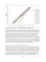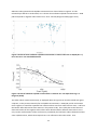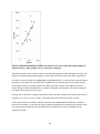
1
SARF085-1
A REPORT COMMISSIONED BY SARF
AND PREPARED BY
Kenny Black
SAMS
Published by the: Scottish Aquaculture Research Forum (SARF)
This report is available at: http://www.sarf.org.uk
Dissemination Statement
This publication may be re-used free of charge in any format or medium. It may only be reused
accurately and not in a misleading context. All material must be acknowledged as SARF copyright
and use of it must give the title of the source publication. Where third party copyright material has
been identified, further use of that material requires permission from the copyright holders concerned.
Disclaimer
The opinions expressed in this report do not necessarily reflect the views of SARF and SARF is
not liable for the accuracy of the information provided or responsible for any use of the content.
Suggested Citation
Title: Sediment sulphide response to organic loading
ISBN: 978-1-907266-63-8
First published: Oct 2014
© SARF 2014
Sediment sulphide response to organic loading
SARF085 - 1
Final Report
Kenny Black
Thom Nickell
March 2014
1
Contents
Contents .................................................................................................................................................. 2
Executive Summary................................................................................................................................. 3
1 Introduction ......................................................................................................................................... 5
Sulphide toxicity .................................................................................................................................. 7
Sulphide as an indicator ...................................................................................................................... 7
2 Measuring sulphide in sediments ........................................................................................................ 8
Calibrating the sulphide ion selective electrode................................................................................. 8
Titration of sodium sulphide ............................................................................................................... 9
Consistency of the calibration ............................................................................................................ 9
Collecting samples in the field for sulphide analysis. ....................................................................... 10
3 Measuring redox in sediments........................................................................................................... 12
4 Field studies in collaboration with fish farm consent monitoring ..................................................... 13
Discussion.......................................................................................................................................... 25
5 Field Studies conducted by SAMS staff .............................................................................................. 26
Field work objectives ........................................................................................................................ 26
Methods ............................................................................................................................................ 26
Results ............................................................................................................................................... 26
Discussion.......................................................................................................................................... 33
6 The effects of SAOB over time ........................................................................................................... 34
Introduction ...................................................................................................................................... 34
The evolution of sulphide concentration in sediments dosed with SAOB ....................................... 35
Discussion.......................................................................................................................................... 38
7 Sulphide model .................................................................................................................................. 39
8 Sulphide module ................................................................................................................................ 44
9 Conclusions ........................................................................................................................................ 45
10 Acknowledgements.......................................................................................................................... 45
11 References ....................................................................................................................................... 46
Appendix 1 - Hargrave comments on SARF085 (December 9, 2013) ................................................... 48
Appendix 2 – derivation of Factor......................................................................................................... 50
2
Executive Summary
Hydrogen sulphide is the toxic product of the natural process of sulphate reduction which is the
dominant anaerobic pathway for the degradation of organic matter in coastal marine sediments.
Elevated hydrogen sulphide pore-water concentrations play a major role in structuring benthic
communities in marine sediments and therefore sulphide concentration should be a good direct
proxy for benthic indicators.
A method for hydrogen sulphide determination in fish farms was developed in Canada in the 1990s
and this method now forms a part of routine monitoring in several other states/countries. In the
present study we examined the utility of hydrogen sulphide as a monitoring tool with respect to
Scottish marine fish farming.
We utilised the protocol for sulphide determination developed in Canada and adapted this for use in
the field. We confirmed the importance of daily electrode calibration.
The practical work had 2 sets of linked components:
1. a) Studies at fish farms piggy-backing on consent monitoring and b) bespoke measurements at
several active fish farms.
2. a) Testing an existing model (RQ) in terms of sulphide predictions and b) developing a sulphide
predicting module for the new AutoDEPOMOD programme
Our studies during consent monitoring gave a set of results that did not precisely match the findings
of previous work but none-the-less indicated the utility of the indicator especially as a potential
screening tool. Our bespoke studies did not yield strong relationships between sulphide
concentration and redox despite these having been found in earlier work. This work has made us
sceptical of the utility of redox measurements except as a very crude indicator. We did establish
relatively good relationships with benthic indicators such as ITI during the consent studies but were
not able to reproduce these during the bespoke studies probably owing to the rather unusual
conditions pertaining in these sediments at the time of our study as discussed later.
The RQ model mostly predicts sulphide concentration to the right order of magnitude. However,
improving the model depends on improving AutoDEPOMOD in 3 ways:
1. Improvement of the determination of “Factor” (defined later) in AutoDEPOMOD which depends
on recoding the model to remember particles after they have hit the seabed.
2. Improvement in resuspension processes. Presently AutoDEPOMOD fails to simulate resuspension
processes at sites with moderate to high current speeds and a key component of the on-going
3
AutoDEPOMOD project is the improvement to the resuspension module. Such improvements will
have a profound effect on the calculation of Factor and thus on the sulphide prediction.
3. The RQ model must be recoded and developed as a new module of the new AutoDEPOMOD. This
will significantly ease its parameterisation as the present RQ model is highly labour intensive with
each run involving a large number of operator manipulations each of which is time consuming and
error prone.
The full delivery of these 3 aspects must await the delivery of the recoded AutoDEPOMOD.
4
1 Introduction
Organic material (OM) from marine cage fish farms (uneaten food, faeces) accumulates on the
seabed around farms with its distribution depending on depth and current speed. The area of initial
deposition (often called the initial footprint) can be changed by the action of near-bed currents in
the process of resuspension, which may carry the material away from the farm thereby reducing
local accumulation. On the seabed, the OM is quickly colonised by bacteria which mediate its
degradation, a process often called remineralisation as the end products are carbon dioxide, water
and inorganic compounds of nitrogen, phosphorus and other minor constituents of the feed. This
remineralisation of highly reduced components of the OM (e.g. lipids and proteins) requires oxidants
(electron acceptors) which are utilised in a sequence of decreasing energy production from oxygen
to sulphate via nitrate and oxidised species of iron and manganese (Fe(III), Mn(IV)). Finally,
methanogenic processes will dominate when these oxidants are exhausted i.e. when the demand for
oxidants exceeds their diffusive supply from overlying sediments and water. Although yielding much
less energy for bacterial respiration than aerobic remineralisation, sulphate is present in sea water at
much higher concentration than the other oxidants and, in coastal sediments, sulphate reduction
(Eq. 1) is generally the dominant mediator of OM remineralisation.
SO42- +CH3OOH → H2S + 2HCO3Equation 1. The reduction of the sulphate ion to hydrogen sulphide during the oxidation of
ethanoic acid to hydrogen carbonate.
Methane can be produced from OM by methanogens (eq. 2) usually deep in the sediment where
sulphate is limiting although, around fish farms, it appears to also be produced in near-surface
sediments (unpublished result, A. Hatton, pers. comm.) - presumably in pockets of sulphate scarcity.
In any case, much of the methane produced can be used as a substrate for sulphate reduction (Eq. 3)
and so the net result is that most OM degrading in sediments will be oxidised via sulphate either
directly or via methane.
CO2 + 4H2 → CH4 + 2H2O a
CH3OOH → CH4 + CO2
b
Equation 2. Methane produced from a) carbon dioxide and hydrogen and b) from methanoic acid.
CH4 + SO42- → HCO3- + HS- + H2O
Equation 3. Anaerobic oxidation of methane.
The demand for oxygen generated by the remineralisation of OM in sediments can be thought of as
being the sum of oxygen utilised directly in aerobic remineralisation plus that consumed during
aerobic re-oxidation of products of anaerobic metabolism plus that consumed by macrofaunal
5
respiration. The re-oxidisable products include hydrogen sulphide, Fe(II) and Mn(II) but also
ammonia– ammonia being formed from protein degradation.
Hydrogen sulphide is present in sediment pore waters as an equilibrium between three species (Eq.
4)
H2S ⇌HS- ⇌ S2-
Equation 4. The equilibrium between sulphide species
Figure 1.1 shows the pH dependence of the speciation (Lewis 2010) which is further discussed in
Section 7 with respect to the method of analysis.
Figure 1.1 pH dependence of sulphide speciation
In sediments, sulphide can react with metals species to form insoluble metal sulphides. Because iron
is usually present in high concentrations in sediments, precipitated iron sulphides are a major
proportion of precipitated metal sulphides (Eq 5).
Fe2+(aq)+ HS- ⇌ FeS(s)+ H+
Equation 5 precipitation of iron “mono”sulphide
Although Equation 5 is presented as a mono-sulphide this material is often non-stoichiometric. This
iron sulphide can be re-oxidised in sediments if appropriate electron acceptors are present or it can
be further reacted to form pyrite. The formation of pyrite can be either via elemental sulphur or via
polysulpide ions (Equations 6 and 7) or, on longer timescales, by sulphide (Hurtgen et al. 1999).
Under aerobic conditions, pyrite is oxidised by oxygen (equation 8), a process that is accelerated in
the presence of Fe(III). Pyrite is stable under anoxic reducing conditions but can be oxidised if
exposed to oxidants such as ferric ion Fe(III) or nitrate ion under some circumstances (Bosch et al.
6
2011), A large proportion of pyrite will become buried and essentially lost to the sulphur cycle
(Hurtgen 2012).
FeS + S0 → FeS2
Equation 6 Anaerobic formation of pyrite through reaction with S0
FeS + Sx2- → FeS2+S(x-1)2Equation 7 Anaerobic formation of pyrite through reaction with polysulphides
FeS2 +H2O + 7O2 → 2Fe2+ + 4H+ + 4SO42Equation 8 Aerobic pyrite oxidation
Sulphide toxicity
Hydrogen sulphide gas is a well-known toxin with the characteristic odour associated with rotten
eggs. Sulphide reacts with iron in respiratory enzymes. It is highly toxic to fish (e.g. Kiemer et al.
1995) and mammals including humans where it has a 5 minute LC50 of about 800 ppm, and a MRL
(minimal risk level) of 0.07 ppm has been derived for acute-duration inhalation 1. Sub-lethal
exposure may lead to long-term damage to brain function.
In sulphidic sediments, some animals have developed detoxification mechanisms that allow them to
survive in quite high sulphide concentrations. In such animals specific oxidising enzymes (e.g.
sulphide: quinone oxidoreductase) are present which convert sulphide to sulphate (Ma et al. 2012).
Sulphide as an indicator
Sulphide was developed as an indicator of benthic status at fish farms by Hargrave et al. (1998) and
by Wildish et al (1999, 2004). These authors spent considerable effort in assessing the method and
developing the protocols for measurement and their works remain as the definitive studies in this
area. Considerable effort was expended to determine cost effective and rapid monitoring methods
and the conclusion was that sulphide and redox measurements together formed a good basis for
monitoring programmes around fish farms. New Brunswick, where much of their work was
conducted, still retains a monitoring protocol largely based around sulphide measurements (Wilson
et al. 2009). Subsequently, several other states/countries adopted sulphide as one of several
indicators to be used during fish farm monitoring.
1
http://www.atsdr.cdc.gov/toxprofiles/tp114-c2.pdf
7
2 Measuring sulphide in sediments
The standard method in use for measuring sulphide in sediments involves the use of the Thermo
Orion Silver/Sulfide electrode (model 9616) which SAMS purchased (10/2011) via Cole Palmer for
£690 plus VAT.
There are several modern protocols (Anon 2010, 2011) for the measurement of sediment sulphide
all of which are derived from Wildish et al. (1999). An important aspect of the method is that
samples and standards are made strongly alkaline by the addition of Sulphide Antioxidant Buffer
(SAOB) which bring the sample to ~pH14 thus driving the sulphide equilibrium partway towards the
S2- species (fig. 1.1) for which the ion selective electrode is specific - more on this in chapter 7.
Sodium sulphide standard solutions must be made up in water purged of oxygen to retard oxidation.
Sodium sulphide is available in technical grades where the degree of hydration may vary and so this
should be quantified by titration against lead perchlorate (see below, Wildish et al. 2004). However,
this step is not reported in other papers which cite this method and may be a source of error if
assumptions on the hydration state of the sodium sulphide are inaccurate.
Calibrating the sulphide ion selective electrode.
The following protocols contain safety indications. It is up to the user to develop safe systems of
work which will keep them safe and are compliant with local chemical safety regulations. If the
method is to be used at sea, careful consideration must be given to ensuring that safety can be
maintained on a moving platform.
1. Purge distilled water with nitrogen gas to reduce the oxygen concentration and store under
nitrogen.
2. Prepare sulphide antioxidant buffer (SAOB) by adding sodium hydroxide (20.0g) and EDTA (17.9 g)
to a 250 cm3 volumetric screw capped flask and making up to the mark with oxygen depleted water.
This can be stored in the fridge under nitrogen. Just before use, add L-ascorbic acid (8.75 cm3).
After adding the ascorbic acid, the solution discolours rapidly and should be used within 3 hours.
Sodium hydroxide solutions are extremely caustic and great care must be taken with them and
appropriate personal protective equipment employed.
3. Weigh accurately approximately 0.024g of sodium sulphide nonahydrate and dilute with 100 cm3
of oxygen depleted water to a nominal concentration of 0.01M (10,000 µM). Dilute aliquots of this
solution serially using 10 cm3 made up to 100 cm3 to give concentrations of 0.001M (1000 µM) and
0.0001M (100 µM). Sodium sulphide is a potent toxin and steps must be taken to avoid inhalation of
fumes or absorption through the skin.
4. The electrode must be filled with Optimum Results A filling solution (Orion #900061) and
acclimatised in distilled water overnight before use as per the manufacturer’s instructions and
connected to an appropriate mV meter. We used the Orion 4 Star portable pH/ISE meter.
8
5. Add an equal volume of SAOB to an aliquot of standard (or sediment sample) in a small screw
capped vial. We used 5 cm3 aliquots. The vial is quickly shaken to ensure thorough mixing and then
the lid removed and the electrode dipped in with a stirring motion. The reading (mV) is recorded
when it stabilises – normally within about one minute. After each measurement, the electrode is
rinsed with deionised (DI) water from a wash bottle and replaced into a beaker of DI water. Before
each measurement the electrode is blotted dry with a clean tissue.
6. The values obtained with the standards are used to construct a straight line between log (10)
concentration and voltage which is subsequently used to determine the concentration of unknown
samples. Calibration must be carried out at least daily, see next section.
Titration of sodium sulphide
A nominal 0.01M (10,000µM) sodium sulphide solution (based on a molar mass of sodium sulphide
nonahydrate of 240.18) is titrated against a 0.1M standard solution of lead perchlorate
Pb(ClO4)2.3H2O (Sigma Aldridge – this material is toxic and an oxidant and must be handled and
disposed of appropriately).
The reaction is:
Na2S + Pb(ClO4)2 → PbS (s) +2NaClO4
We used the sulphide macroelectrode to monitor the reaction which gives a sharp drop in voltage at
the endpoint. The result of this is that the technical sodium sulphide that we used has a hydration
value of approximately 4.3 – much lower than the nominal value of 9. The consequence of this is
that nominal values must be multiplied by approximately 1.546 to yield correct values for sulphide
concentration.
Consistency of the calibration
All of the calibrations were linear (r2 >0.997) however the equation of the line did vary somewhat
over time (Table 2.1, Figure 2.1).
Table 2.1 Mean and standard deviations of the r2, gradient and intercepts of calibration lines (Fig
2.1).
mean
Stdev
r2
0.999
0.000625
Intercept
-26.9
3.76
Gradient
0.0338
0.00403
9
Figure 2.1 Calibrations of the ion selective sulphide electrode on separate days
Although the range of the calibrations at the lower end of the scale appears wider than at the top
this is merely a consequence of the log scale. At the lower end the range is between 848 and 878
mV which equates to 153 to 157 µM respectively the difference being about 2.5% of the range. At
the top end the range is between 912 and 923 mV which corresponds to 15125 and 15631 µM
respectively, the difference being about 3.3% of the range. However, considered from the
perspective of the sulphide analysis, the concentration range across the calibrations is large. For
example, consider a measurement of 880 mV. An analyst looking at figure 2.1 and without the
benefit of a recent calibration could only conclude that the sulphide concentration was between 160
and 1500 µM while a reading of 912mV would imply a concentration between 500 and 15000 µM.
This confirms that for accurate analysis contemporaneous electrode calibration is essential and also
that consistently waiting for the electrode reading to stabilise is very important as small differences
in reading potentially make large differences in the concentration determined.
Collecting samples in the field for sulphide analysis.
We found that the easiest way to collect samples from sediment grabs was using 5 cm3 plastic
syringes with rubber piston seals. The Luer-slip end of each syringe was carefully cut off to produce
a small coring device from the syringe. Only grabs which retain surficial water should be sampled.
Two hands are typically required to suck up sediment from the top 2 cm of the sediment surface
only using a 45 degree angle to avoid including deeper sediments while avoiding any air bubbles.
There are various ways of doing this and reproducibility is somewhat affected by the type of
sediment and the density of subsurface infauna. Keeley et al. (2013) simplified the procedure by
coring vertically to 4.5 cm with their syringe but, as this means that the results will likely
10
overestimate sulphide concentration, they will not be comparable to measurements taken using the
original method (Wildish et al. 1999) and so we do not recommend this procedure. Once the core is
taken it is adjusted to exactly 5 cm3 volume and sealed. We found that the most effect seal was a
second rubber piston seal taken from a separate syringe. This can be pressed into the open end of
the syringe while gently retracting the piston to give a tight fit without trapping any air.
Samples are then either measured immediately or stored at 4˚C for a maximum of 72 h before
analysis - they should not be frozen (Wildish et al. 1999).
11
3 Measuring redox in sediments
Redox measurements have been an important component of marine monitoring for a very long time
(Whitfield 1969). However, redox is not without its problems.
Regarding redox as a useful indicator around fish farms, Brooks and Mahnken (2003) concluded:
“The literature also suggests a great deal of variation for redox readings in sediments from a single
sample station. No information was obtained that would help partition the variance into instrument,
method, technician or true environmental compartments. Brooks (2001a) reported long seek-times
resulting in difficulty in establishing an appropriate endpoint using OrionTM redox probes and
hypothesized that the 6.5mm wide membrane was sampling a range of redox conditions unless it
was inserted vertically into the sediments at a precise (±0.5 mm) depth (see Meijer and Avnimelech,
1999). Same sample triplicate redox potential data collected in 2000 and 2001 had mean coefficients
of variation (CV) of 39.5%, which was twice the sulphide CV of 17.7%. For these and other reasons,
Brooks (2001a) recommended that redox potential continue to be evaluated as part of the salmon
farm waste monitoring program in British Columbia, but that it not be used as a trigger for biological
monitoring.”
We measured redox using a Thermo Scientific platinum tipped glass electrode (gel filled,
KDCMPTB11) during fieldwork from the SAMS vessel. During consent monitoring surveys where we
accompanied fish farm staff, we relied on their measurements using standard SEPA protocols.
A variety of redox protocols is given in the literature and we examined some of these in this project,
described in section 5.
12
4 Field studies in collaboration with fish farm consent monitoring
We accompanied fish farm staff on routine consent monitoring surveys at 10 sites (labelled A – J,
Table 4.1). Prior to the work starting we proposed and agreed with the farmers to keep the farm
identities anonymous. This strategy was adopted so that we could gather sulphide concentration
data alongside data routinely collected for compliance monitoring and thus maximise the use of
project resources. Thus we were able to compare our sulphide measurements to the variety of
measurements currently demanded by SEPA’s monitoring protocols.
Benthic data generated by consent monitoring were extracted from the statutory returns to SEPA
provided by the fish farmers once sample analysis was complete. Benthic samples were triplicate
0.045 m2 van Veen grabs per station. As all diversity indices in SEPA returns are calculated on total
numbers per station, and not mean values per station, all indices have been recalculated after
removal of any presence/absence data and means per station calculated. ITI values have also been
treated as mean per station, not totals, and calculated in Excel using the latest available SEPA
assigned values.
All other diversity indices (number of taxa, individual abundance, Pielou’s J’, Shannon H’ (loge),
Brillouin, Fisher, Margalef’s d, and Simpson’s 1-λ’) were calculated in PRIMER v6 (Clarke & Gorley
2001). In the following figures, curves have been fitted using the trendline function in MS Excel with
a view to maximising the value of R2.
Physical sediment data were also obtained from the same SEPA returns. LOI and PSA data were not
averaged as only single values were presented. Redox values (corrected) were taken as the mean of
two values from 2 cm sediment depth.
Table 4.1 Sample location data.
Site
Biomass on site (t)* Date of survey
A
720
05/06/2012
B
1350
06/06/2012
C
848
07/06/2012
D
553
20/06/2012
E
1194
16/08/2012
F
1324
17/08/2012
G
1194
17/08/2012
H
1287
27/09/2012
I
850
08/01/2013
J
928
09/01/2013
* Not maximum consented biomass
Biotic relationships
We attempted to identify potentially useful relationships between biotic descriptors, such as indices
of diversity, and sediment sulphide concentrations. We consider first the relationship between ITI
13
and sulphide concentration (henceforth [S]). The cluster of low ITI values (<15) in the present study
at [S] <1000 µM (Figure 4.1) isn’t present in the Hargrave (2010) data (Figure 4.2). Out of 10 stations
that have much lower ITI scores than Hargrave (2010) would predict from the [S], 4 were cage edge
stations – unsurprisingly, none were reference stations. None were from farms C, F and J and these
data are plotted separately in Figure 4.3. All the other sites had one or more stations where there
was some deviation from the Hargrave relationship.
Figure 4.1 Plot of mean sediment sulphide concentration vs mean Infaunal Trophic Index (ITI).
Error bars are ± one standard deviation.
14
Figure 4.2 Plot of mean sediment sulphide concentration vs Infaunal Trophic Index (ITI), adapted
from Fig. 5 in Hargrave (2010).
Figure 4.3 [S] and ITI for stations at sites C, F and J.
The associations between number of taxa (S) and sulphide in the present study (Fig. 4.4) and the
study by Hargrave (2010) (Fig. 4.5) are quite similar – observe that the x-axis scales are different
between the two figures.
The relationships between log [S] and the other benthic indictors fall into 2 types: a) those like S (Fig.
4.4) where the log relationship appears somewhat linear (d Fig 4.6; α Fig. 4.7) and b) those like ITI
(Fig. 4.1, 4.3) where the relationship is relatively stable before a large change at [S] around 1000 µM
(N, J’, Brillouin, Simpson and Shannon Figs. 4.8 – 4.12).
15
Figure 4.4 Plot of mean sediment sulphide concentration vs mean number of taxa (S). Error bars
are ± one standard deviation.
Figure 4.5 Plot of sediment sulphide concentration vs total number of taxa from Fig. 5 in Hargrave
(2010).
16
Figure 4.6 Plot of mean sediment sulphide concentration vs mean Margalef’s diversity (d). Error
bars are ± one standard deviation.
Figure 4.7 Plot of mean sediment sulphide concentration vs mean Fisher’s diversity α. Error bars
are ± one standard deviation.
17
Figure 4.8 Plot of mean sediment sulphide concentration vs mean number of individuals (N) per
grab (0.1 m2). Error bars are ± one standard deviation.
Figure 4.9 Plot of mean sediment sulphide concentration vs mean Pielou’s evenness (J'). Error bars
are ± one standard deviation.
18
Figure 4.10 Plot of mean sediment sulphide concentration vs mean Brillouin’s diversity. Error bars
are ± one standard deviation.
Figure 4.11 Plot of mean sediment sulphide concentration vs mean Simpson’s diversity (1- λ').
Error bars are ± one standard deviation.
19
Figure 4.12 Plot of mean sediment sulphide concentration vs mean Shannon’s diversity (H'). Error
bars are ± one standard deviation.
Figure 4.13 Plot of sediment sulphide concentration vs Shannon diversity (H') from Fig. 5 in
Hargrave (2010).
The plots of sulphide vs Shannon diversity H' obtained presently (Figure 4.12) and those of Hargrave
(2010) (Figure 4.13) show that most of the points in the present study fall below the curve proposed
by Hargrave i.e. H’ is lower than would be expected from the sulphide value –again observe the
differences in x-axis scale between the two figures.
Abiotic relationships
20
Sediment redox potential and sulphide concentration are shown below in Figure 4.14. This
relationship is difficult to understand, as it is known that sediment sulphide concentrations > 1500
µM correspond to negative redox values from -25 to -210 mV (Hargrave 2010) (Figure 4.15).
Figure 4.14 Plot of mean sediment sulphide concentration vs mean redox at 2 cm depth (Eh-2 cm).
Error bars are ± one standard deviation.
Figure 4.15 Plot of sediment sulphide concentration vs redox at 0-2 cm depth from Fig. 1 in
Hargrave (2010).
The redox values measured presently, as displayed above in Figure 4.14 must be viewed with great
suspicion, as they remain relatively flat at sulphide concentrations > 1000 µM, and do not become
more negative, as would be expected from numerous data sets from other fish farm sites – and from
the benthic data collected at these stations in the present study. This is likely due to the failure of
electrode used as all the negative redox values were obtained with a different electrode at site H
only. Further reinforcement of this conclusion is provided by the low biotic indices obtained at these
same sulphide values, which show impact that is not reflected in the redox values. Thus
21
unfortunately this dataset has not provided us with good evidence of the relationship between [S]
and Eh.
Figure 4.16 provides some evidence of a relationship between [S] and organic matter (LOI, loss on
ignition
Figure 4.16 Plot of mean sediment sulphide concentration vs organic matter content (LOI).
22
Figure 4.17. Plot of mean sediment sulphide concentration vs large sediment particle fraction
(PSA>2 mm).
Figure 4.18 Plot of mean sediment sulphide concentration vs fine sediment particle fraction
(PSA<63 µm).
As might be expected, [S] is not obviously linked to the large sediment particle size fraction (Figure
5.17).
23
The distributions of [S] vs LOI (Figure 4.16) and fine sediments (< 63 µm) (Figure 4.19) have some
similarity, which is not surprising, as the organic matter measured by LOI is generally associated with
the smaller particle sizes (eg. silt).
Figure 4.19 The relationship between LOI% and particles <63µm
Although the relations observed between benthic indicators and sulphide concentration are
somewhat different from Hargrave (2010) who used a much larger dataset, some of the indicators
performed quite well with respect to the R2 values of fitted curves with ITI, Shannon, Brillouin,
Simpson and Pielou performing better than the others (Table 4.2). The fact that ITI provided the
highest R2 of those tested is perhaps fortuitous given the utility of that indicator in the development
of DEPOMOD.
Table 4.2 R2 values for fitted curves of benthic indicators vs [S]
Indicator
R2
ITI
0.636
Shannon
0.606
Brillouin
0.597
Simpson
0.587
Pielou
0.565
Fishers
0.483
Margalef
0.477
S
0.472
N
0.274
24
Discussion
On the whole, sulphide concentrations were low with only 6 stations having mean values in excess of
1500 µM (table 4.3)
Table 4.3 Mean [S] greater than 1500µM
Site
E
G
J
F
I
I
Station
CE
CE
CE
CE
AZE
CE
Mean Sulphide µM
4627
3432
2847
2219
1995
1969
Stdev
735
1267
490
486
2947
1103
Although good news for the environment, this is less than ideal for the purposes of this study where
we would have benefited from having samples from a wider proportion of the benthic impact
spectrum. Nonetheless several benthic indicators produced relationships which are rational and can
be fitted quite well to simple curves. This supports the generally held view that the sulphide
indicator is quite a good proxy of benthic impact and therefore a useful candidate for a rapid
screening method for fish farm monitoring.
The relationships between redox and sulphide were, in contrast, neither rational nor useful. We
believe that this is much more likely to be a consequence of the difficulties in taking reproducible
redox measurements than a problem with the sulphide methodology. On balance we agree with
Brooks’ comments quoted above (section 4) and would be unhappy about according much weight to
the precise numeric value obtained using redox. However, as it is easy to measure, it is still worth
doing provided the results are not over-interpreted. Perhaps the classification of redox in circa 100
mV ranges as per Hargrave et al (2008, Fig. 5) is the appropriate level of information from redox. In
contrast to sulphide we do not think that the evidence supports the use of redox as a rapid screening
tool on its own despite its obvious simplicity of use in the field.
25
5 Field Studies conducted by SAMS staff
Field work objectives
1. To improve understanding of the relationship between redox and sulphide concentration in fish
farm sediments.
2. To improve understanding of both sulphide and redox with respect to benthic indicators.
Methods
Four fish farms (K, L, M, N) were sampled for sulphide and redox from RV Seol Mara. Sites K and L
were also sampled for benthos. At each site, stations were selected to maximise the expected
benthic impact by tying up to various cages, with one nominally intermediate impact station taken
while moored to a grid buoy.
All sulphide determinations were made within a few minutes of collection using the sulphide
macroelectrode. On each day the electrode was calibrated on board using prepared standards
prepared on shore in deoxygenated water stored under nitrogen in screw-capped vials. SAOB was
made up in advance with the exception of the ascorbic acid which was added immediately prior to
use.
Redox measurements were taken with a Thermo Scientific Eh electrode (gel filled, KDCMPTB11).
This was checked against Zobell’s solution between each measurement batch. The electrode
showed a clear response to reducing conditions indicating that it was functioning correctly. The
precise way the electrode was used to measure redox varied between days and is outlined and
discussed in the results section. Redox measurements were corrected by adding 220 mV as
appropriate for this electrode at approximately 10°C.
At sites K and L, two benthic samples per station were taken by van Veen grab (0.1 m2), sieved over a
1 mm mesh and preserved in buffered formalin solution. Any grab showing signs of leakage through
the top flap (indicating over-penetration) was discarded. At site M, samples for sulphide and redox
profile were made from Craib cores. After the redox profile was made, the core was partly extruded
such that the sediment surface was near the top of the core tube allowing syringe samples to be
taken for sulphide determination.
Results
At site K, triplicate samples of both sulphide and redox were obtained from each grab. Sulphide
samples from the top 2 cm were taken first in cut off syringes and sealed with rubber caps and
analysed immediately. The redox electrode was placed into the sediment to a depth of
approximately 2 cm and a value measured when the reading approached stability – after up to 5
min. However, as is typical for redox probes (Pearson & Stanley 1979), the instrument rarely
achieved complete stability and the decision to record the reading was somewhat subjective. Three
measurements were made from each grab. The expected plot of declining redox with increasing
sulphide concentration was not however observed (Figure 5.1).
26
Figure 5.1 Triplicate measurements of both total sulphide and redox (-2 cm) at several stations
around site K. Error bars show ±1 standard deviation.
On the face of it, there appears to be a trend of increasing redox with [S] – quite counter to theory
and observations of other workers. However, given the variability of redox measurements, too much
should not be read in to this as the redox measurements are all rather similar, as are all but one of
the sulphide concentrations.
At Site L a very similar protocol was used and the plot of redox with total sulphide yields only a weak
trend of decreasing redox with increasing sulphide with a large amount of variation (Figure 5.2).
Again the range of redox values is very low with wide error bars so too much cannot be read into
this.
27
Figure 5.2 Triplicate measurements of both total sulphide and redox (-2 cm) at several stations
around site L. Error bas show ±1 standard deviation. The regression line shows a very weak trend
of decreasing redox with increasing sulphide.
At site M, cores were taken instead of grabs as benthos was no longer required. This enabled redox
profiles to be measured more easily. The redox probe was marked at 1 cm intervals and a
measurement was made at each interval after 60 s or when the measurement had become stable.
After completion of the redox profile, the sediment was extruded to near the top of the core
without major disturbance and sulphide samples were taken in the normal manner from the
superficial sediments. There was only sufficient undisturbed sediment for 2 sulphide samples.
The redox profiles obtained were plausible based on experience (Figure 5.3) indicating correct
functioning of the redox probe. However, comparison of redox at any depth with sulphide
concentration does not yield a significant trend – Figure 5.4 shows total sulphide concentration
versus redox potential at -2 cm. Although the redox values occupy a much larger range than at the
previous sites, all but one of the measurements is positive.
28
Figure 5.3 Redox profiles from 5 stations at site M.
Figure 5.4. Redox at -2cm depth versus total sulphide concentration at Site M. Error bars are ± one
standard deviation.
Redox profiles were also made from cores at site N are given for the 5 stations in figure 5.5.
29
Figure 5.5. Redox profiles at Site N.
Figure 5.5 shows rapidly changing redox with depth at -2 cm so we additionally plot the relationship
between sulphide and redox at -4 cm (Figs. 5.6 and 5.7). Neither figure shows a strong trend of
decreasing redox with increasing sulphide concentration.
Figure 5.6 Relationship between sulphide concentration and Eh (-2 cm, mV, corrected) at site N.
Error bars are ± one standard deviation.
30
Figure 5.7 Relationship between sulphide concentration and Eh (-4 cm, mV, corrected) at site N.
Error bars are ± one standard deviation.
All of the stations at site N had a mean concentration of sulphide < 1500 µM and positive Eh.
Relationships with biotic data
The biotic data from sites K and L were generally quite species rich even where abundances were
high (Figure 5.12) – the minimum number of species at any station was 7 and the maximum at cage
edge stations was 78. The three stations which were sampled from the grid buoys adjacent to the
cages had numbers of species of 78 (Site L) and 59 and 39 (Site K) indicating that these near cage
stations were not highly impacted.
Figure 5.12 Number of species S versus animal abundance N (per 0.1 m2 grab)
31
Figure 5.13 The relationships between sulphide concentration (left) and redox (-2 cm, right) for
four benthic indices (Shannon diversity, Simpson index, AMBI and animal abundance N (per 0.1 m2
grab and ITI) for each grab taken from sites K and L.
32
The presentation of benthic data with both sulphide and redox (Fig. 5.13) showed no obvious
correlation. For example, samples with ITI <1 had a wide range of both sulphide and redox values.
The benthic samples may however have been atypical. Especially at site K, grabs appeared to have a
cohesive carpet of worms and their tubes overlying grey (not black) sediment with little smell of
sediment. One hypothesis is that these stations were exceptional in that the very high densities of
small worms together with the relatively strong tidal bottom currents prevented high accumulation
of OM and the associated low redox and high sulphide concentrations.
Discussion
We believe that the redox probe used was functioning correctly although it is clear that redox is a
somewhat subjective measure prone to probe contamination issues (Wildish et al. 2004). We had no
reason to doubt the sulphide measurements. The calibrations were good and in general the
sulphide electrode stabilised quite quickly (ca. 60 s). It is clear from the data gathered during this
work that we have not been able to reproduce the trends in redox and sulphide plots shown by
Hargrave (2010).
After this work was completed, Barry Hargrave kindly commented on methods used and suggested
some methods that would improve the quality of the relationship between sulphide and redox. This
correspondence is reproduced in full in appendix 1.
A feature of this work is that all of the sites in this study were much less impacted than expected in
terms of the benthic response. Although focussing at cages edges we found that most stations have
relatively high diversity in terms of species richness even when total abundance was high (40, 80k
individuals m-2, Fig. 5.12). This low range of sulphide and redox conditions encountered makes
interpretation more difficult than if the full range of redox and sulphide values given in Hargrave’s
dataset (Hargrave 2010).
The work in this section is consistent with the conclusions of section 4 in that sulphide could be a
useful screening tool. If developed as such a tool, it would be sensible to set an action criteria for
cage edge stations at 1500 µM as this is the threshold identified by Hargrave et al (2008) as an
important transition between oxic and hypoxic conditions. All of the station in sites K and L were
below this threshold and thus would not warrant a full benthic investigation and this seems
reasonable given their high abundance and their generally oxic visual appearance.
It has been suggested that there might also be an upper limit for sulphide where we could predict
that there will be so few fauna that it would not be worth full faunal analysis. In the work reported
in chapter 4, the station with the highest mean sulphide concentration (4627 µM) had very low
mean ITI (0.09), low mean number of species (4) and low mean abundance (768 ind. m-2). It is
reasonable to expect from this and from Hargrave et al (2008) that values above 6000 µM would be
essential azoic. However, as the labour and hence cost of benthic analysis of near-azoic samples is
very small, it might be in the farmer’s interests to do this analysis anyway especially if there was
visual evidence of significant numbers of worms.
33
6 The effects of SAOB over time
Introduction
A paper by Brown et al (2011) considered the standard method of sulphide detection by
macroelectrode (Wildish et al. 1999) as used in aquaculture monitoring in North America and also in
this project. An extract from the abstract is given below:
“…we report that on the timescale of field measurements, the accepted protocol can lead to
significant bias of free sulphide measurements, with orders of magnitude higher concentration
detected in the buffered sediment–porewater slurry than in porewater samples isolated and
analysed separately. Laboratory experiments with model marine sediments and analysis of sediment
composition indicate that this bias is likely introduced by the dissolution of particulate sulphides
and/or sulphur present in the sediments under the intense alkaline conditions of the protocol.”
Essentially, they hypothesize that just as the addition of acids can release hydrogen sulphide from
solid phase FeS (an intermediate of pyrite FeS2) – so called acid volatile sulphides (AVS), the addition
of a strong alkali (OH-) competes with the sulphide in authigenic (i.e. formed in situ) FeS (and other
metal sulphides) with the release of free sulphide ion S2-. In the standard method (Wildish et al.
2004), just prior to measurement, sediments are treated with sulphide anti-oxide buffer (SAOB), the
main component of which is sodium hydroxide having a very high pH (~pH 14). The buffer is added
to increase the ionic strength of the sample and to ensure that the dissolved sulphide equilibrium
(H2S,↔HS-↔S2-) is driven to the right so that much of the sulphide is in the S2- form and therefore
measurable by the macro-electrode which is S2- selective.
Using the pK1 and pK2 values for this equilibrium from Lewis (2010) the proportion of each chemical
species as it varies with pH can be determined (Fig. 1.1). The implication from this figure is that only
a small proportion of the total sulphide is present at pH 14.
Essentially, Brown et al (2011) maintain that the use of high pH SAOB means that pore-water
concentrations of dissolved sulphide species are much higher than they would be at natural in situ
pH. Although it is the pore-water hydrogen sulphide concentration that is toxic to infaunal animals,
the authors do not dispute the validity of empirical relationships derived between biological indices
and sulphide as measured using the standard high pH method:
“This bias does not necessarily invalidate the use of empirical relationships between sulphide
determined by the existing method and measures of benthic invertebrate taxa richness and diversity
that are applied as a proxy for alterations in benthic habitat proximal to fish farms in response to
organic matter loading” (e.g., Brooks 2001).
Correspondence between Kenny Black and Barry Hargrave, one of the pioneers of the standard
methodology yielded the following:
34
“Sediments with <700 µM S could have extremely low free S but even if all of the measured S is due
to solubilized metal-S complexes, concentrations below this threshold are still considered to be
characteristic of oxic conditions. This is consistent with the macrofauna species richness data. On the
other hand, if brief exposure of hypoxic sediments (1300 to 3000 µM S) to SAOB increases free S
concentrations by solubilizing metal-S complexes which are then measured along with true free S,
we still know that species richness decreases dramatically and opportunistic species become
dominant in this concentration range. At higher concentrations (>4500 µM and especially >6000 µM
S) measured with SAOB treatment, we know that macrofauna biodiversity is highly impacted even
though at these high levels perhaps even more S of the total measured is derived from the
solubilized fraction.”
However, in their discussion Brown et al. (2011) state that:
“ modifications to the protocol to minimize or eliminate the observed bias would likely improve the
accuracy and precision of measurements thus strengthening the predictive power of empirical
relationships and allow for direct comparison of field measurements of free sulphide with laboratory
studies that report the toxicity of free sulphide to marine organisms.”
Brown et al. (2011) detail 2 modifications to the standard method that would give accurate porewater sulphide measurements:
“ we recommend two courses of action to address the issue of metal sulphide dissolution and
eliminate or minimize the potential for such dissolution to bias measurements of free sulphide in
marine sediments: (1) to obviate the use of SAOB by conducting measurements of sediment-free
sulphide at natural pH, using paired measurement of S2- and pH; (2) isolation of porewater from the
collected sediments, followed by immediate analysis with the current MOE protocol or preservation
with zinc acetate for later analysis in the laboratory.”
Finally, Barry Hargrave comments (26/04/13):
“The industry and provincial departments of Aquaculture & Fisheries [Canada] (west and east coasts)
have been informed of the findings in the [Brown] paper and the limitations implied in the
application of AgS electrodes for measuring 'free' S. The low cost of measurements, portability of the
equipment for use in the field and rapid return of results as measurements are made has resulted in
the method being widely adopted as a 'standard' measure in assessing benthic conditions in soft
sediments in environmental monitoring programs on both coasts.”
The evolution of sulphide concentration in sediments dosed with SAOB
We also consider the relationship between sulphides measured by the standard method and a new
operational class of sulphur TARS – Total Alkali Released Sulphur – obtained after long equilibration
of sediment samples and SAOB (see below).
After the initial sulphide measurement, additional samples from Site N treated with SAOB were kept
for 48h at 10˚C in the dark prior to re-measuring sulphide concentration. The relationship between
initial (0 h) sulphide concentration and that 48 h later is given in Figure 6.1.
35
Figure 6.1 Relationship between sulphide concentration at 0 h and +48 h after adding SAOB. The
regression line(r2 = 0.51, gradient = 2.15, y-intercept = 1262 mV).
Samples from Site N show a large increase in [S] after 48 h (Figure 6.2), but subsequent analysis of a
third set of samples showed [S] dropping to much lower than initial values after 504 h (Figure 6.4).
In order to further consider post-SAOB addition sulphide behaviour, on two occasions (Set A and Set
B) samples were taken from enriched tanks at SAMS and re-measured after various time intervals.
Set A (Figure 6.3) has a variable response at ~340 h with most samples much higher than at 0h,
except sample 2a which changed little. By 1512 h all samples had dropped in [S] concentration but
two were still more than at 0 h (1a, 3b).
In Set B, and in contrast to samples from Site N, some of the 48 h samples are actually lower than at
0h (Figure 7.5: 1a, 2a, 2,c 3c). At 1224 h, all samples were much lower that at 0 h and 48 h.
Further experiments are needed to better understand post-SAOB addition behaviour in both the
tanks and in the field. In particular we need to determine whether this method can be used to infer
the concentration of particle-associated labile metal sulphides that may be oxidisable during
sediment recovery.
36
Figure 6.2 Change in [S] in Site N samples incubated at 10C after addition of SAOB
Figure 6.3 Change in [S] in tank samples incubated at 10C after addition of SAOB, Set A
37
Figure 6.4. Change in [S] in tank samples incubated at 10C after addition of SAOB, Set B
Discussion
Even without the findings of Brown et al (2011) that much of the sulphide measured is not free
sulphide but sulphide generated by stripping from metal sulphide precipitates at high pH, the
evidence of figure 1.1 is that only a small proportion of total sulphide would be in the sulphide ion
(S2- ) form anyway and its concentration would likely depend on the actual pH achieved.
Any changes to the standard methodology will require a lot of field validation against benthic data in
order to establish the true relationships between pore-water free sulphide and benthic response.
The pragmatic decision taken in Canada to retain the standard method despite its analytical
shortcomings is based on their long track record of using this in the field. This need not be a
significant influence in the Scottish context as we have no such heritage. However, revising the
whole basis of the method seems to be unwarranted for the pragmatic reasons given by Hargrave
above. He has shown empirical relationships between whatever this method measures and benthic
impact and there is no reason to doubt or disregard that large body of evidence. On the other hand,
these doubts about the underlying science of the method could give cause for pause before
introducing this method to the Scottish aquaculture context.
Our own work on longer incubations is at present inconclusive and further experimentation is
required to understand the evolution of sulphur chemistry with long exposure to low pH and
whether this might be useful as on indicator of non-pyrite reduced sulphur in fish farm sediments.
38
7 Sulphide model
The RQ model (Black et al. 2011) was run for all of the SARF Recovery project stations and the
outputs compared to relationships derived by Hargrave (2010). Hargrave’s data were inferred from
our relatively small dataset (Cromey et al. 2002) rather than a specific study to relate sulphide
concentration to ITI. Hargrave’s sources were from sites in production rather than during the
recovery phase. With this caveat in mind, the relationships between modelled sulphide and
Hargrave’s equations show some similarities (Fig. 7.1) which is encouraging given that the RQ
model’s parameterisation is presently un-optimised.
Figure 7.1 The relationship between modelled sulphide and ITI at peak biomass and after some
recovery from the SARF Benthic Recovery project, together with empirically derived relationships
(Hargrave 2010).
Factors were obtained for each station of the consent surveys (section 4) and run through the RQ
model using the default settings to produce relationship between modelled and measured sulphide
measurements. (Fig 7.2, 7.3). The derivation of Factor is given in appendix 2.
39
Figure 7.2 Modelled [S] µM using RQ on the default settings established in the benthic recovery
project versus measured sulphide from consent surveys.
40
Figure 7.3 As figure 7.2 but excluding the two highest predicted sulphide values. The straight line
represents the 1:1 relationship.
41
Figure 7.4 The relationship between Factor and RQ model predicted [S]
Predicted [S] depends heavily on Factor (fig. 7.4). Factor depends entirely on AutoDEPOMOD
correctly predicting post-resuspension accumulation at a particular site. Given this and that
measured sulphide is a relatively good proxy for benthic impact as measured by ITI (fig 4. 1) it
follows that where the predicted sulphide is much greater than the measured sulphide this could
simply be due to poor predictive ability in AutoDEPOMOD rather than in the RQ model. But the
situation is more complicated than that as can be seen if we follow the outlier at the bottom right on
figure 7.4. This point represents the site A cage edge station which has a measured [S] of 866 µM
and a predicted [S] of 9560 µM. Thus the RQ model over-predicts the sulphide concentration by an
order of magnitude. However, considering the relationship between measured sulphide and ITI (Fig.
4.1) we find that this station has a greater benthic impact (ITI = 2.84) than would be expected by the
measured [S]. Thus uncertainties about the relationship between predicted and measured [S] are
caused by uncertainties in both values.
Let’s now consider the 2 predictions excluded from figure 7.3 i.e. sites F and G cage edge stations.
These have predicted [S] of 16,340 and 64,520 and corresponding measured [S] of 2,219 and 3,432
µM respectively. An immediate thought is that the RQ model is somehow not tuned properly for
relatively high impact stations but consideration of cage edge stations for sites E (4,620 predicted ,
4627 measured ) and C (6,470 predicted, 94 measured) and I (6,790 predicted, 1,969 measured)
reveals a more complex picture (Table 7.1). In all cases the ratio of predicted/measured [S] is greater
42
or equal to unity, i.e. the predictions are higher than the measurements. In 6 out of the 9 sites the
ratio is less than an order of magnitude but in the worst example (site C) the ratio is nearly 70. In
the case of site C, the predicted sulphide value is plausible but the measured sulphide values are
very hard to rationalise – these cage edge stations have[S] an order of magnitude lower than at the
reference stations in the same loch.
Table 7.1 Predicted and measured [S] for cage edge stations at sites A-J together with the ratio
between predicted and measured.
Site
A
B
C
D
E
F
G
H
I
J
[S] µM
9,560
650
6,470
2,580
4,620
16,340
64,520
1,310
6,790
6,320
Measured µM
866
367
94
716
4,627
2,219
3,432
1,085
1,969
2,847
Stdev
668
20
15
148
735
486
1,267
160
1,103
490
Ratio
11.0
1.8
68.7
3.6
1.0
7.4
18.8
1.2
3.4
2.2
In order to improve the predictive ability of the RQ model two issues must be addressed:
1. Improvement of the determination of Factor. At present this is imprecise as AutoDEPOMOD does
not remember the provenance of the carbon (i.e. whether food or faeces) after it hits the seabed
and subsequent erosion is based on mass wasting rather than movement of remembered particles.
This means that the precise nature of the OM left on the bed is not modelled and as the two
components have different decay rates in case where erosion is important this may be important in
the sulphide prediction. It is likely that the revised AutoDEPOMOD will remember particles at least
until they are resuspended and thus this inaccuracy will be resolved.
2. Improvement in resuspension processes. Presently AutoDEPOMOD fails to simulate resuspension
processes at sites with moderate to high current speeds and a key component of the ongoing
AutoDEPOMOD project is the improvement to the resuspension module. Such improvements will
have a profound effect on the calculation of Factor and thus on the sulphide prediction.
Taken together these 2 aspects are likely to be much more important than optimising
parameterisation of the RQ model which therefore cannot proceed until these issues are addressed
in the present AutoDEPOMOD revision project.
43
8 Sulphide module
As mentioned in the previous section, in order to address the issues of determination of Factor,
AutoDEPOMOD needs significant revision much of which has to take place anyway in order to
improve the predictive ability of the model. Once this has been done then the RQ model can be
recoded and developed as a new module. This will significantly ease its parameterisation as the
present RQ model is highly labour intensive with each run involving a large number of operator
manipulations each of which is time consuming not to mention tedious.
This work will be presented in a supplementary report.
44
9 Conclusions
This work has explored the potential utility of sulphide concentration as an indicator in Scottish
seabed monitoring requirements for marine fish farms. This work builds on a very considerable
body of work conducted over many years by colleagues on the east coast of Canada. Sulphide is
used as a component of monitoring requirements in several countries/states including British
Columbia, Maine and New Zealand and is the main monitoring method in New Brunswick.
Despite some safety issues when dealing with sulphide, we were able to develop a method that
allows calibration of the sulphide ion-selective electrode in the field using standards prepared in the
lab. This allows analysis either on board the sampling vessel or immediately on return to shore
obviating any issues relating to sample storage. The method is relatively easy to perform and, apart
from the initial cost of the electrode and meter, cheap. It has the considerable advantage over redox
in that the electrode stabilises quite quickly.
Recent studies have shown that the change in pH involved in adding sulphide antioxidant buffer
causes drastic changes to the dissolved sulphide concentration which means that what is measured
has little clear relation to the conditions at in situ pH. Never-the-less, empirical relationships
between sulphide and benthic indicators remain valid especially if the indicator is used in screening
format. In this case the precise result is unimportant – what is more important is whether the result
breaks the trigger value for carrying out a full faunal analysis.
There is some justification for setting a trigger value at 1500 µM as this is recognised as a threshold
above which hypoxic conditions and hence significantly perturbed benthos are likely. However,
thought needs to be given as to where to apply such a screening tool spatially as we do not have
enough evidence to provide a trigger value at the edge of the AZE. One interim solution would be to
invoke a full benthic analysis at all stations if this criterion is reached at any of them. The derivation
of an appropriate AZE edge screening trigger would best be informed by the results of future
monitoring.
10 Acknowledgements
The authors are indebted to Barry Hargrave for his advice and correspondence, to the fish farmers
who offered us access to their sites and their data and to the crew of RV Seol Mara.
45
11 References
Anon (2010) Standard operating practices for the environmental monitoring of the marine finfish
cage aquaculture industry in New Brunswick. In. New Brunswick Department of Environment
Anon (2011) Nova Scotia Aquaculture Environmental Monitoring Program: Standard operating
procedures for the environmental monitoring of marine aquaculture in Nova Scotia. In. Nova
Scotia Fisheries and Aquaculture
Black KD, Cromey CJ, Nickell TD (2011) SARF030 Final Report: Benthic Recovery Project. In. SARF
Bosch J, Lee K-Y, Jordan G, Kim K-W, Meckenstock RU (2011) Anaerobic, Nitrate-Dependent
Oxidation of Pyrite Nanoparticles by Thiobacillus denitrificans. Environ Sci Technol 46:20952101
Brooks K (2001) An evaluation of the relationship between salmon farm biomass, organic inputs to
sediments, physicochemical changes associated with those inputs and the infaunal response
- with emphasis on total sediment sulfides, total volatile solids, and oxidation-reduction
potential as surrogate endpoints for biological monitoring, Vol. Aquatic Environmental
Sciences, 644 Old Eaglemount Road, Port Townsend, Washington
Brooks KM, Mahnken CVW (2003) Interactions of Atlantic salmon in the Pacific northwest
environment II. Organic wastes. Fish Res 62:255-293
Brown KA, McGreer ER, Taekema B, Cullen JT (2011) Determination of Total Free Sulphides in
Sediment Porewater and Artefacts Related to the Mobility of Mineral Sulphides. Aquatic
Geochemistry 17:821-839
Clarke KR, Gorley RN (2001) PRIMER v5: User manual/tutorial. PRIMER-E, Plymouth, 91 pp.
Cromey CJ, Nickell TD, Black KD (2002) DEPOMOD - modelling the deposition and biological effects of
waste solids from marine cage farms. Aquaculture 214:211-239
Hargrave BT (2010) Empirical relationships describing benthic impacts of salmon aquaculture.
Aquaculture Environment Interactions 1:33-46
Hargrave BT, Doucette LI, Phillips GA, Milligan TG, Wildish DJ (1998) Biogeochemical observations to
assess benthic impacts of organic enrichment from marine aquaculture in the Western Isles
region of the Bay of Fundy, 1995. Can Data Rep Fish Aquat Sci 1031:iv + 50
Hargrave BT, Holmer M, Newcombe CP (2008) Towards a classification of organic enrichment in
marine sediments based on biogeochemical indicators. Mar Pollut Bull 56:810-824
Hurtgen MT (2012) The Marine Sulfur Cycle, Revisited. Science 337:305-306
Hurtgen MT, Lyons TW, Ingall ED, Cruse AM (1999) Anomalous enrichments of iron monosulfide in
euxinic marine sediments and the role of H(2)S in iron sulfide transformations: Examples
from Effingham Inlet, Orca Basin, and the Black Sea. Am J Sci 299:556-588
Keeley NB, Forrest BM, Macleod CK (2013) Novel observations of benthic enrichment in contrasting
flow regimes with implications for marine farm monitoring and management. Mar Pollut Bull
66:105-116
Kiemer MCB, Black KD, Lussot D, Bullock AM, Ezzi I (1995) The effects of chronic and acute exposure
to hydrogen sulfide on Atlantic salmon (Salmo salar L). Aquaculture 135:311-327
Lewis AE (2010) Review of metal sulphide precipitation. Hydrometallurgy 104:222-234
Ma Y-B, Zhang Z-F, Shao M-Y, Kang K-H, Zhang L-T, Shi X-L, Dong Y-P (2012) Function of the anal sacs
and mid-gut in mitochondrial sulphide metabolism in the echiuran worm Urechis unicinctus.
Marine Biology Research 8:1026-1031
Pearson TH, Stanley SO (1979) Comparative measurement of redox potential of marine sediments as
a rapid means of assessing the effects of organic pollution. Mar Biol 53:371-379
Whitfield M (1969) REDOX POTENTIAL AS AN OPERATIONAL PARAMETER IN ESTUARINE STUDIES.
Limnology and Oceanography 14:547-558
46
Wildish DJ, Akagi HM, Hamilton N, Hargrave BT (1999) A recommended method for monitoring
sediments to detect organic enrichment from mariculture in the Bay of Fundy. Canadian
Technical Report of Fisheries and Aquatic Sciences 2286:31
Wildish DJ, Akagi HM, Hargrave BT, Strain PM (2004) Interlaboratory calibration of redox potential
and total sulfide measurements in interfacial marine sediments and the implications for
organic enrichment assessment. Can Tech Rep Fish Aquat Sci 2546:iii + 25 p.
Wilson AM, Magill SH, Black KD (eds) (2009) Review of environmental impact assessment and
monitoring in salmon aquaculture., Vol. FAO, Rome
47
Appendix 1 - Hargrave comments on SARF085 (December 9, 2013)
1. The report states that "Only grabs which retain surficial water should be sampled". In Section 5
for sampling at Site K it is stated that "The redox electrode was placed into the sediment to a depth
of approximately 2 cm and a value measured when the reading approached stability – after up to
5 min". If a redox electrode is inserted vertically or obliquely into surface sediment in a grab
containing water, supernatant water can flow into the sediment around the electrode barrel to the
depth to which the probe is inserted. In addition, any vibration or movement of the electrode
creates a cavity around the Pt tip which then may fill with overlying water. It is almost impossible to
ensure that the electrode does not move if it is held in position by hand. Eh potentials typically
measured in oxygenated seawater (+300 to +350 mV) are similar to the range of values measured at
some stations in the report. If supernatant water was present around the tip of the electrode during
Eh measurements potentials would almost always be positive. Excessive potential drift would also be
characteristic of oxic conditions in supernatant water.
3. An additional problem, discussed at the beginning of Section 3 and in Brooks (2001a), concerns
the effect of probe placement and geometry on the Eh measurements when steep vertical gradients
are present in surface sediment layers. If an Eh electrode with a flat Pt disc is used and it is inserted
vertically the potential is measured on a single depth horizon (e.g. at 1 cm). The statement in Section
4 "Redox values (corrected) were taken as the mean of two values from 2 cm sediment depth."
implies that the redox electrode was inserted vertically to 2 cm depth to make a reading. Samples
for S measurement, on the other hand, were collected by withdrawing a volume of sediment
encompassing a range of depth layers between 0 and 2 cm. S is therefore measured in a volume of
sediment while Eh is measured at a fixed depth. The measurements are therefore not made on the
same sample.
5. The original Eh-S protocol (Wildish et al 1999) described using core tubes (about 30 cm long) with
a series of 1 cm dia holes drilled in a spiral fashion and sealed with duct tape. The cores were
inserted into sediment by a diver and closed at each end with a rubber bung. Immediately after
collection supernatant water was carefully siphoned off without removing sediment or disturbing
the sediment surface. Then a scalpel was used to cut into the tape over the first hole (usually
between 0-1 cm) below the sediment surface. The dia of Eh combination electrode used was slightly
smaller than the hole in the core tube and if the duct tape was tight against the probe no leakage of
sediment occurred.
6. The Eh probe was immediately inserted laterally into the sediment to the centre of the core. The
Pt electrode was clamped or otherwise held stationary in the horizontal position to make Eh
potential reading. After 2 to 3 min downward drift usually decreased to <10 mV/min and a reading
was made. If the drift rate did not stabilize (as often the case in oxic sediments) the potential was
recorded after 5 min. Potentials stabilize more rapidly in reduced sediment where sulfate reduction
is the major reaction. No single redox couple controls Eh in oxidized sediments and potentials due to
mixed oxidation-reduction reactions are not at equilibrium and thus stable potentials are usually not
obtained.
48
8. After a reading, the Eh electrode was withdrawn, wiped of excess sediment and immediately
placed in the next hole down the core. A cut-off 5 mL plastic syringe was used as a subcorer in the
first hole. The barrel was pushed into the sediment as the syringe plunger was held back, which I
think is the same procedure described in your report for obtaining syringe samples for S
measurements. This provides a volume of mixed sediment from across the same depth layer into
which the Eh probe was inserted. Eh and S measurements can therefore be considered
representative of whatever depth layer was sampled.
9. A change to the protocol was implemented after the intercalibration workshop in 2004 (Wildish et
al 2004). We knew that core sampling using divers was not possible in deep water and the use of
divers added cost to sample collection. In addition most monitoring programs only required that
surface sediment be collected. Profiles of variables in sediment cores were not needed for
monitoring conditions of recent sedimentation under and around farm sites. Thus sample collection
method were modified to use grabs (e.g. a 0.025 m2 Van Veen).
10. When a partially filled grab was returned with a visibly undisturbed sediment surface,
supernatant water was carefully removed by siphon and short cores (15 cm long) were inserted into
the exposed sediment. These had the tape-covered holes as in the longer diver-collected cores and
the hole closest to the sediment surface used for sampling (usually the 0-2 cm layer). A cut-off
syringe was used to extract a horizontal sample across the width of the core using the full extension
of the syringe barrel. The syringes were sealed with air-tight plastic caps and stored on ice for
transfer to the lab.
11. Analyses for Eh and S usually occurred with 6 hr of collection. The scale on the 5 mL syringe was
used to extrude 2 mL into a beaker and immediately the Eh probe was inserted with the electrode
clamped to prevent movement. The sediment was wet since pore water was transferred from the
syringe with the sediment. The water is necessary to form an electron bridge between the Pt tip and
the reference filling solution. If insufficient water is present no stable Eh potentials can be read and
drift is usually in the positive direction. The potential was recorded after 2-3 min (if drift was <10
mV/min) or 5 min as above. This sample could be further processed for water content (% weight loss
on drying) and organic matter (% weight loss on ashing) if the monitoring protocol required this
data.
11. As the Eh probe was stabilizing, a second 2 mL aliquot of sediment was extruded into a small
beaker and 2 mL of SAOB was immediately added for S measurements. The electrode could be used
to gently mix the buffer-sediment slurry and a stable potential was usually obtained in 1-2 min when
a reading was made.
12. The modified procedure for grab-collected sediment provided measurements of S and Eh on the
same sediment sample. One sample could processed for Eh and S in less than 5 min. Vertical profiles
could be obtained to a the limited depth contained in the shortened cores if desired. It was often
possible to remove more than one core per grab to provide information on within core variation. For
monitoring purposes, however, three grabs were usually collected at one station with one short core
removed from each grab for Eh-S measurements to give spatial representation of variation at a site.
49
Appendix 2 – derivation of Factor
What is Factor
The RQ model gains station specific information on the amount of carbon remaining at a particular
station after resuspension through the use of parameter derived from DEPOMOMD output as
follows – Factor. Factor is a scalable constant for each station at a site but varies between stations
and between sites. The use of Factor means that calculations of sulphide concentration are only
made at specific stations using DEPOMOD outputs in a standalone model (RQ) rather than being run
within DEPOMOMD as is envisaged for the new DEPOMOD version being completed in 2014.
Overview of procedure
Run AutoDepomod using sulphide sample date cage and benthic station positions ->Copy projects ->
Run Partrack/Resus -> Run RQM with feed data -> Calculate Factor
Run AutoDepomod
1) This procedure assumes that there has been a change in cage layout since the time of
original modelling, which may have been pre-AutoDepomod.
2) Run AutoDepomod from the desktop
3) Create new project from the pull-down menu, giving name that is consistent with
project files for site
4) Select Edit Cages option
5) When the Excel spreadsheet opens containing the default orientation and cage setup,
use cage edge position from benthic monitoring report to define cage group position.
Cage type, number and diameter (circumference) can be obtained from the monitoring
report. Orientation may be the same as the original modelling, but may only be available
from the farmer, as may cage separation distance. Once correct positions (in eastings
and northings) and other dimensions listed above have been entered, the cage group
should be centred in the left hand button under the group schematic to the left of the
information cells. Cage edge positions can be converted from lat/long or National Grid
reference to eastings and northings by a number of online coordinate converters. At this
stage, the site name.ini file in the gridgen directory from the original site modelling file
should be opened using notepad++ and DataAreaXMin and DataAreaYMin noted as they
must form the bottom left (i.e. origin) values for the displayed grid. Use the move cage
buttons in the x and y planes until the origin values match the DataAreaXMin and
DataAreaYMin values from the site name.ini file.
6) If cage group layout/cage edge positions match the description in the monitoring report,
then click exit. Wait until the Surfer generated plot has loaded; exit Surfer.
7) At this point the current-data folder must be placed from the original site modelling files
into the newly created partrack folder within the directory C:\Sepa Consent\DATA\site
name\depomod\ partrack. The site name.bln, site name.csv, and site name.ini files from
the original modelling files must be copied from the gridgen folder to the new gridgen
folder in C:\Sepa Consent\DATA\site name\depomod\ gridgen. In the Grid and Cage
Setup pane of AutoDepomod, the files should be listed in the white windows as C-map
50
files. The minimum E and N values from the site name.ini file should match the C-map
limit values. If these values are not displayed, press Edit Cages again, and exit when the
cage layout spreadsheet has loaded, and the Surfer generated plot has loaded; exit
Surfer.
8) Click the Model Parameters tab and the correct Current files should be displayed in the
white windows. Click Perform Single Run, and wait until the model has finished running.
Click Exit, and when the main AutoDepomod window returns, click Exit.
Copy Projects
1) Create a folder to copy the projects to and copy the required projects there.
2) Find and replace the original project location:
a. Using notepad++ open the find in files dialog (Search -> Find in Files)
b. Enter the original location in the find what text box e.g. C:\SEPA Consent\Data
c. Enter the new location in the replace with text box e.g. C:\Sulphides
d. Filters should be set to *.*
e. Directory should be set to the new location
f. Press OK and OK on the pop up confirmation dialog
3) The directory, files and the entries there in must be consistent, so enter each project and
carry out the following:
a. Ensure that there is a depomod folder between the ‘site name’ directory and the
gridgen, partrack, resus, etc folders(should match the entry in the
site name -BcnstFI-N-x.cfg file).
b. Ensure that the ‘site name’ directory name e.g. Aird Taranaish and the name of grid,
partrack .cfg files etc agree. If not rename directory and use notepad++ find in files
to apply the change to every file below the ‘site name’ directory.
4) Enter the resus folder and delete all the contents
5) Enter the partrack folder and:
a. Delete every file (excluding the current-data folder) except for one set of benthic
analysis files 2 (e.g. site name -BcnstFI-N-25.cfg and site name -BcnstFI-N-25.cfh) 3
b. Create empty text files file named output.txt and outappend.txt 4
c. Reset the benthic analysis files numbering to 1 e.g. site name -BcnstFI-N-1.cfg and
site name -BcnstFI-N-1.cfh
d. Use notepad++ find in files to apply the numbering change in the partrack directory
2
Choose the lowest numbered file
If these files do not exist they must be created by running the model once in Autodepomd
4
These files are required to avoid a file not found error when using DEPOMOD partrack
3
51
Running Partrack and Resus
1) Open the command prompt (start -> run, type cmd into the dialog and press return)
2) In the command window enter CD C:\SEPA Consent\depomod and press return
3) Run Partrack:
a. In the command window enter part12.exe –v 5
b. File -> open -> Input data configuration file
c. Select the site name -BcnstFI-N-1.cfg file
d. Under fish characteristics heading in the particle information sections click change
e. In the waste allocation data dialog select Carbon radio button on the right hand side
and click accept data
f. Click run model (the partrack progress bar dialog will appear)
g. Important copy the site name -BcnstFI-N-1.inp file from the partrack to the resus
directory
h. Do not save the new configuration file
i. Close the input data configuration file window
j. Close the partrack window
4) Run Resus:
a. In the command window enter resus12.exe –V
b. File -> new -> Input data configuration file
c. Ensuring you are in the correct sites directory structure load the
site name -BcnstFI-N-1.inp from the resus directory 6
d. For the output file click save as, change the filter in the browser to all files (*.*)
select the site name -BcnstFI-N-1.inp change the extension to .out and click SAVE
e. Select the resus check box (on the left hand side) 7
f. Run the model
g. A dialog will appear asking for a file name for the configuration file. Change the filter
in the browser to all files (*.*) select the site name -BcnstFI-N-1.inp change the
extension to .cfg and click OK (the resus progress bar dialog will appear)
h. Close the input data configuration file window
i. Close the resus window
j. Copy the file site name -BcnstFI-N-1.sur in the resus directory to site name -BcnstFIN-1-Monthly.csv also in the resus directory.
5
The partrack application will start with the window minimised
If the file is not in the resus directory copy it from the partrack directory and try again
7
If the model runs very quickly it is possible that the resus may not have been checked
6
52
Calculate Factor
The procedure is split into two parts. Firstly, the total carbon flux is calculated, this procedure only
needs to be carried out once. Secondly, the flux at each spatial position sampled is obtained. These
quantities together provide the Factor that is required to calculate the flux at the locations for a
given feed input, the flux value will vary with location.
Obtain total Carbon Flux
1) In the partrack folder open the site name -BcnstFI-N-1.cfg file
a. On line 28 note the 2nd value (this assumes a constant input of food) 89
b. Calculate 365/12 times the above value 10
2) In the command window enter RQM.exe
a. Click View Feed Input Data
b. In the Husbandry data dialog Set the number of months to 12 using the controls on
the right
c. Enter the value calculated in step 1.b above 12 times into the feed input per month
textboxes.
d. In the file to save parameters for this scenario click save as,
i. change the filter in the browser to all files (*.*)
ii. navigate to the correct site name folder and into the resus directory
iii. select the site name -BcnstFI-N-1.inp change the extension to .ini
iv. now go up one directory level and create a directory called rqm and enter it
v. click OK to save the .ini file in the rqm directory
e. Select and copy (Ctrl+c) the entire path and file name in the file to save parameters
for this scenario text box and paste (Ctrl+v) into the Results file dialog (do not click
save) and change the extension to .csv
f. Click run, the model will run and write its output to the rqm directory
3) Use a spread sheet to calculate:
Where Cfood and Cfaeces are obtained from columns D and E respectively of the RQM output
site name -BcnstFI-N-1.csv file in the RQM directory and n is the number of months (12 in
this example).
4) Enter the CTotal value into a spread sheet, this value will be used to obtain the Factor required
to be enter into RQM in the final step
8
This is assumed to be the feed input in Kg per day (as indicated in AutoDEPOMOD)
This is not necessarily the consented feed input and may appear to be very large but since a ratio is taken the
magnitude of the input is assumed to be irrelevant. The actual feed input will be used once the Factor has
been calculated
10
This will give feed in Kg per month
9
53
Obtain spatial Carbon Flux11
1) Open surfer (short cut on desktop)
2) File->Open and navigate to the resus directory, open the site name -BcnstFI-N-1-Monthly.csv
created after running resus (the file will open in a spread sheet style window)
a. Change the heading in column C from g carbon/m2.yr to g carbon/(m2.Month)
b. Select column C
c. Data->Transform a dialog will open change the transform equation to C = C/12, and
change the entry in first row to 2 and ensure last row is 1522 (assuming a 39x39)
d. Data->Transform in two separate operations use the same dialog to add the
required off sets to the x and y data columns (transform equations
A=A+xOffsetValue and B=B+yOffsetValue). The offset values can be obtained from
the site name.ini file in the gridgen directory as DataAreaXMin and DataAreaYMin.
e. Click OK
f. File->Save
g. Close the spread sheet style window
3) Select an empty plot document (if a plot document is not visible use File->New->Plot
Document)
4) Grid->Data, open the site name -BcnstFI-N-1-Monthly.csv just saved
a. Select Kriging interpolation 12
b. Change both Spacing entries to 1 (assuming a 39x39 grid), the spacing value should
now be 951
c. Click OK
d. Close the generated report file
5) File->Open site name -BcnstFI-N-1-Monthly.grd
a. Clicking in the map and using the arrow keys to navigate, the spatial sample values
(Clocal)can now be obtained and recorded in the spread sheet along with Ctotal. The
spread sheet should be set up to produce Factor as Clocal/Ctotal
After every model run
Every run of AutoDepomod will leave a file on C:\ called input.reo, and two files on C:\SEPA
Consent\depomod called outappend.txt and output.txt. The latter two files will also be created after
every run of the old Depomod programme. These files must be deleted before each new model run,
or there may well be problems running Depomod. Also, some AutoDepomod runs leave a curious
double \\ path specifier in .cfg files in the gridgen and partrack folders. This \\ doesn’t seem to cause
AutoDepomod much bother, but it causes Depomod to crash. Use notepad++ to tidy these up after
AutoDepomod runs and before Depomod runs. Sometimes, the mere deletion of troubling files is
not sufficient to prevent Depomod crashing; it may be necessary to empty the Recycling bin, and
restart the computer after each run to avoid crashes.
11
The spatial value could also be obtained using the sampling stations option of the gridgen application, this is
-2
-1
-2
-1
not described here, however the values obtained will be in g carbon.m .year and not g carbon.m .month .
12
The actual interpolation is not important as long as the method used is a ‘perfect interpolator’. This is
because the data is going to be re-sampled at it existing sample points.
54 | P a g e
Charity Registration: SC035745
Company Registration: SC267177