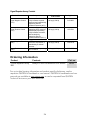
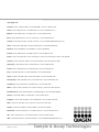
1
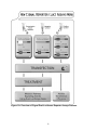
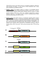
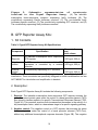
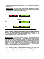
January 2011 Cignal Reporter Assay Handbook For cell-based pathway activity assays Sample & Assay Technologies QIAGEN Sample and Assay Technologies QIAGEN is the leading provider of innovative sample and assay technologies, enabling the isolation and detection of contents of any biological sample. Our advanced, high-quality products and services ensure success from sample to result. QIAGEN sets standards in: Purification of DNA, RNA, and proteins Nucleic acid and protein assays microRNA research and RNAi Automation of sample and assay technologies Our mission is to enable you to achieve outstanding success and breakthroughs. For more information, visit www.qiagen.com. Product Use Limitations Cignal Reporter Assay Kits are intended for molecular biology applications. These products are not intended for the diagnosis, prevention, or treatment of a disease. CONTENTS I. Introduction 4 II. Product Contents and Descriptions 8 III. Additional Materials Required 12 IV. Protocol 13 A. Before you begin 13 B. Generalized Transfection Protocols 14 C. Co-transfection Protocol for siRNA + Reporter Assay 16 D. Co-transfection Protocol for shRNA + Reporter Assay 19 E. Co-transfection Protocol for Expression Vector + Reporter Assay 22 F. Transfection and Treatment Protocol for Reporter Assay + Small Molecules/Organic Compounds 25 G. Transfection and Treatment Protocol for Reporter Assay + Peptide/Recombinant Protein 28 H. Scaling up Transfection Experiments 31 Appendix: Cignal Reporter Assay Kits and Controls 32 Ordering information 34 I. Introduction The Cignal Reporter Assays are designed for accurate, sensitive and quantitative assessment of the activation of signal transduction pathways. SABiosciences has developed a series of inducible reporter constructs that encode a reporter gene under the control of a basal promoter element (TATA box) joined to tandem repeats of specific transcriptional response elements (TRE; Figures 1A and 1B). Transcription factor activities can act as readouts for the intracellular status of many signal transduction pathways. Our constructs are specifically engineered for measuring changes in activity (both increases and decreases) of these signaling pathways. Each of the Cignal reporter assays is available in a dual-luciferase format. In addition, six of the Cignal reporter assays are also available as GFP reporter constructs. These include the CRE, SRE, AP-1, NFkB, SMAD, and TCF/LEF Cignal Reporter Assays. The Cignal reporter assays are valuable tools for deciphering gene function, as well as determining the mechanism of action of proteins, peptides, ligands, and small molecule compounds. Each of the dual-luciferase formatted reporters encodes for the mammalian codonoptimized, non-secreted form of the firefly luciferase gene, carrying a proteindestabilizing sequence. Cells rapidly degrade the destabilized form of the firefly luciferase protein and hence the background luciferase activity (noise level) is greatly reduced. Due to low background activity, the magnitude of the response that can be measured (signal to noise ratio) as well as the speed of measuring changes in transcription are enhanced. The Cignal dual-luciferase reporter assays provide outstanding reproducibility, sensitivity, specificity, and signal to noise ratio. They are extremely useful for carrying out endpoint pathway regulation assays. The Cignal GFP reporter assays enable you to monitor the dynamics of pathway activation on living cells with single cell resolution. The Cignal GFP reporter constructs utilize the Monster Green™ Fluorescent Protein reporter gene. Monster GFP is encoded by an improved synthetic version of the green fluorescent protein gene. This GFP expression cassette has been codon optimized to maximize mammalian cell expression and also utilizes an optimized Kozak sequence to increase translation efficiency. The synthetic GFP is an ideal fluorescent reporter, providing high-level fluorescence and minimal cytotoxicity. Moreover, the synthetic GFP gene is resistant to photobleaching. In addition, most consensus sequences for transcription factor binding have been removed from the synthetic GFP gene in order to minimize aberrant transcription and improve the reliability of the GFP as an accurate reporter. The spectral properties of the synthetic GFP are slightly red-shifted compared to other commercially available GFPs. Peak excitation occurs at 505nm, with a shoulder at 480nm; peak emission occurs at 515nm. 4 Benefits of Cignal Reporter Assays • PERFORMANCE: Both the dual-luciferase and GFP reporter systems provide exceptional sensitivity, reproducibility, specificity, and signal to noise ratio • VERSATILITY: Can monitor signal transduction pathway activity utilizing the dual-luciferase reporter system in an endpoint format assay, or measure pathway activation dynamics on live cells using the GFP reporter system • CONVENIENCE: Transfection-ready constructs, including positive and negative controls, coupled with a transient reporter system, enable rapid analysis of signal transduction pathway regulation. 5 Figure 1A: Overview of Cignal Dual-Luciferase Reporter Assays Process. 6 Figure 1B: Overview of Cignal GFP Reporter Assays Process. 7 II. Product Descriptions A. Contents and Dual-Luciferase Reporter Assay Kits: 1. Kit Contents Table 1: Cignal Reporter Assay Kit Specifications Component Reporter Negative control Positive control Concentration and total volume Specification A mixture of an inducible transcription factor responsive firefly luciferase reporter and (100 ng/μl; 500 µl) constitutively expressing Renilla construct (40:1). A mixture of non-inducible firefly luciferase reporter and constitutively expressing (100 ng/μl; 500 µl) Renilla construct (40:1). A mixture of a constitutively expressing GFP construct, constitutively expressing firefly (100 ng/μl; 250 µl) luciferase construct, and constitutively expressing Renilla luciferase construct (40:1:1). NOTE: These constructs are transfection-grade and are ready for transient transfection. These constructs are specifically designed to inhibit transformation and are NOT MEANT for introduction and amplification in bacteria. 2. Description: Each Cignal Reporter Assay Kit includes the following components: 1. Reporter: The reporter is a mixture of inducible transcription factor responsive construct and constitutively expressing Renilla luciferase construct (40:1). The inducible transcription factor-responsive construct encodes the firefly luciferase reporter gene under the control of a basal promoter element (TATA box) joined to tandem repeats of a specific Transcriptional Response Element (TRE; Figure 2A). This construct monitors both increases and decreases in the activity of a key transcription factor, which is a downstream target of a specific signaling pathway. The constitutively expressing Renilla construct encodes the Renilla luciferase reporter gene under the control of a CMV immediate early enhancer/promoter 8 (Figure 2B) and acts as an internal control for normalizing transfection efficiencies and monitoring cell viability. It is also useful to confirm transfection and to verify active luciferase in the transfected culture. 2. Negative control: The negative control is a mixture of non-inducible reporter construct and constitutively expressing Renilla luciferase construct (40:1). The noninducible reporter construct encodes firefly luciferase under the control of a basal promoter element (TATA box), without any additional transcriptional response elements (Figure 2C). The negative control is critical to identifying specific effects and determining background reporter activity. 3. Positive control: The positive control is a constitutively expressing GFP construct (Figure 2D), pre-mixed with a constitutively expressing firefly luciferase construct (Figure 2E), and a constitutively expressing Renilla luciferase construct (Figure 2B) (40:1:1). The positive control is necessary for visual confirmation of transfection. It is also useful for transfection optimization studies. The expression of the GFP from the positive control construct can be monitored by fluorescence microscopy using an excitation filter of 470 ± 20 nm (470 / 40 nm) and an emission filter of 515 nm (long pass). A. B. Tandem repeats of TREs TATA box Firefly Luc CMV immediate early enhancer/promoter Renilla Luc TATA box C. Firefly Luc D. CMV immediate early enhancer/promoter MGFP E. CMV immediate early enhancer/promoter Firefly Luc 9 Figure 2: Schematic representation of constructs involved in the Cignal Reporter Assay. (A) The inducible transcription factor-responsive construct expressing firefly luciferase, (B) The constitutively expressing Renilla luciferase construct, (C) The non-inducible firefly luciferase reporter construct, (D) The constitutively expressing GFP construct, and (E) The constitutively expressing firefly luciferase construct. B. GFP Reporter Assay Kits: 1. Kit Contents Table 2: Cignal GFP Reporter Assay Kit Specifications Component Reporter Specification Concentration and total volume An inducible transcription factor responsive GFP reporter (100 ng/μl; 500 µl) Negative control A GFP reporter construct in which GFP expression is controlled by a minimal (100 ng/μl; 500 µl) promoter Positive A constitutively expressing GFP construct control (100 ng/μl; 250 µl) NOTE: These constructs are transfection-grade and are ready for transient transfection. These constructs are specifically designed to inhibit transformation and are NOT MEANT for introduction and amplification in bacteria. 2. Description: Each Cignal GFP Reporter Assay Kit includes the following components: 1. Reporter: The inducible transcription factor-responsive GFP reporter encodes the green fluorescent protein gene under the control of a basal promoter element (TATA box) joined to tandem repeats of a specific Transcriptional Response Element (TRE; Figure 3A). This construct monitors both increases and decreases in the activity of a key transcription factor, which is a downstream target of a specific signaling pathway 2. Negative control: The negative control is a GFP reporter that encodes the green fluorescent protein under the control of a basal promoter element (TATA box), without any additional transcriptional response elements (Figure 3B). The negative 10 control is critical to identifying pathway-specific effects and determining background reporter activity. 3. Positive control: The positive control is a constitutively expressing GFP construct (Figure 3C). The positive control is necessary for visual confirmation of transfection. It is also useful for transfection optimization studies. A. Tandem repeats of TREs TATA box GFP TATA box B. C. GFP CMV immediate early enhancer/promoter GFP Figure 3: Schematic representation of constructs involved in Cignal GFP Reporter Assay. (A) The inducible transcription factor-responsive reporter expressing GFP, (B) The GFP reporter controlled by a minimal promoter (negative control), (C) The constitutively expressing GFP construct (positive control). IMPORTANT NOTE: There are a few reports in the literature of the CMV regulatory element being activated by certain stimuli (see below). We recommend that you confirm that the stimulus used in each Cignal reporter assay does not induce the CMV regulatory element, in order to confirm that the CMV-Renilla construct is the appropriate normalization construct for your experiment. This can be done empirically by testing the impact of your stimulus on the Cignal positive control reporters, which are each under the control of the CMV enhancer/promoter cassette. If your stimulus is one of the very few reported activators of the CMV regulatory element, we advise using an alternative reporter as an internal control. • W. Bruening, B. Giasson, W. Mushynski, and H. D. Durham. 1998. Nucleic Acids Research 26(2):486-489. Activation of stress-activated MAP protein kinases up-regulates expression of transgenes driven by the cytomegalovirus immediate/early promoter 11 • Madhu S. Malo, Moushumi Mozumder, Alexander Chen, Golam Mostafa, Xiao Bo Zhang, Richard A. Hodin. 2006. Analytical Biochemistry 350:307309. pFRL7: An ideal vector for eukaryotic promoter analysis III. Additional Materials Required: Mammalian cell line cultured in the appropriate growth medium Cell culture medium and standard cell culture supplies 96-well tissue culture plates Multi-channel pipettor and pipettor reservoirs Transfection reagent [We recommend Attractene Transfection Reagent (QIAGEN, cat. no. 301005), however, other transfection reagents work equally well] Polystyrene test tubes (BD FALCON, Cat # 352099) Opti-MEM® I Reduced Serum Medium (Invitrogen, Cat. No. 31985-062) Fetal bovine serum (FBS) Non-essential amino acids (NEAA) (Invitrogen, Cat. No. 11140-050) Penicillin/Streptomycin Hemacytometer Dual-Luciferase® Assay System o Dual-Luciferase® Reporter Assay System (Promega, Cat. No. E1910) This system requires cell lysis, and is well-suited for the rapid quantitation of both luciferase reporters when using luminometers with reagent auto-injectors. o Dual-Glo® Luciferase Assay System (Promega, Cat. No. E2920) This system is used to assay for both luciferase reporters on intact cells in growth medium. This system can be used with any luminometer, including those without reagent auto-injectors. 96-well white opaque flat bottom microtiter plate 12 Luminometer FACS, flow cytometer, fluorescent microscope, or fluorometer IV. Protocol: A. Before you begin: 1. Cell line selection: The Cignal Reporter Assay may be used with various mammalian cell lines. Cell lines show a great deal of variation in the levels of signaling proteins. The transcriptional activator activities in the cell line used will determine the sensitivity of the assay. A cell line should be selected based on the functionality of the signal transduction pathway under investigation, as well as for the “transfectability” of the cell line (see below). 2. Transfection reagent selection: We recommend the use of Attractene Transfection Reagent (cat. no. 301005) as a transfection reagent. The Cignal Reporter Assay, however, also performs equally well with other transfection reagents such as Lipofectamine 2000 (Invitrogen, Cat. No. 11668-027), or FuGENE 6 (Roche, Cat. No. 1815091). When using alternative transfection reagents, please refer to the manufacturer’s instructions on the use of those reagents. 3. Optimization of transfection conditions: The sensitivity of the Cignal Reporter Assay depends on the transfection efficiency. The transfection efficiency, in turn, primarily depends upon cell line used. Therefore, it is very important to optimize the transfection conditions for each cell type under study. Variables to consider, when optimizing the transfection conditions include cell density, cell viability, amount of DNA, ratio of DNA to transfection reagent, transfection complex formation time, and transfection incubation time (see the detailed protocols for our recommendations). The positive control construct included with each Cignal Reporter Assay can be used for determining the optimal transfection conditions. 4. Optimization of assay condition: The response rate in the Cignal Reporter Assay depends on the assay conditions (conditions of the experimental treatment). To obtain maximum response given by any stimulus, perform dosing and time-course studies. The optimal amount of stimulus and the time of treatment must be obtained empirically for each experiment (see different protocols for our recommendations). 5. Important recommendations for best results: A. Perform all transfections in triplicate to minimize variability among treatment groups. B. Include positive and negative controls in each experiment to obtain reliable results. 13 C. Use low-passage cells that are actively growing and are greater than 90% viable, for maximal transfection efficiencies. D. Do not add antibiotics to media during transfection, as this may cause cell death. E. Take care to always seed the same number of cells in each well, in order to maximize the reproducibility of your experiment. F. Serum induces various signaling pathways, leading to cross-talk and high background. Therefore, use reduced amounts of serum (0.5%) in the assay medium during the experimental treatment to minimize these serum effects. B. Generalized Transfection Protocols: We recommend using reverse transfection protocols with the Attractene Transfection Reagent throughout the Cignal Reporter Assays User Manual. This is due to the time savings and improved reproducibility of using this method, compared to traditional forward transfection methods. However, Cignal Reporter Assays also work well with traditional forward transfection methods and transfection reagents from other vendors. Below are general protocol overviews for the Cignal Reporter Assays, using either reverse or forward transfection approaches. 1. Reverse Transfection Protocol Overview (1 DAY PROCEDURE) DAY 1 Prepare nucleic acid mixtures in appropriate ratios. This may include any of the following combinations, depending upon the experimental design (we recommend carrying out each transfection condition in triplicate): Experimental transfection i. Cignal Reporter + test nucleic acid (expression plasmids, shRNA plasmids, or siRNAs) 14 Control transfections ii. Cignal Reporter + negative control for test nucleic acid iii. Cignal Negative Control + test nucleic acid (expression plasmids, shRNA plasmids, or siRNAs) iv. Cignal Negative Control + negative control for test nucleic acid v. Cignal Positive Control Dilute Attractene into Opti-MEM Add diluted Attractene to nucleic acid mixtures, incubate at room temperature for 20 minutes Trypsinize (if necessary), count, and suspend cells to appropriate density Aliquot transfection complexes into wells Immediately seed cells to each well * * For detailed information on the transfection conditions, and treatment of cultures post-transfection, refer to the application-specific protocols within this user manual. 2. Traditional Transfection Protocol Overview (2 DAY PROCEDURE) DAY 1 Trypsinize (if necessary), count, and suspend cells to appropriate density Seed cells into multiwell plate(s) DAY 2 Prepare nucleic acid mixtures in appropriate ratios. This may include any of the following combinations, depending upon the experimental design (we recommend carrying out each transfection condition in triplicate): 15 Experimental transfection i. Cignal Reporter + test nucleic acid (expression plasmids, shRNA plasmids, or siRNAs) Control transfections ii. Cignal Reporter + negative control for test nucleic acid iii. Cignal Negative Control + test nucleic acid (expression plasmids, shRNA plasmids, or siRNAs) iv. Cignal Negative Control + negative control for test nucleic acid v. Cignal Positive Control Dilute Attractene Transfection Reagent into appropriate medium (If you are using a transfection reagent other than Attractene Transfection Reagent follow their manufacturer’s protocol for transfection) Add diluted transfection reagent to nucleic acid mixtures, incubate at room temperature for 20 minutes Aliquot transfection complexes into wells containing overnight cell cultures C. Co-transfection Protocol for siRNA + Reporter Assay The following protocol is designed to reverse transfect adherent cell line, HEK-293H, using Attractene Transfection Reagent (cat. no. 301005) in a 96-well plate format. The Cignal Reporter Assay works well with transfection reagents from other vendors. If you are using a transfection reagent other than Attractene Transfection Reagent follow their manufacturer’s protocol for optimizing transfection. The Cignal Reporter Assay also works well using traditional forward transfection protocols. Moreover, if you are using plates or wells of different size, adjust the components in proportion to the surface area (see section IV.H). This is just a general guideline; the optimal conditions/amounts should be optimized according to the cell type and the study requirements. Read the protocol completely before starting the experiment. IMPORTANT: (1) Do not add antibiotics to media during transfection as this causes cell death. 16 Table 2: Guidelines for setting up co-transfections of siRNA and Cignal Reporter Assays. Table 2 represents the total components needed, on a per well basis, for each condition to be tested. Note that individual components must be added sequentially, as instructed in the protocol. # 1 2 3 4 5 Cignal Reporter (per well) Cignal Negative Control (per well) Cignal Positive Control (per well) 100 ng (1.0 μl) 100 ng (1.0 μl) Specific siRNA (per well) Negative Control siRNA (per well) Opti-MEM Nucleic Acid Diluent (per well) Attractene (per well) Opti-MEM Attractene Diluent (per well) 2 pmol 25 μl 0.6 μl 25 μl 25 μl 0.6 μl 25 μl 25 μl 0.6 μl 25 μl 25 μl 0.6 μl 25 μl 25 μl 0.6 μl 25 μl 2 pmol 100 ng (1.0 μl) 100 ng (1.0 μl) 2 pmol 2 pmol 100 ng (1.0 μl) Time of transfection (hours) 48 h or 72 h 1. The recommended experimental setup, on a per well basis, follows. Please note that we recommend setting up multiple replicates for each condition, and preparing transfection cocktail volumes sufficient for transfecting multiple wells. In addition, we advise always taking 5-10% extra amounts of nucleic acid, Opti-MEM® serum-free culture medium, and Attractene to compensate for pipettor error when setting up transfection cocktails (steps 1 through 4). Add 25 µl of Opti-MEM® to each of 5 polystyrene tubes, along with the following: Experimental transfection 1 μl (100 ng) Cignal reporter + 2 pmol sequence-specific siRNA Control transfections 1 μl (100 ng) Cignal reporter + 2 pmol negative control siRNA 1 µl (100 ng) Cignal negative control + 2 pmol sequence-specific siRNA 1 µl (100 ng) Cignal negative control + 2 pmol negative control siRNA 1 µl (100 ng) Cignal positive control Mix each transfection cocktail gently. 2. Prepare an Attractene dilution for 5 tubes (mentioned in step 1) by dispensing 3 µl of Attractene into 125 µl of Opti-MEM® serum-free culture medium (for every well dilute 0.6 μl of Attractene in 25 µl of Opti-MEM® serum-free culture medium) in a polystyrene test tube. Mix gently and set the tube at room temperature for 5 minutes. 3. After the 5 minute incubation, add 25 µl of diluted Attractene into each of the five tubes containing 25 µl of the diluted nucleic acids (1:1 ratio) as detailed in Table 2. 4. Mix gently and incubate for 20 minutes at room temperature to allow complex formation to occur. 5. Meanwhile, wash cells* in a culture dish once with Dulbecco’s PBS without calcium and magnesium, and treat with 1-3 ml trypsin-EDTA for 2-5 minutes at 37ºC in a 17 humidified atmosphere containing 5% CO2. Suspend the cells in 7-9 ml of Opti-MEM® containing 5% of fetal bovine serum, then centrifuge the cells down, remove the supernatant, and resuspend the cells to 4 x 10⁵ cells/ml in Opti-MEM® containing 5% of fetal bovine serum and 1% NEAA**. To ensure reproducible transfection results, it is important to accurately measure the cell density with a hemacytometer or an automated cytometry device. 6. After the 20 minute incubation for complex formation is completed, aliquot 50 µl of specific constructs-siRNA-Attractene complexes into the appropriate wells. 7. Add 100 µl of prepared cell suspension (4 x 10⁵ cells/ml in Opti-MEM® containing 5% of fetal bovine serum) to each well containing constructs-siRNA-Attractene complexes. This gives a final volume of 150 µl. Mix gently by rocking the plate back and forth. 8. Incubate cells at 37°C in a 5% CO2 incubator for 16-24 hours. 9. After 16-24 hours of transfection, change the medium to complete growth medium (DMEM with 10% FBS, 0.1mM NEAA, 1mM Sodium pyruvate, 100 U/ml penicillin and 100 µg/ml streptomycin). 10. To study the effect of knockdown, we recommend harvesting cells 48 or 72 hours after transfection to perform dual-luciferase assay. 12. The luciferase assay can be developed by using Dual-Luciferase Reporter Assay System from Promega (Cat. No. 1910). Follow the manufacturer’s protocol for developing the assay. Expression of the Monster GFP reporter can be monitored via FACS, flow cytometry, fluorescent microscopy, or standard fluorometry. The spectral properties of the Monster Green Fluorescent Protein are slightly red-shifted compared to other commercially available GFP reporters. We recommend using the standard FACS settings of an argon laser (488 nm excitation) and filters of 530+15 nm (530/30 nm) for emission. When analyzing GFP expression via fluorescent microscopy or standard fluorometry, we recommend using standard fluoroisothiocyanate (FITC) filters [excitation of 470+20 nm and an emission filter of 515 nm (long pass)]. *Cells that have been passed 1:3 or 1:4 the day before are generally more easily transfected than cells that have reached a confluent state at the time of use. ** In most cases, cells grow well in Opti-MEM® serum-reduced growth medium with 35% FBS due to extra growth factors and nutrients supplied in Opti-MEM®. Cell should reach ~50-90% confluence once attached to the wells, otherwise increase the cell numbers. 18 D. Co-transfection Protocol for shRNA + Reporter Assay The following protocol is designed to reverse transfect adherent cell line, HEK-293H, using Attractene Transfection Reagent (cat. no. 301005) in a 96-well plate format. The Cignal Reporter Assay works well with transfection reagent from other vendors. If you are using transfection reagent other than Attractene Transfection Reagent follow their manufacturer’s protocol for transfection. The Cignal Reporter Assay also works well using traditional forward transfection protocols. Moreover, if you are using plates or wells of different size, adjust the components in proportion to the surface area (see section IV.H). This is just a general guideline; the optimal conditions/amounts should be adjusted according to the cell type and study requirements. Read the protocol completely before starting the experiment. IMPORTANT: (1) Do not add antibiotics to media during transfection as this causes cell death. Table 3: Guidelines for setting up co-transfections of a shRNA vector and Cignal Reporter Assay. Table 3 represents the total components needed, on a per well basis, for each condition to be tested. Note that individual components must be added sequentially, as instructed in the protocol. # 1 2 3 4 5 Cignal Reporter (per well) Cignal Negative Control (per well) Cignal Positive Control (per well) 100 ng (1.0 μl) 100 ng (1.0 μl) Specific shRNA (per well) Negative Control shRNA (per well) Opti-MEM Nucleic Acid Diluent (per well) Attractene (per well) Opti-MEM Attractene Diluent (per well) 200 ng 25 μl 0.6 μl 25 μl 25 μl 0.6 μl 25 μl 25 μl 0.6 μl 25 μl 25 μl 0.6 μl 25 μl 25 μl 0.6 μl 25 μl 200 ng 100 ng (1.0 μl) 100 ng (1.0 μl) 200 ng 200 ng 100 ng (1.0 μl) Time of transfection (hours) 48 h or 72 h 1. The recommended experimental setup, on a per well basis, follows. Please note that we recommend setting up multiple replicates for each condition, and preparing transfection cocktail volumes sufficient for transfecting multiple wells. In addition, we advise always taking 5-10% extra amounts of nucleic acid, Opti-MEM® serum-free culture medium, and Attractene to compensate for pipettor error when setting up transfection cocktails (steps 1 through 4). Add 25 µl of Opti-MEM® to each of 5 polystyrene tubes, along with the following: Experimental transfection 1 µl (100 ng) Cignal reporter + 200 ng sequence-specific shRNA Control transfections 1 µl (100 ng) Cignal reporter + 200 ng negative control shRNA 19 1 µl (100 ng) Cignal negative control + 200 ng sequence-specific shRNA 1 µl (100 ng) Cignal negative control + 200 ng negative control shRNA 1 µl (100 ng) Cignal positive control Mix each transfection cocktail gently. 2. Prepare an Attractene dilution for 5 tubes (mentioned in step 1) by dispensing 3 µl of Attractene into 125 µl of Opti-MEM® serum-free culture medium (for every well dilute 0.6 μl of Attractene in 25 µl of Opti-MEM® serum-free culture medium) in a polystyrene test tube. Mix gently and set the tube at room temperature for 5 minutes. 3. After the 5 minute incubation, add 25 µl of diluted Attractene into each of the five tubes containing 25 µl of diluted constructs (1:1 ratio) as detailed in Table 3. 4. Mix gently and incubate for 20 minutes at room temperature to allow complex formation to occur. 5. Meanwhile, wash cells* in a culture dish once with Dulbecco’s PBS without calcium and magnesium, and treat with 1-3 ml trypsin-EDTA for 2-5 minutes at 37ºC in a humidified atmosphere containing 5% CO2. Suspend the cells in 7-9 ml of Opti-MEM® containing 5% of fetal bovine serum, then centrifuge the cells down, remove the supernatant, and resuspend the cells to 4 ×10⁵ cells/ml in Opti-MEM® containing 5% of fetal bovine serum and 1% NEAA**. To ensure reproducible transfection results, it is important to accurately measure the cell density with a hemacytometer or automated cytometry device. 6. After the 20 minute incubation for complex formation is completed, aliquot 50 µl of specific constructs-shRNA-Attractene complexes into the appropriate wells. 7. Add 100 µl of prepared cell suspension (4 ×10⁵ cells/ml in Opti-MEM® containing 5% of fetal bovine serum) to each well containing construct-shRNA-Attractene complexes. This gives a final volume of 150 µl. Mix gently by rocking the plate back and forth. 8. Incubate cells at 37°C in a 5% CO2 incubator for 16-24 hours. 9. After 16-24 hours of transfection, change the medium to complete growth medium (DMEM with 10% FBS, 0.1mM NEAA, 1mM Sodium pyruvate, 100 U/ml penicillin and 100 µg/ml streptomycin). 10. To study the effect of knockdown, we recommend harvesting cells 48 or 72 hours after transfection to perform dual-luciferase assay. 11. The luciferase assay can be developed by using Dual-Luciferase Reporter Assay System from Promega (Cat. No. 1910). Follow the manufacturer’s protocol for developing the assay. Expression of the Monster GFP® reporter can be monitored via FACS, flow cytometry, fluorescent microscopy, or standard fluorometry. The spectral properties of the Monster Green Fluorescent Protein are slightly red-shifted compared to other commercially 20 available GFP reporters. We recommend using the standard FACS settings of an argon laser (488 nm excitation) and filters of 530+15 nm (530/30 nm) for emission. When analyzing GFP expression via fluorescent microscopy or standard fluorometry, we recommend using standard fluoroisothiocyanate (FITC) filters [excitation of 470+20 nm and an emission filter of 515 nm (long pass)]. *Cells that have been passed 1:3 or 1:4 the day before are generally more easily transfected than cells that have reached a confluent state at the time of use. **In most cases, cells grow well in Opti-MEM® serum reduced growth medium with 3%5% FBS due to extra growth factors and nutrients supplied in Opti-MEM®. Cell should reach ~50-90% confluence once attached to the wells, otherwise increase the cell numbers. 21 E. Co-transfection Protocol for Expression Vector + Reporter Assay The following protocol is designed to reverse transfect adherent cell line, HEK-293H, using Attractene Transfection Reagent (cat. no. 301005) in a 96-well plate format. The Cignal Reporter Assay works well with transfection reagent from other vendors. If you are using transfection reagent other than Attractene Transfection Reagent follow their manufacturer’s protocol for transfection. The Cignal Reporter Assay also works well using traditional forward transfection protocols. Moreover, if you are using plates or wells of different size, adjust the component in proportion to the surface area (see section IV.H). This is just a general guideline; the optimal conditions/amounts should be adjusted according to the cell type and the study requirements. Read the protocol completely before starting the experiment. IMPORTANT: (1) Do not add antibiotics to media during transfection as this causes cell death. Table 4: Guidelines for setting up co-transfections of an expression vector and Cignal Reporter Assay. Table 4 2 3 4 5 6 7 8 9 a Opti-MEM Nucleic Acid Diluent (per well) Attractene (per well) Opti-MEM Attractene Diluent (per well) 100 ng 100 ng 25 μl 0.6 μl 25 μl 200 ng 100 ng 100 ng 200 ng 100 ng (1.0 μl) 100 ng (1.0 μl) 100 ng (1.0 μl) 100 ng (1.0 μl) 100 ng 100 ng 200 ng 100 ng 100 ng 200 ng 100 ng (1.0 μl) 25 μl 0.6 μl 25 μl 25 μl 0.6 μl 25 μl 25 μl 0.6 μl 25 μl 25 μl 0.6 μl 25 μl 25 μl 0.6 μl 25 μl 25 μl 0.6 μl 25 μl 25 μl 0.6 μl 25 μl 25 μl 0.6 μl 25 μl Time of transfection (hours) Carrier a DNA Experimental Vector with Gene of Interest (per well) Cignal Negative Control (per well) Cignal Positive Control (per well) 100 ng (1.0 μl) 100 ng (1.0 μl) 100 ng (1.0 μl) 100 ng (1.0 μl) Experimental Vector Without Insert (per well) 1 Cignal Reporter (per well) # represents the total components needed, on a per well basis, for each condition to be tested. Note that individual components must be added sequentially, as instructed in the protocol. 32 h - 48 h Carrier DNA means any empty plasmid, such as a pUC or a pBR plasmid. 1. The recommended experimental setup, on a per well basis, follows. Please note that we recommend setting up multiple replicates for each condition, and preparing transfection cocktail volumes sufficient for transfecting multiple wells. In addition, we advise always taking 5-10% extra amounts of nucleic acid, Opti-MEM® serum-free 22 culture medium, and Attractene to compensate for pipettor error when setting up transfection cocktails (steps 1 through 4). Add 25 µl of Opti-MEM® to each of 9 polystyrene tubes, along with the following: Experimental transfections 1 μl (100 ng) Cignal reporter + 100 ng experimental vector expressing gene of interest + 100 ng carrier DNA 1 μl (100 ng) Cignal reporter + 200 ng experimental vector expressing gene of interest Control transfections 1 μl (100 ng) Cignal reporter + 100 ng negative control expression vector + 100 ng carrier DNA 1 μl (100 ng) Cignal reporter + 200 ng negative control expression vector 1 µl (100 ng) Cignal negative control + 100 ng experimental vector expressing gene of interest + 100 ng carrier DNA 1 µl (100 ng) Cignal negative control + 200 ng experimental vector expressing gene of interest 1 µl (100 ng) Cignal negative control + 100 ng negative control expression vector + 100 ng carrier DNA 1 µl (100 ng) Cignal negative control + 200 ng negative control expression vector 1 µl (100 ng) Cignal positive control Mix each transfection cocktail gently. 2. Prepare an Attractene dilution for 9 tubes (mentioned in step 1) by dispensing 5.4 µl of Attractene into 225 µl of Opti-MEM® serum-free culture medium (for every well dilute 0.6 μl of Attractene in 25 µl of Opti-MEM® serum-free culture medium) in a polystyrene test tube. Mix gently and set the tube at room temperature for 5 minutes. 3. After the 5 minute incubation, add 25 µl of diluted Attractene into each of the nine tubes containing 25 µl of diluted constructs (1:1 ratio) as detailed in Table 4. 4. Mix gently and incubate for 20 minutes at room temperature to allow complex formation to occur. 5. Meanwhile, wash cells* in a culture dish once with Dulbecco’s PBS without calcium and magnesium, and treat with 1-3 ml trypsin-EDTA for 2-5 minutes at 37ºC in a humidified atmosphere containing 5% CO2. Suspend the cells in 7-9 ml of Opti-MEM® containing 5% of fetal bovine serum, then centrifuge the cells down, remove the supernatant, and resuspend the cells to 4 ×10⁵ cells/ml in Opti-MEM® containing 5% of fetal bovine serum and 1% NEAA**. To ensure reproducible transfection results, it is important to accurately measure the cell density with a hemacytometer or automated cytometry device. 23 6. After the 20 minute incubation for complex formation is completed, aliquot 50 µl of specific complexes into the appropriate wells. 7. Add 100 µl of prepared cell suspension (4 ×10⁵ cells/ml in Opti-MEM® containing 5% of fetal bovine serum) to each well containing construct-vector-Attractene complexes. This gives a final volume of 150 µl. Mix gently by rocking the plate back and forth. 8. Incubate cells at 37°C in a 5% CO2 incubator for 16-24 hours. 9. After 16-24 hours of transfection, change the medium to complete growth medium (DMEM with 10% FBS, 0.1mM NEAA, 1mM Sodium pyruvate, 100 U/ml penicillin and 100 µg/ml streptomycin). 11. To study the effect of the gene product, we recommend harvesting cells 32 hours or 48 hours after transfection to perform the dual-luciferase assay. 12. The luciferase assay can be developed by using Dual-Luciferase Reporter Assay System from Promega (Cat. No. 1910). Follow the manufacturer’s protocol for developing the assay. Expression of the Monster GFP reporter can be monitored via FACS, flow cytometry, fluorescent microscopy, or standard fluorometry. The spectral properties of the Monster Green Fluorescent Protein are slightly red-shifted compared to other commercially available GFP reporters. We recommend using the standard FACS settings of an argon laser (488 nm excitation) and filters of 530+15 nm (530/30nm) for emission. When analyzing GFP expression via fluorescent microscopy or standard fluorometry, we recommend using standard fluoroisothiocyanate (FITC) filters [excitation of 470+20 nm and an emission filter of 515nm (long pass)]. *Cells that have been passed 1:3 or 1:4 the day before are generally more easily transfected than cells that have reached a confluent state at the time of use. **In most cases, cells grow well in Opti-MEM® serum reduced growth medium with 3%5% FBS due to extra growth factors and nutrients supplied in Opti-MEM®. Cell should reach ~50-90% confluence once attached to the wells, otherwise increase the cell numbers. 24 F. Transfection and Treatment Protocol for Reporter Assay + Small Molecules/Organic Compounds The following protocol is designed to reverse transfect adherent cell line, HEK-293H, using Attractene Transfection Reagent (cat. no. 301005) as a transfection reagent in 96well plate format. The Cignal reporter assay works well with transfection reagent from other vendors. If you are using transfection reagent other than Attractene follow their manufacturer’s protocol for transfection. The Cignal Reporter Assay also works well using traditional forward transfection protocols. Moreover, if you are using plates or wells of different size, adjust the component in proportion to the surface area (see section IV.H). This is just a general guideline; the optimal conditions/amounts should be adjusted according to the cell type and the study requirements. Read the protocol completely before starting the experiment. IMPORTANT: (1) Do not add antibiotics to media during transfection as this causes cell death. Table 5: Guidelines for studying the effect of small molecules/organic compounds. Table 5 represents the total components needed, on a per well basis, for each condition to be tested. Note that individual components must be added sequentially, as instructed in the protocol. # 1 2 3 4 5 6 7 8 9 a Cignal Reporter (per well) Cignal Negative Control (per well) Cignal Positive Control Construct (per well) 100 ng (1.0 μl) 100 ng (1.0 μl) 100 ng (1.0 μl) 100 ng (1.0 μl) Small Molecule/ Organic Compound (per well) Opti-MEM DNA diluent (per well) Attractene (per well) Opti-MEM Attractene diluent (per well) 25 μl 0.6 μl 25 μl a 25 μl 0.6 μl 25 μl 10X 25 μl 0.6 μl 25 μl 100X 25 μl 0.6 μl 25 μl 25 μl 0.6 μl 25 μl 1X 25 μl 0.6 μl 25 μl 10X 25 μl 0.6 μl 25 μl 100X 25 μl 0.6 μl 25 μl 25 μl 0.6 μl 25 μl 1X 100 ng (1.0 μl) 100 ng (1.0 μl) 100 ng (1.0 μl) 100 ng (1.0 μl) 100 ng (1.0 μl) Time of Transfection (hours) 30 h or 42 h 1X is a smallest appropriate amount of small molecule or organic compound expected to modulate signaling pathway. 1. The recommended experimental setup, on a per well basis, follows. Please note that we recommend setting up multiple replicates for each condition, and preparing transfection cocktail volumes sufficient for transfecting multiple wells. In addition, we advise always taking 5-10% extra amounts of nucleic acid, Opti-MEM® serum-free 25 culture medium, and Attractene to compensate for pipettor error when setting up transfection cocktails (steps 1 through 4). Set up three polystyrene tubes, as follows Experimental transfections Tubes 1 - 4: 100 µl Opti-MEM® + 4 μl (400 ng) Cignal reporter (4 volumes for conditions 1 to 4 of Table 5; for every well dilute 1 μl (100 ng) of Cignal reporter in 25 µl of Opti-MEM® serum-free culture medium) Control transfections Tubes 5 - 8: 100 µl Opti-MEM® + 4 μl (400 ng) Cignal negative control (4 volumes for conditions 5 to 8 of Table 5; for every well dilute 1 μl (100 ng) of Cignal reporter in 25 µl of Opti-MEM® serum-free culture medium) Tube 9: 25 µl Opti-MEM® + 1μl (100 ng) Cignal positive control Mix each transfection cocktail gently. 2. Prepare an Attractene dilution for 9 tubes (mentioned in step 1) by dispensing 5.4 µl of Attractene into 225 µl of Opti-MEM® serum-free culture medium (for every well dilute 0.6 μl of Attractene in 25 µl of Opti-MEM® serum-free culture medium) in a polystyrene test tube. Mix gently and set the tube at room temperature for 5 minutes. 3. After the 5 minute incubation, add 100 µl of diluted Attractene to the two tubes (mentioned in step 1) containing equal volume (100 µl) of diluted Cignal reporter, and add 25 µl of diluted Attractene into the positive control tube containing 25 µl of diluted constructs (1:1 ratio) as detailed in Table 5. 4. Mix gently and incubate for 20 minutes at room temperature to allow complex formation to occur. 5. Meanwhile, wash cells* in culture dish once with Dulbecco’s PBS without calcium and magnesium, and treat with 1-3 ml trypsin-EDTA for 2-5 minutes at 37 ºC in a humidified atmosphere containing 5% CO2. Suspend the cells in 7-9 ml of Opti-MEM® containing 5% of fetal bovine serum, then centrifuge the cells down, remove the supernatant, and resuspend the cells to 4 ×10⁵ cells/ml in Opti-MEM® containing 5% of fetal bovine serum and 1% NEAA**. To ensure reproducible transfection results, it is important to accurately determine the cell density with a hemacytometer or an automated cytometry device. 6. After the 20 minute incubation for complex formation is completed, aliquot 50 µl of specific complexes into the appropriate wells. 7. Add 100 µl of prepared cell suspension (4 ×10⁵ cells/ml in Opti-MEM® containing 5% of fetal bovine serum) to each well containing constructs- Attractene complexes. This gives a final volume of 150 µl. Mix gently by rocking the plate back and forth. 26 8. Incubate cells at 37°C in a 5% CO2 incubator for 16 hours. 9. After 16 hours of transfection, change medium to assay medium (Opti-MEM® containing 0.5% of fetal bovine serum, 1% NEAA, 100 U/ml Penicillin and 100 µg/ml Streptomycin). 10. After 24 hours of transfection, treat the cells, as described in Table 5, with 1×, 10× and 100× amount of small molecule or organic compound (1× is the lowest appropriate amount of small molecule or organic compound expected to modulate the signaling pathway). 11. To study the effect of small molecule or organic compound, we recommend harvesting cells 6 hours or 18 hours after treatment to perform dual-luciferase assay. 12. The luciferase assay can be developed by using Dual-Luciferase Reporter Assay System from Promega (Cat. No. 1910). Follow the manufacturer’s protocol for developing the assay. Expression of the Monster GFP reporter can be monitored via FACS, flow cytometry, fluorescent microscopy, or standard fluorometry. The spectral properties of the Monster Green Fluorescent Protein are slightly red-shifted compared to other commercially available GFP reporters. We recommend using the standard FACS settings of an argon laser (488 nm excitation) and filters of 530+15 nm (530/30 nm) for emission. When analyzing GFP expression via fluorescent microscopy or standard fluorometry, we recommend using standard fluoroisothiocyanate (FITC) filters [excitation of 470+20 nm and an emission filter of 515 nm (long pass)]. *Cells that had been passed 1:3 or 1:4 the day before are generally more easily transfected than cells that have reached a confluent state at the time of use. **In most cases, cells grow well in Opti-MEM® serum reduced growth medium with 3%5% FBS due to extra growth factors and nutrients supplied in Opti-MEM®. Cell should reach ~50-90% confluence once attached to the wells, otherwise increase the cell numbers. 27 G. Transfection and Treatment Protocol for Reporter Assay + Peptide/Recombinant Protein The following protocol is designed to reverse transfect adherent cell line, HEK-293H, using Attractene Transfection Reagent (cat. no. 301005) in a 96-well plate format. The Cignal reporter assay works well with transfection reagent from other vendors. If you are using transfection reagent other than Attractene follow their manufacturer’s protocol for transfection. The Cignal Reporter Assay also works well using traditional forward transfection protocols. Moreover, if you are using plates or wells of different size, adjust the component in proportion to the surface area (see section IV.F). This is just a general guideline; the optimal conditions/amounts should be adjusted according to the cell type and the study requirements. Read the protocol completely before starting the experiment. IMPORTANT: (1) Do not add antibiotics to media during transfection as this causes cell death. Table 6: Guidelines for studying the effect of a peptide or recombinant protein. Table 6 represents the total components needed, on a per well basis, for each condition to be tested. Note that individual components must be added sequentially, as instructed in the protocol. # 1 2 3 4 5 6 7 8 9 a Cignal Reporter (per well) Cignal Negative Control (per well) Cignal Positive Control (per well) 100 ng (1.0 μl) 100 ng (1.0 μl) 100 ng (1.0 μl) 100 ng (1.0 μl) Peptide or Recombinant Protein (per well) Opti-MEM DNA Diluent (per well) Attractene (per well) Opti-MEM Attractene diluent (per well) 25 μl 0.6 μl 25 μl a 25 μl 0.6 μl 25 μl 10× 25 μl 0.6 μl 25 μl 100× 25 μl 0.6 μl 25 μl 25 μl 0.6 μl 25 μl 1× 25 μl 0.6 μl 25 μl 10× 25 μl 0.6 μl 25 μl 100× 25 μl 0.6 μl 25 μl 25 μl 0.6 μl 25 μl 1× 100 ng (1.0 μl) 100 ng (1.0 μl) 100 ng (1.0 μl) 100 ng (1.0 μl) 100 ng (1.0 μl) Time of Transfection (hours) 30 h or 42 h 1X is a smallest appropriate amount of interfering peptide/recombinant protein/growth factor expected to modulate signaling pathway. 1. The recommended experimental setup, on a per well basis, follows. Please note that we recommend setting up multiple replicates for each condition, and preparing transfection cocktail volumes sufficient for transfecting multiple wells. In addition, we advise always taking 5-10% extra amounts of nucleic acid, Opti-MEM® serum-free 28 culture medium, and Attractene to compensate for pipettor error, when setting up transfection cocktails (steps 1 through 4). Set up three polystyrene tubes, as follows: Experimental transfections Tubes 1 – 4: 100 µl Opti-MEM® + 4 μl (400 ng) Cignal reporter (4 volumes for conditions 1 to 4 of Table 6; for every well dilute 1 μl (100 ng) of Cignal reporter in 25 µl of Opti-MEM® serum-free culture medium) Control transfections Tubes 5-8: 100 µl Opti-MEM® + 4 μl (400 ng) Cignal negative control (4 volumes for conditions 5 to 8 of Table 6; for every well dilute 1 μl (100 ng) of Cignal reporter in 25 µl of Opti-MEM® serum-free culture medium) Tube 9: 25 µl Opti-MEM® + 1μl (100 ng) Cignal positive control Mix each transfection cocktail gently. 2. Prepare an Attractene dilution for 9 tubes (mentioned in step 1) by dispensing 5.4 µl of Attractene into 225 µl of Opti-MEM® serum-free culture medium (for every well dilute 0.6 μl of Attractene in 25 µl of Opti-MEM® serum-free culture medium) in a polystyrene test tube. Mix gently and set the tube at room temperature for 5 minutes. 3. After the 5 minute incubation, add 100 µl of diluted Attractene to the two tubes (mentioned in step 1) containing equal volume (100 µl) of diluted Cignal reporter, and add 25 µl of diluted Attractene into the positive control tube containing 25 µl of diluted constructs (1:1 ratio) as detailed in Table 6. 4. Mix gently and incubate for 20 minutes at room temperature to allow complex formation to occur. 5. Meanwhile, wash cells* in culture dish once with Dulbecco’s PBS without calcium and magnesium, and treat with 1-3 ml trypsin-EDTA for 2-5 minutes at 37 ºC in a humidified atmosphere containing 5% CO2. Suspend the cells in 7-9 ml of Opti-MEM® containing 5% of fetal bovine serum, then centrifuge the cells down, remove the supernatant, and resuspend the cells to 4 ×10⁵ cells/ml in Opti-MEM® containing 5% of fetal bovine serum and 1% NEAA**. To ensure reproducible transfection results, it is important to accurately determine the cell density with a hemacytometer or an automated cytometry device. 6. After the 20 minute incubation for complex formation is completed, aliquot 50 µl of specific complexes into the appropriate wells. 29 7. Add 100 µl of prepared cell suspension (4 ×10⁵ cells/ml in Opti-MEM® containing 5% of fetal bovine serum) to each well containing constructs-Attractene complexes. This gives a final volume of 150 µl. Mix gently by rocking the plate back and forth. 8. Incubate cells at 37°C in a 5% CO2 incubator for 16 hours. 9. After 16 hours of transfection, change medium to assay medium (Opti-MEM® containing 0.5% of fetal bovine serum, 1% NEAA, 100 U/ml Penicillin and 100 µg/ml Streptomycin). 10. After 24 hours of transfection, treat the cells, as described in Table 12, with 1x, 10x and 100x amount interfering peptide/recombinant protein/growth factor (1x is an smallest appropriate amount of small molecule or organic compound expected to modulate signaling pathway). 11. To study the effect of interfering peptide/recombinant protein/growth factor, we recommend harvesting cells 6 hours or 18 hours after treatment to develop luciferase assay. 12. The luciferase assay can be developed by using Dual-Luciferase® Reporter Assay System from Promega (Cat. No. 1910). Follow the manufacturer’s protocol for developing the assay. Expression of the Monster GFP reporter can be monitored via FACS, flow cytometry, fluorescent microscopy, or standard fluorometry. The spectral properties of the Monster Green Fluorescent Protein are slightly red-shifted compared to other commercially available GFP reporters. We recommend using the standard FACS settings of an argon laser (488 nm excitation) and filters of 530+15 nm (530/30nm) for emission. When analyzing GFP expression via fluorescent microscopy or standard fluorometry, we recommend using standard fluoroisothiocyanate (FITC) filters [excitation of 470+20 nm and an emission filter of 515 nm (long pass)]. *Cells that had been passed 1:3 or 1:4 the day before are generally more easily transfected than cells that have reached a confluent state at the time of use. ** In most cases, cells grow well in Opti-MEM® serum reduced growth medium with 3%5% FBS due to extra growth factors and nutrients supplied in Opti-MEM®. Cell should reach ~50-90% confluence once attached to the wells, otherwise increase the cell numbers. 30 H. Scaling up transfection experiments: To transfect cells in different tissue culture formats, vary the amounts of constructs, number of cells, and volume of Attractene and medium used in proportion to the surface area, as shown in the Table 7. The parameters shown in Table 7 are standardized for HEK-293H cells. Use these parameters as a starting point to optimize transfections for your cell line of interest. Table 7. Reagent amounts for different size culture vessels Type of Plate Surface Area 2 (cm per well) 96-well transfecting cells in siRNA / shRNA Vector or Gene Expression Vector (per Well) 2 pmol / 200 ng Starting Volume of Attractene (μl / well) Starting Volume of Attractene (μl / well) Volume of Cell Suspension (μl / well) Starting No. of Adherent Cells (per Well) Volume of Opti-MEM Medium (μl) 0.3 Starting amount of construct (ng / well) 100 0.6 0.3 100 40,000 2 X 25 48-well 0.95 150 1.8 0.8 250 130,000 2 X 50 5 pmol / 500 ng 24-well 1.9 250 3.2 1.6 500 250,000 2 X 50 10 pmol / 750 ng 12-well 3.8 500 6.4 3.2 1000 500,000 2 X 100 20 pmol / 1.5 μg 6-well 9.4 1000 16.0 8.0 2500 1500,000 2 X 250 50 pmol / 4.0 μg 35 mm 8.0 1000 16.0 8.0 2500 1500,000 2 X 250 50 pmol / 4.0 μg 5000 3.0 X 10 6 2 X 500 100 pmol / 8.0 μg 9.0 X 10 6 2 X 1500 60 mm 100 mm 21 55 2000 5000 36.0 90.0 18.0 45.0 15000 (15 a 300 pmol / 25 μg ml) a 2X means one volume of Opti-MEM® medium for diluting constructs and another volume of Opti-MEM® medium for diluting Attractene. For any other troubleshooting or technical questions about the Cignal Reporter Assay, please call one of our Technical Support representatives at 1-888-503-3187 or 301-682-9200 or email at [email protected]. 31 Appendix: Cignal Reporter Assay Kits Dual luciferase Cat. No. MGFP Cat. No. Pathway Transcription Factor Amino Acid Deprivation ATF4/ATF3/ATF2 CCS-8034L Androgen Androgen Receptor CCS-1019L Antioxidant Response Nrf2 & Nrf1 CCS-5020L ATF6 ATF6 CCS-9031L C/EBP C/EBP CCS-001L CCS-1001G cAMP/PKA CREB CCS-002L CCS-002G Cell Cycle E2F/DP1 CCS-003L DNA Damage p53 CCS-004L Early Growth Response EGR1 CCS-8021L ER Stress CBF/NF-Y/YY1 CCS-2032L Estrogen Receptor Estrogen Receptor (ER) CCS-005L GATA GATA CCS-1035L Glucocorticoid Receptor Glucocorticoid Receptor (GR) CCS-006L Heat Shock Response HSF CCS-4023L Heavy Metal Response MTF1 CCS-5033L Hedgehog Gli CCS-6030L Hepatocyte Nuclear Factor 4 HNF4 CCS-3039L Hypoxia HIF-1 CCS-007L Interferon Regulation IRF-1 CCS-7040L Interferon Type I STAT1/STAT2 CCS-008L Interferon Gamma STAT1/STAT1 CCS-009L KLF4 KLF4 CCS-4036L Liver X Receptor LXRa CCS-0041L 32 CCS-3021G CCS-0033G CCS-008G CCS-9036G MAPK/ERK Elk-1/SRF CCS-010L CCS-010G MAPK/JNK AP-1 CCS-011L CCS-011G MEF2 MEF2 CCS-7024L c-Myc Myc/Max CCS-012L Nanog Nanog CCS-7037L NFκB NFκB CCS-013L CCS-013G Notch RBP-Jκ CCS-014L CCS-1014G Oct4 Oct4 CCS-0025L Pax6 Pax6 CCS-3042L PI3K/AKT FOXO CCS-1022L PKC/Ca++ NFAT CCS-015L PPAR PPAR CCS-3026L Progesterone Progesterone Receptor CCS-6043L Retinoic Acid Receptor RAR CCS-016L Retinoid X Receptor RXR CCS-9044L Sox2 Sox2 CCS-0038L SP1 SP1 CCS-6027L STAT3 STAT3 CCS-9028 TGFβ SMAD2/SMAD3/SMAD4 CCS-017L Vitamin D VDR CCS-2029L Wnt TCF/LEF CCS-018L CCS-018G Xenobiotic AhR CCS-2045L CCS-7045G 33 CCS-6022G CCS-1027G CCS-017G Cignal Reporter Assay Controls Control Construct Cignal Negative Control (LUC) Cignal Positive Control (LUC) Cignal Negative Control (GFP) Cignal Positive Control (GFP) Components Concentration And Volume A mixture of non-inducible firefly luciferase reporter construct and constitutively expressing Renilla luciferase construct (40:1). A mixture of a constitutively expressing GFP construct, constitutively expressing firefly luciferase construct, and constitutively expressing Renilla luciferase element (40:1:1). A GFP reporter construct in which GFP expression is controlled by a minimal promoter A constitutively expressing GFP construct Catalog Number 100 ng/µl; 500 µl CCS-NCL 100 ng/µl; 250 µl CCS-PCL 100 ng/µl; 500 µl CCS-NCG 100 ng/µl; 250 µl CCS-PCG Ordering Information Product Contents Cat. no. Cignal Reporter Assay Kits Assays in dual-luciferase or GFP format Varies For up-to-date licensing information and product-specific disclaimers, see the respective QIAGEN kit handbook or user manual. QIAGEN kit handbooks and user manuals are available at www.qiagen.com or can be requested from QIAGEN Technical Services or your local distributor. 34 Trademarks: QIAGEN® (QIAGEN Group). Limited License Agreement Use of this product signifies the agreement of any purchaser or user of the Cignal Reporter Assay Kits to the following terms: 1. The Cignal Reporter Assay Kits may be used solely in accordance with the Cignal Reporter Assay Handbook and for use with components contained in the Kit only. QIAGEN grants no license under any of its intellectual property to use or incorporate the enclosed components of this Kit with any components not included within this Kit except as described in the Cignal Reporter Assay Handbook and additional protocols available at www.qiagen.com. 2. Other than expressly stated licenses, QIAGEN makes no warranty that this Kit and/or its use(s) do not infringe the rights of third-parties. 3. This Kit and its components are licensed for one-time use and may not be reused, refurbished, or resold. 4. QIAGEN specifically disclaims any other licenses, expressed or implied other than those expressly stated. 5. The purchaser and user of the Kit agree not to take or permit anyone else to take any steps that could lead to or facilitate any acts prohibited above. QIAGEN may enforce the prohibitions of this Limited License Agreement in any Court, and shall recover all its investigative and Court costs, including attorney fees, in any action to enforce this Limited License Agreement or any of its intellectual property rights relating to the Kit and/or its components. . For updated license terms, see www.qiagen.com Firefly and/or Renilla Luciferase and Monster Green Limited Use Label License READ THIS FIRST BEFORE OPENING PRODUCT For research use only. The terms of the limited license conveyed with the purchase of this product are as follows: Researchers may use this product in their own research and they may transfer derivatives to others for such research use provided that at the time of transfer a copy of this label license is given to the recipients and the recipients agree to be bound by the conditions of this label license. Researchers shall have no right to modify or otherwise create variations of the nucleotide sequence of the luciferase gene or Monster Green® gene except that Researchers may: (1) clone heterologous DNA sequences at either or both ends of said luciferase or Monster Green® gene so as to create fused gene sequences provided that the coding sequence of the resulting luciferase or Monster Green gene has no more than four deoxynucleotides missing at the affected terminus when compared to the intact luciferase or Monster Green® gene sequence, and (2) insert and remove nucleic acid sequences in furtherance of splicing research predicated on the inactivation or reconstitution of the luminescent activity of the encoded luciferase. In addition, Researchers must do one of the following: (1) use luminescent assay reagents purchased from Promega Corporation for all determinations of luminescence activity resulting from the research use of this product and its derivatives; or, (2) contact Promega Corporation to obtain a license for the use of the product and its derivatives. No other use or transfer of this product or its derivatives is authorized without the express written consent of Promega Corporation including, without limitation, Commercial Use. Commercial Use means any and all uses of this product and derivatives by a party for monetary or other consideration and may include, but is not limited to use in: (1) product manufacture; and (2) to provide a service, information or data; and/or resale of the product or its derivatives, whether or not such product or derivatives are resold for use in research. With respect to such Commercial Use, or any diagnostic, therapeutic or prophylactic uses, please contact Promega Corporation for supply and licensing information. If the purchaser is not willing to accept the conditions of this limited use statement, SABiosciences is willing to accept the return of the unopened product and provide the purchaser with a full refund. However, in the event the product is opened, then the purchaser agrees to be bound by the conditions of this limited use statement. The above license relates to Promega Corporation patents and/or patent applications on improvements to the luciferase and Monster Green® gene. United States Patent No. 5,292,658 licensed from Millipore Corporation. Dual-Glo, Dual-Luciferase and Monster Green are trademarks of Promega Corporation. Opti-MEM is a registered trademark of Life Technologies. © 2011 QIAGEN, all rights reserved. www.qiagen.com Australia Orders 1-800-243-800 Fax 03-9840-9888 Technical 1-800-243-066 Austria Orders 0800-28-10-10 Fax 0800-28-10-19 Technical 0800-28-10-11 Belgium Orders 0800-79612 Fax 0800-79611 Technical 0800-79556 Brazil Orders 0800-557779 Fax 55-11-5079-4001 Technical 0800-557779 Canada Orders 800-572-9613 Fax 800-713-5951 Technical 800-DNA-PREP (800-362-7737) China Orders 86-21-3865-3865 Fax 86-21-3865-3965 Technical 800-988-0325 Denmark Orders 80-885945 Fax 80-885944 Technical 80-885942 Finland Orders 0800-914416 Fax 0800-914415 Technical 0800-914413 France Orders 01-60-920-926 Fax 01-60-920-925 Technical 01-60-920-930 Offers 01-60-920-928 Germany Orders 02103-29-12000 Fax 02103-29-22000 Technical 02103-29-12400 Hong Kong Orders 800 933 965 Fax 800 930 439 Technical 800 930 425 Ireland Orders 1800 555 049 Fax 1800 555 048 Technical 1800 555 061 Italy Orders 800-789-544 Fax 02-334304-826 Technical 800-787980 Japan Telephone 03-6890-7300 Fax 03-5547-0818 Technical 03-6890-7300 Korea (South) Orders 080-000-7146 Fax 02-2626-5703 Technical 080-000-7145 Luxembourg Orders 8002-2076 Fax 8002-2073 Technical 8002-2067 Mexico Orders 01-800-7742-639 Fax 01-800-1122-330 Technical 01-800-7742-436 The Netherlands Orders 0800-0229592 Fax 0800-0229593 Technical 0800-0229602 Norway Orders 800-18859 Fax 800-18817 Technical 800-18712 Singapore Orders 1800-742-4362 Fax 65-6854-8184 Technical 1800-742-4368 Spain Orders 91-630-7050 Fax 91-630-5145 Technical 91-630-7050 Sweden Orders 020-790282 Fax 020-790582 Technical 020-798328 Switzerland Orders 055-254-22-11 Fax 055-254-22-13 Technical 055-254-22-12 UK Orders 01293-422-911 Fax 01293-422-922 Technical 01293-422-999 USA Orders 800-426-8157 Fax 800-718-2056 Technical 800-DNA-PREP (800-362-7737) 1062766 01/2011 Sample & Assay Technologies