1
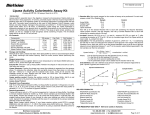
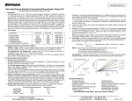
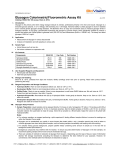
BioVision
For research use only
rev. 02/13
Starch Colorimetric/Fluorometric Assay Kit
(Catalog #K647-100; 100 assays; Store at -20°C)
II.
Introduction:
Starch is a complex carbohydrate consisting of a large number of glucose units. All plants
contain starch, present as amylose, (linear α-1, 4 linked polymer) and amylopectin, (highly α1, 6 branched α-1, 4 polymer). Starch generally contains 0-25% amylose and 75–100%
amylopectin. The BioVision Starch Assay Kit provides an easy, convenient method to
measure starch levels in a variety of samples. In the assay, starch is hydrolyzed to glucose
which is oxidized to generate color (λmax= 570 nm) and fluorescence (Ex/Em = 535/587 nm).
The assay can detect starch at 0.0004 to 2 mg/ml.
Kit Contents:
Components
Hydrolysis Buffer
Development Buffer
OxiRed Probe
Hydrolysis Enzyme Mix
Development Enzyme Mix
Starch Standard (2.0 mg/ml)
K647-100
Cap Code
Part Number
25 ml
25 ml
0.4 ml
Lyophilized
Lyophilized
100 µl
NM
WM
Red
Blue
Green
Yellow
K647-100-1
K647-100-2
K647-100-3A
K647-100-5
K647-100-6
K647-100-7
III.
Storage and Handling:
Store kit at –20°C, protect from light and moisture. Warm Buffers to room temperature before
use. Briefly centrifuge all small vials prior to opening. Read entire protocol before the assay.
IV. Reagent Preparation and Storage Conditions:
OxiRed Probe: Ready to use as supplied. Warm up >18°C to melt frozen DMSO before use.
Mix well, store at –20°C, protect from light and moisture.
Hydrolysis Enzyme Mix, Development Enzyme Mix: Dissolve with 220 µl Hydrolysis Buffer.
Vortex gently to dissolve. Keep on ice. Store at –20°C. Stable for at least two months.
V. Starch Assay Protocol:
1. Standard Curve Preparations:
Colorimetric: Dilute Starch Standard to 0.2 mg/ml by adding 10 µl of the Standard to 90 µl of
distilled water, mix well. Add 0, 2, 4, 6, 8, 10µl into a series of wells. Adjust volume to 50 µl/
well with Hydrolysis Buffer to generate 0, 0.4, 0.8, 1.2, 1.6 and 2.0 µg/well of the Starch
Standard.
Fluorometric: Dilute Starch Standard to 0.02 mg/ml by adding 10 µl of the Standard to 990 µl
of distilled water, mix well. Add 0, 2, 4, 6, 8, 10 µl into a series of wells. Adjust volume to 50
µl/well with Hydrolysis Buffer to generate 0, 0.04, 0.08, 0.12, 0.16 and 0.2 µg/well of Starch
Standard.
2. Sample Preparation:
Depending on your assay purpose (quantitation, mw distribution, compartmentalization, etc.),
prepare starch samples according to established protocols1-4.
A. Soluble Starch Extraction: Grind up 5-10 mg sample, wash off any free glucose and small
oligosaccharides with 1 ml 90% ethanol, warm to 60°C for 5 minutes with occasional
vortexing. Centrifuge at 10,000g for 2 minutes. Decant the supernatant. Repeat the wash
twice. Soluble starch can be extracted with 1 ml H2O and heating on a boiling water bath for 5
minutes. Spin at 10,000g for 2 minutes to remove insoluble materials. The supernatant is
soluble starch.
B. Resistant Starch Extraction: After extracting soluble starch, extract the water insoluble
pellet with 1 ml 10N KOH, heat on boiling water bath for 5 minutes. Neutralize with 1 ml 10M
H3PO4 slowly. Spin at 10,000g for 2 minutes to remove insoluble materials. The supernatant
is resistant starch.
C. Total Starch Extraction: After the 90% ethanol wash (Step A), extract the washed sample
directly with 10N KOH/H3PO4 as per the procedure for resistant starch (B). The supernatant is
total starch. For starch sample testing: Take 20 µl of the extracted starch, add 180 µl of
Hydrolysis Buffer, mix. Add up to 50 µl of the diluted sample or buffer (blank) to test wells.
Adjust the volume to 50 µl with Hydrolysis Buffer. For unknown samples, we suggest testing
several doses of the sample to ensure the readings are within the standard curve.
BioVision Incorporated
155 S. Milpitas Boulevard, Milpitas, CA 95035 USA
3. Hydrolysis*:
Colorimetric
Fluorometric
Hydrolysis Enzyme Mix
2 µl
1 µl
Mix well; incubate for at least 30 minutes at room temperature to hydrolyze starch.
*Note: Glucose generates background. Glucose control is done without the hydrolysis enzyme
(add equal
volume of Buffer). Glucose background can be subtracted from sample reading.
4. Development:
Mix enough reagents for the number of samples and standards. For each well, prepare a total 50
µl Reaction Mix.
Colorimetric
Fluorometric
Development Buffer
46 µl
48.7 µl
Development Enzyme Mix
2 µl
1.0 µl
OxiRed Probe
2 µl
0.3 µl
Add 50 µl of Development Mix to each well containing Starch Standard or samples.
5. Incubate at room temperature for 30 minutes, protect from light.
6. Measure colorimetrically (OD=570 nm) or fluorometrically (Ex/Em 535/587 nm).
7. Calculation: Correct background by subtracting the value of the 0 starch control from all sample
readings (Note: The background can be significant and must be subtracted). Plot standard curve
µg/well vs. OD. Apply sample readings to the standard curve to get the amount of starch in the
sample wells. The starch concentration in the test samples:
C = Ay/Sv (µg/µl or mg/ml)
Where:
Ay is the amount of starch (µg) in your sample from the standard curve.
Sv is the sample volume (µl) added to the sample well. Multiply by the dilution factors.
Starch molecular size: ~ 60,000 glucose molecules (MW ~106-107daltons).
glucose y = 0.651x - 0.027
y = 0.700x - 0.006
corn
1.2
potato
rice
OD 570 nm
I.
wheat
0.8
y = 0.648x - 0.012
y = 0.653x - 0.003
y = 0.690x - 0.002
0.4
0
0
0.4
0.8
1.2
1.6
2
Starch (µg/well)
Figure 1. Starch Standard Curve: Different types of pure starch were extracted with 10N
KOH/H3PO4 as described following the kit protocol.
VII. References:
1) Quantitative Isolation and Dispersion of Starch from Corn Kernels without Degradation,
James P. McGuire, Stig R. Erlander, Starch - Stärke, Vol 18 No. 11, (2006) 342 – 346.
2) Overview of Laboratory Isolation of Starch from Plant Materials,Thava Vasanthan, Current
Protocols in Food Analytical Chemistry, UNIT E2.1 (2001), John Wiley & Sons, Inc.
3) A rapid micro-starch quantitation method for potato callus and its application with potato
tubers, J. L. Varns and J. R. Sowokinos, Journal American Journal of Potato Research Vol. 51,
No. 12(1974).
4) Critical study of a procedure for the assay of starch in ligneous plants",Gomez, L., Rubio E.,
Lescourret F., Journal of the Science of Food and Agriculture, vol. 83, no. 11 (2003) 1114-1123.
FOR RESEARCH USE ONLY! Not to be used on humans.
Tel: 408-493-1800 | Fax: 408-493-1801
www.biovision.com | [email protected]
Page 1 of 2
BioVision
rev. 02/13
For research use only
GENERAL TROUBLESHOOTING GUIDE:
Problems
Cause
Solution
Assay not working
• Use of ice-cold buffer
• Buffer must be at room temperature
• Omission of a step in the protocol
• Refer and follow the data sheet precisely
• Plate read at incorrect wavelength
• Check the wavelength in the data sheet and the filter settings of the instrument
• Fluorescence: Black plates (clear bottoms) ; Luminescence: White plates ; Colorimeters:
Clear plates
• Use of a different 96-well plate
Samples with erratic readings
• Use of an incompatible sample type
• Refer data sheet for details about incompatible samples
• Samples prepared in a different buffer
• Refer data sheet for instructions
• Samples were not deproteinized (if indicated in datasheet)
• Use the 10 kDa spin cut-off filter or PCA precipitation as indicated
• Cell/ tissue samples were not completely homogenized
• Samples used after multiple free-thaw cycles
Lower/ Higher readings in Samples
and Standards
Readings do not follow a linear
pattern for Standard curve
Unanticipated results
• Use Dounce homogenizer (increase the number of strokes); observe for lysis under
microscope
• Aliquot and freeze samples if needed to use multiple times
• Presence of interfering substance in the sample
• Troubleshoot if needed, deproteinize samples
• Use of old or inappropriately stored samples
• Use fresh samples or store at correct temperatures till use
• Improperly thawed components
• Thaw all components completely and mix gently before use
• Use of expired kit or improperly stored reagents
• Always check the expiry date and store the components appropriately
• Allowing the reagents to sit for extended times on ice
• Always thaw and prepare fresh reaction mix before use
• Incorrect incubation times or temperatures
• Refer datasheet & verify correct incubation times and temperatures
• Incorrect volumes used
• Use calibrated pipettes and aliquot correctly
• Use of partially thawed components
• Thaw and resuspend all components before preparing the reaction mix
• Pipetting errors in the standard
• Avoid pipetting small volumes
• Pipetting errors in the reaction mix
• Prepare a master reaction mix whenever possible
• Air bubbles formed in well
• Pipette gently against the wall of the tubes
• Standard stock is at an incorrect concentration
• Always refer the dilutions in the data sheet
• Calculation errors
• Recheck calculations after referring the data sheet
• Substituting reagents from older kits/ lots
• Use fresh components from the same kit
• Measured at incorrect wavelength
• Check the equipment and the filter setting
• Samples contain interfering substances
• Troubleshoot if it interferes with the kit
• Use of incompatible sample type
• Refer data sheet to check if sample is compatible with the kit or optimization is needed
• Sample readings above/below the linear range
• Concentrate/ Dilute sample so as to be in the linear range
Note: The most probable list of causes is under each problem section. Causes/ Solutions may overlap with other problems.
BioVision Incorporated
155 S. Milpitas Boulevard, Milpitas, CA 95035 USA
Tel: 408-493-1800 | Fax: 408-493-1801
www.biovision.com | [email protected]
Page 2 of 2