1
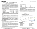
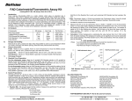
FOR RESEARCH USE ONLY!
Glycogen Colorimetric/Fluorometric Assay Kit
rev. 9/13
(Catalog # K646-100; 100 assays; Store at -20°C)
I.
Introduction:
Glycogen is the primary short term energy storage molecule in animals, synthesized primarily in the liver and muscle. Glycogen is a
branched glucose polymer, in α-1,4 linkage, with branching via α-1,6 linkage. Abnormal ability to utilize glycogen is found in diabetes
and in several genetic glycogen storage diseases. The BioVision Kit is an easy and convenient assay to measure glycogen levels in
biological samples. In the assay, glucoamylase hydrolyzes the glycogen to glucose which is then specifically oxidized to produce a
product that reacts with OxiRed probe to generate color (OD 570 nm) and fluorescence (Ex/Em = 535/587 nm). The assay can detect
glycogen 0.0004 to 2 mg/ml.
II.
Application:
• Measurement of Glycogen in various tissues/cells
• Analysis of metabolism and cell signaling in various cells
III.
Sample Type:
• Animal tissues such as liver etc.
• Cell culture: adherent or suspension cells
IV.
Kit Contents:
Components
K646-100
Cap Code
Part Number
Hydrolysis Buffer
Development Buffer
OxiRed Probe
Hydrolysis Enzyme Mix
Development Enzyme Mix
Glycogen Standard (2.0 mg/ml)
25 ml
25 ml
0.2 ml
lyophilized
lyophilized
100 µl
NM
WM
Red
Blue
Green
Yellow
K646-100-1
K646-100-2
K646-100-3A
K646-100-5
K646-100-6
K646-100-7
V.
User Supplied Reagents and Equipment:
• 96-well plate with flat bottom
• Multi-well spectrophotometer
VI.
Storage and Handling:
Store kit at –20°C, protected from light and moisture. Briefly centrifuge small vials prior to opening. Read entire protocol before
performing the assay.
VII.
Reagent Preparation and Storage Conditions:
• Hydrolysis Buffer: Warm to room temperature before use. Store at –20°C or 4°C.
• Development Buffer: Warm to room temperature before use. Store at –20°C or 4°C.
• OxiRed Probe: Ready to use as supplied. Warm to room temperature to melt frozen DMSO before use. Mix well, store at –20°C.
Protect from light and moisture. Use within 2 months.
• Hydrolysis Enzyme Mix: Reconstitute with 220 µl of Hydrolysis Buffer. Vortex gently to dissolve. Keep on ice. Store at –20°C. Use
within two months.
• Development Enzyme Mix: Reconstitute with 220 µl of Development Buffer. Vortex gently to dissolve. Keep on ice. Store at –20°C.
Reagents are stable for at least two months
VIII.
Glycogen Assay Protocol:
1. Sample Preparation: Liquid samples can be assayed directly. For tissue or cells, homogenize 106 cells or 10 mg tissue with 200 µl
dH₂O on ice. Boil the homogenates for 10 min. to inactivate enzymes. Spin the boiled samples at 18,000 x g for 10 min. to remove
insoluble material; the supernatant is ready for assay. Add 2-50 µl samples to a 96-well plate. Adjust the volume to 50 µl/well with
Hydrolysis Buffer.
Notes:
a. For unknown samples, we suggest performing a pilot experiment & testing different sample dilutions to ensure the readings are
within the Standard Curve range.
b. Glycogen can be metabolized very rapidly in some tissues after death (within a min.), therefore special care must be taken to
minimize glycogen loss when taking tissue samples, such as freezing samples immediately and keeping cold while working.
c. For samples having glucose background, prepare parallel well(s) containing same amount of sample as in the test well as
background control (see section 3).
d. Endogenous compounds may interfere with the reaction. To ensure accurate determination of Glycogen in the test samples, we
recommend spiking samples with a known amount of Standard (0.8 µg).
e. There are varieties of methods for extraction of glycogen from tissues1-4 depending upon the type of tissue or type of information
desired. We strongly recommend consulting the literature to determine the best method for your purposes. However, for
convenience a few methods taken from literature are described on page 3.
155 S. Milpitas Blvd., Milpitas, CA 95035 USA | T: (408)493-1800 F: (408)493-1801 | www.biovision.com | [email protected]
FOR RESEARCH USE ONLY!
2. Standard Curve Preparation: For colorimetric assay: dilute the Glycogen Standard to 0.2 mg/ml by adding 10 µl of the Standard to
90 µl of distilled water, mix well. Add 0, 2, 4, 6, 8, 10 µl to a series of wells. Adjust volume to 50 µl/well with Hydrolysis Buffer to
generate 0, 0.4, 0.8, 1.2, 1.6 and 2.0 µg per well of the Glycogen Standard. For fluorometric assay: Dilute the Glycogen Standard to
0.02 mg/ml by adding 10 µl of the Standard to 990 µl of distilled water, mix well. Add 0, 2, 4, 6, 8, 10 µl to a series of wells. Adjust
volume to 50 µl/well with Hydrolysis Buffer to generate 0, 0.04, 0.08, 0.12, 0.16 and 0.2 µg per well of the Glycogen Standard.
3. Hydrolysis: Add Hydrolysis Enzyme mix to Standards and samples and mix well.
Colorimetric
Hydrolysis Enzyme Mix
2 µl
Fluorometric
1 µl
Incubate for 30 min. at room temperature.
Note: Glucose generates background readings. If glucose is present in your sample, you may do a glucose control without the
addition of hydrolysis enzyme to determine the level of glucose background in your sample. The glucose background can then be
subtracted from glycogen readings.
4. Reaction Mix: Mix enough reagents for the number of samples and Standards to be performed: For each well, prepare a total 50 µl
Reaction Mix.
Colorimetric
46 µl
2 µl
2 µl
Development Buffer
Development Enzyme Mix
OxiRed Probe
Fluorometric
48.7 µl
1.0 µl
0.3 µl
Add 50 µl of the Reaction Mix to each well containing the Glycogen Standard or samples, mix well.
5. Measurement: Incubate the reaction for 30 min. at room temperature, protected from light. Measure absorbance (OD 570 nm) or
fluorescence (Ex/Em = 535/587 nm).
6. Calculation: Correct background by subtracting the 0 glycogen Standard from all readings (Note: The background can be significant
and must be subtracted). Plot Glycogen Standard Curve. Apply sample readings to the standard curve to get B µg of glycogen in the
sample wells.
Sample Glycogen concentration (C) = B/V X D µg/µl
Where: B is the amount of glycogen from Standard Curve (µg)
V is the sample volume added into the reaction well (µl)
D is the sample dilution factor
Note: For spiked samples, correct for any sample interference by subtracting the sample reading from spiked sample reading.
ODsample corrected
* Glycogen Spike (µg)
For spiked samples, Glycogen amount in sample well (B) = OD
sample glycogen Stdcorrected-ODsamplecorrected
6
7
Glycogen molecular size: ~ 60,000 glucose molecules (MW ~10 -10 daltons).
Glucose Molecular Weight: 180.16.
8000
1.2
1
0.8
0.6
0.4
0.2
0
6000
4000
2000
y = 0.664x + 0.009
y = 39234x + 40.68
0.4
0.8
1.2
1.6
Glycogen Amount (µg)
2
2
1.5
1
0.5
0
0
0
Glycogen (µg/µl)
(c)
(b)
RFU
OD (570 nm)
(a)
0
0.04
0.08
0.12
0.16
0.2
Rat Liver
Glycogen Amount (µg)
Figure: Glycogen Standard Curve: colorimetric (a), fluorometric (b). c) Representative Glycogen Standard Curve in the presence of
rat liver lysate. Sample of rat liver was homogenized with deionized water (0.05 mg liver/µl water). Homogenate was boiled for 10
min., centrifuged for 10 min. at 18000 x g and supernatant was collected. Samples (5 µl of 10 times diluted) were spiked with
glycogen (0.8 µg) and assayed according to the protocol. Assays were performed following the kit protocol.
IX.
Related Products:
Glucose Colorimetric/Fluorometric Assay Kit (K606)
PicoProbe™ Glucose Fluorometric Assay Kit (K688)
Glucose-6-Phosphate Colorimetric Assay Kit (K657)
Glucose Uptake Fluorometric Assay Kit (K666)
Glucose and Sucrose Colorimetric/Fluorometric Assay Kit (K616)
Glucose Dehydrogenase Activity Colorimetric Assay Kit (K786)
Glucose Colorimetric Assay Kit II (K686)
Glucose-1-Phosphate Colorimetric Assay Kit (K697)
Phosphoglucomutase Colorimetric Assay Kit (K774)
Glucose Uptake Colorimetric Assay Kit (K676)
Maltose and Glucose Colorimetric/Fluorometric Assay Kit (K618)
Glucose Oxidase Activity Assay Kit (K788)
FOR RESEARCH USE ONLY! Not to be used on humans.
155 S. Milpitas Blvd., Milpitas, CA 95035 USA | T: (408)493-1800 F: (408)493-1801 | www.biovision.com | [email protected]
FOR RESEARCH USE ONLY!
Sample Preparation:
There are a variety of methods for extraction of glycogen from tissues depending upon a) the type of tissue the glycogen is to be
extracted form and b) the type of information desired. The gentlest procedure is the method referred in reference 1, which maintains the
molecular weight of the glycogen so that analysis of the molecular distribution is possible.
A rapid method useful for small tissue samples is detailed in reference 4. Basically a small sample of tissue is homogenized in 50
volumes of distilled water, diluted appropriately and immediately used in the assay. Since endogenous glucose will be a significant
factor utilizing this method, a glucose background control must be conducted where the sample is directly placed in development buffer
with development enzyme mix (without prior treatment with the hydrolysis reagents). If the sample will not be immediately assayed, it
should be placed in a capped, vented microcentrifuge tube and boiled for 5 min to inactivate any enzyme activities present and stored
at -20°C until assayed. Samples from high content tissues (liver, muscle) prepared in this way should have sufficient glycogen such that
5-25 µl aliquots will give a clearly measureable colorimetric signal. If the sample is from low content tissues, either take a large aliquot
(50µl) for the colorimetric assay or a proportionally smaller aliquot (10-25µl) in the fluorometric assay.
Caveats:
1) in some tissues such as neural tissue, very rapid rates of anaerobic metabolism continue after death causing rapid declines in
glucose to undetectable levels within a few seconds. Utilization of glycogen follows and large decreases in glycogen content are seen
within less than a minute. Thus accurate measurement of glycogen in such tissues requires very rapid quenching of metabolic activity
such as freeze clamp or immediate removal of tissue to liquid nitrogen followed by grinding in the liquid nitrogen and storage at -20 or 80°C until used.
2) In some samples i.e., Saccharomyces, the glycogen is distributed between soluble and insoluble pools. It is not clear that both pools
are completely hydrolyzed
If the sample to be analyzed is sufficiently large (a few hundred milligrams to grams of tissue), a more quantitative method is as follows:
Take tissue or cells to a final content of 30-50% in 30% KOH. Heat to 100°C for 2 hours, cool and add 2 volumes of 95% ethanol. This
will precipitate the crude glycogen. Centrifuge and collect the precipitate. Dissolve/suspend the precipitate in a minimal amount of
distilled water and acidify to pH 3 with HCL (5N). Reprecipitate with 1 volume of ethanol. Repeat wash/acidification/precipitation 2 more
times, then wash precipitate with ethanol and dry. This procedure removes the vast majority of the glucose background with minimal
effect on the glycogen. The dried material can be weighed and dissolved/suspended in hydrolysis buffer for analysis.
X.
References:
1) E.Bueding and S.A. Orrell (1964) A Mild Procedure for the Isolation of Polydisperse Glycogen from Animal Tissues. J. Biol.Chem.
239, 12, pp 4018-4020
2) R. H. Dalrymple, R. Hamm (1973) A method for the extraction of glycogen and metabolites from a single muscle sample. Intl J of
Food Sci & Tech, 8, 4 pp 439-444
3) G. Cappeln, F. Jessen (2002) ATP, IMP, and Glycogen in Cod Muscle at Onset and During Development of Rigor Mortis Depend on
the Sampling Location. J. Food Sci. 67, #3, pp 991-995
4) Huijing, F. (1970) A Rapid Enzymic Method For Glycogen Estimation In Very Small Tissue Samples., Clin. Chim. Acta. 30, pp 567572.
5) Monique Rousset, etc. (1981) Presence of Glycogen and Growth related Variations in 58 Cultured Human Tumor Cell Lines. Cancer
Research. 41, 1165-1170.
155 S. Milpitas Blvd., Milpitas, CA 95035 USA | T: (408)493-1800 F: (408)493-1801 | www.biovision.com | [email protected]