1
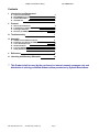
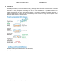
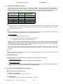
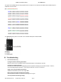
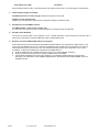
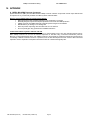
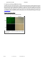
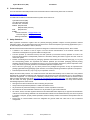
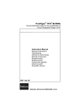
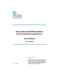
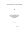
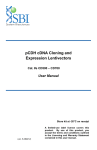
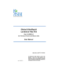
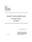
miRZip™ Lentivirus Pool of Anti- MicroRNAs Cat. # MZIPPLVA-1 User Manual Grow bacterial stock on receipt Make plasmid DNA for experiments (ver. 2010-03-16) A limited-use label license covers this product. By use of this product, you accept the terms and conditions outlined in the Licensing and Warranty Statement contained in this user manual. miRZip™ Pooled Virus Library Cat. # MZIPPLVA-1 Contents I. Introduction and Background A. Purpose of this Manual B. MicroRNA Background C. Product Description D. Intended Use 2 2 2 3 II. Protocol A. General Considerations B. Transduction of miRZip Virus Library C. Phenotypic Selection D. Recovery of miRZip Effector shRNA Sequences III. Troubleshooting 4 5 5 6 7 IV. Appendix A. miRZip anti-microRNA constructs B. Production and Titering of miRZip Virus Library C. List of Kit Components D. Related Products E. Technical Support F. Safety Guidelines 9 11 13 13 14 14 V. References 15 VI. Licensing and Warranty Statement 16 ** This Product shall be used by the purchaser for internal research purposes only and distribution is strictly prohibited without written permission by System Biosciences. 888-266-5066 (Toll Free) 650-968-2200 (outside US) Page 1 System Biosciences (SBI) User Manual I. Introduction and Background A. Purpose of this Manual This manual provides information about the design and production of the MiRZip™ virus library and detailed instructions on its use. The MiRZip™ virus library contains all of SBI’s microRNA precursor clones as a pooled, ready-to-infect, VSV-G pseudotyped lentiviral preparation. For more detailed information regarding the individual clones that comprise the library, please refer to the user manual for the MZIPxxx-PA-1 product line, this is available online at www.systembio.com. Before using this reagent and associated materials provided in this kit, please read the entire manual. B. MicroRNA Background The term microRNA (miRNA) describes a novel class of small, non-coding RNAs which regulate gene expression posttranscriptionally by disrupting translation or directing cleavage of complementary mRNAs. These 17-26 nucleotide (nt) single-stranded miRNA molecules are synthesized as primary transcripts (pri-miRNA) that are often polycistronic, containing a small number of clustered miRNA units. Following transcription, and while the transcript still remains nuclear, the Drosha RNAse III nuclease processes the pri-miRNA into ~70nt stem loop precursors (pre-miRNA). These pre-miRNA molecules are transported to the cytoplasm by a complex of proteins which includes the dsRNA binding protein Exportin-5, where they are processed to their final mature form by another RNAse III nuclease, Dicer (Lee, 2002, Yi, 2003). It is here in the cytoplasm that mature miRNAs ultimately affect the protein levels of their target mRNAs by binding to complementary regions, and either inhibiting translation, or directing mRNA cleavage (Kim, 2005), see FIG 1. FIG1. Processing of microRNA precursors. The MiRZip library will suppress the mature microRNA forms. C. Product Description The MiRZip™ virus library contains a pool of SBI’s anti-microRNA shRNA clones in a ready-to-infect lentiviral preparation. Each virus within the pool will knockdown an individual microRNA . Also included in this kit are a positve control virus with a scramble hairpin (MZIP000-VA-1) to assess transduction efficiency in your cells of interest and the oligonucleotide primers that may be used to identify the anti-microRNA shRNA sequence responsible for the observed phenotypes. Page 2 ver. 1-2010.03.16 www.systembio.com miRZip™ Pooled Virus Library Cat. # MZIPPLVA-1 D. Intended use The MiRZip™ virus library is a tool that enables the study of phenotypic effects associated with the knockdown of individual microRNAs. The lentivirus preparation is pseudotyped with VSV-G that allows for broad cellular tropism. Hard-to-transfect mammalian cell lines, primary cells, non-dividing cells and even whole animal studies are possible. Transduced cells exhibiting the phenotypes of interest are isolated by selection or sorting. The microRNA or microRNAs responsible for generating the phenotypes of interest may be recovered through simple genomic PCR using lentivector-specific primers followed by direct sequencing of shRNA inserts. FIG 2. An overall experimental workflow for virus transduction, phenotypic selection is shown. 888-266-5066 (Toll Free) 650-968-2200 (outside US) Page 3 System Biosciences (SBI) User Manual II. Protocol A. General considerations: • The transduction efficiency of the MiRZip™ library varies significantly for different cells and experimental conditions. In order to optimize transduction conditions, we recommend that you use the positive transduction control MZIP000-VA-1 virus provided in this kit to determine the optimal MOI for your cells of interest (see below). • Determining the Optimal MOI MOI is defined as the number of transducing units per cell. A number of factors can influence determination of an optimal MOI including the nature of your mammalian cell (actively- versus non-dividing), its transduction efficiency, your application of interest. If you are transducing the MiRZip virus library into the your cells for the first time, we recommend using a range of MOIs (e.g. 1, 2, 5, 10) to determine the MOI required to obtain optimal expression for your particular application. Efficiency of transduction of your cell type with the positive control virus (MZIP000-VA-1) or the MiRZip virus library can be monitored by copGFP expression. • Expression of the anti-microRNA shRNAs can be measured directly at about 48-72 hours after transduction. For more information, refer to the QuantiMir small RNA quantitation system’s User manual (www.systembio.com). Design a forward primer for the “anti-sense” of the microRNA of interest for miRZips. • Transient versus Stable screen? Depending on the nature of your phenotypic screen, you may use the MiRZip virus library to transiently or stably transduce your cells. Tranduced cells can be sorted by flow cytometry based upon copGFP expression or stable selection for puromycin. • Biosafety considerations. Although these viral particles are replication-incompetent, they can infect mammalian cells and integrate into the host cell genome. For detailed biosafety information see Appendix F. Please follow all relevant Federal, State and Local regulations for working with BSL-2 class viruses. Page 4 ver. 1-2010.03.16 www.systembio.com miRZip™ Pooled Virus Library B. Cat. # MZIPPLVA-1 Transduction of miRZip Virus Library The following protocol provides procedures for transduction of the MiRZip virus library into target cells and is optimized for adherent cell lines such as HeLa or MCF7 in a 6-well plate format. Use these guidelines as a starting point for determining optimal conditions for your cells and experiments. To use a different culture dish, adjust the number of cells, volumes of reagents and quantities of virus in proportion to the surface area of the dish/well (see Table below). Tissue Culture Dish 100mm 60mm 35mm 6 well 12 well 24 well 96 well Surface area per well (cm2) 56 20 8 9.4 3.8 1.9 0.3 Suggested medium volume per well (ml) 10 4 2 2 1 0.5 0.2 Day 1. 1. 5 Plate target cells in a 6-well plate at a density of 1x10 cells per well 24 hours prior to viral infection in 2 ml of complete medium with serum and antibiotics. Incubate cells at 37°C with 5% CO2 overnight. Typically cells will be 30-50% confluent at the time of infection. Day 2. 2. Dilute an appropriate amount (depending on the optimal MOI) of virus into 0.5 ml of complete medium. Add Polybrene (example 200x TransDux™, SBI cat# LV850A-1) to a final concentration of 1x. IN THIS EXAMPLE : 100,000 cells/well were seeded on Day 1. Assume doubling overnight, yields 200,000 cells/well on Day 2. To infect at an MOI of 5, use 1 x 10⁶ IFUs virus per well. Note: Polybrene® is a polycation that neutralizes charge interactions to increase binding between the pseudoviral capsid and the cellular membrane. The optimal concentration of Polybrene depends on cell type and may need to be empirically determined (usually in the range of 2-10 μg/ml). Excessive exposure to Polybrene (>12 hr) can be toxic to some cells. We recommend using SBI’s TransDux infection reagent. 3. Remove the culture medium from cells. Infect target cells by adding the viral stock dilutions to the wells. For one well (mock well control) add 0.5 ml of D-MEM medium with TransDux or Polybrene. Incubate cells at 37°C with 5% CO2 overnight. Day 3. 4. Remove the culture medium and replace with 2 ml of complete medium (without TransDux or Polybrene). Incubate the cells at 37°C with 5% CO2 overnight. C. Phenotypic Selection Day 4 to 6: By 48 to 72 hours post-transduction, copGFP expression will be apparent in infected cells. ● Transient transduction screen: Begin your phenotypic analysis and select cells exibiting the desired phenotypes. ● Stable transduction screen: Begin your FACS sort for copGFP or puromycin selection for positive cells to enrich for stably transduced cells. Establish a stably-transduced cell population before performing your phenotypic analysis. D. Recovery of Effector MicroRNA Clone Sequences Materials required and not supplied in kit: For Purification of genomic DNA • DNeasy Tissue Kit (QIAGEN, Cat. # 69504 or equivalent) For PCR Amplification • Standard PCR kit (TITANIUM Taq PCR Kit, Clontech Cat# 639210 or equivalent) 888-266-5066 (Toll Free) 650-968-2200 (outside US) Page 5 System Biosciences (SBI) • • User Manual Thermal Cycler (DNA Engine, MJ Research, Cat. # PTC-200 or equivalent) 2.5% 1X TAE Agarose gel 1. Purify Genomic DNA from Selected Cells from MiRZip Phenotypic Screen Prepare genomic DNA from selected cells according to manufacturer’s recommendations. Measure the yield of DNA by spectrophotometer. You should expect to isolate 5-10 μg of genomic DNA from approximately 1×10⁶ cells. Dilute the DNA sample in deionized water to a concentration of 0.2 μg/μl. 2. An sample PCR reaction for microRNA effector clone sequence amplification is provided below. A. Prepare a PCR Master Mix for all reaction tubes, plus one additional tube. Combine the following components in the order shown: 41 5 1 1 1 1 50 μl μl μl μl μl μl μl Deionized H2O 10X PCR buffer 50X dNTP mix (10 mM of each dNTP) ZipLV Primer Mix (10 μM of each primer) Purified Genomic DNA (~200ng) 50X Taq DNA polymerase Total volume Note: Include no DNA negative control. B. Mix contents by vortexing, and spin the tube briefly in a microcentrifuge. C. Perform PCR amplification according to these guidelines: • • • • 94°C for 2 min (94°C for 30 sec; 60°C for 30 sec) for 25 cycles 68°C for 1 min 15°C hold D. When the program is completed, analyze a 10-μl sample from each tube and suitable DNA size marker (50 bp-2 kb) on a 1.5% agarose/EtBr gel in 1X TAE. Note that each anti-microRNA shRNA clone within the library collection varies in length. Therefore the expected amplicon size may range from 200 to 240 bp (see sample data on the following page). E. Page 6 Sequence the effector anti-microRNA shRNA amplicons recovered after gel purification using either the Forward or Reverse PCR primer (PCR primer sequences are provided in Appendix C.) ver. 1-2010.03.16 www.systembio.com miRZip™ Pooled Virus Library Cat. # MZIPPLVA-1 The region of the miRZip lentivector being amplified and an example of a successful anti-microRNA effector shRNA sequence amplification is shown below. Fwd 5’-TGCATGTCGCTATGTGTTCTGGGA-3’ Rev 3’-AATCTGGTCTAGACTCGGACCCTC-5’ ATCGATGAAC GCTGACGTCA TCAACCCGCT CCAAGGAATC GCGGGCCCAG TAGCTACTTG CGACTGCAGT AGTTGGGCGA GGTTCCTTAG CGCCCGGGTC TGTCACTAGG CGGGAACACC CAGCGCGCGT GCGCCCTGGC AGGAAGATGG ACAGTGATCC GCCCTTGTGG GTCGCGCGCA CGCGGGACCG TCCTTCTACC CTGTGAGGGA CAGGGGAGTG GCGCCCTGCA ATATTTGCAT GTCGCTATGT GACACTCCCT GTCCCCTCAC CGCGGGACGT TATAAACGTA CAGCGATACA GTTCTGGGAA ATCACCATAA ACGTGAAATG TCTTTGGATT TGGGAATCTT CAAGACCCTT TAGTGGTATT TGCACTTTAC AGAAACCTAA ACCCTTAGAA ATAAGTTCTG TATGAGACCA CTTGGATCCG NNNNNNNNNN NNNNNNNNNN TATTCAAGAC ATACTCTGGT GAACCTAGGC NNNNNNNNNN NNNNNNNNNN NNNNNNCTTC CTGTCAGANN NNNNNNNNNN NNNNNNNNNN NNNNCTTTTT NNNNNNGAAG GACAGTCTNN NNNNNNNNNN NNNNNNNNNN NNNNGAAAAA GAATTCnnnn CCAATTCTTC GATTCTGCTT TTTGCTTCTA CTGGGTCTCT CTTAAGnnnn GGTTAAGAAG CTAAGACGAA AAACGAAGAT GACCCAGAGA CTGGTTAGAC CAGATCTGAG CCTGGGAGCT CTCTGGCTAA CTAGGGAACC GACCAATCTG GTCTAGACTC GGACCCTCGA GAGACCGATT GATCCCTTGG CACTGCTTAA GCCTCAATAA AGCTTGCCTT GAGTGCTTCA AGTAGTGTGT GTGACGAATT CGGAGTTATT TCGAACGGAA CTCACGAAGT TCATCACACA The NNNNNNNN string refers to the location of the individual miRZip anti-microRNA shRNA. III. Troubleshooting 1. Poor Infection Efficiency Too high a volume of infecting supernatant Keep the volume as low as possible to achieve maximal adsorption of viral particles to the cells. The assay is carried out too early Normally, the maximal expression of integrated provirus is expected to develop by 48 hours after infection. However, some cells display delayed expression. Try the assay at a later time, such as 72 or 96 hours. Target cell line may be difficult to transduce Optimize the transduction protocol. Use a higher MOI. Wrong amount of Polybrene added during titration Add and optimize Polybrene concentration in the range of 4-10 μg/ml. Loss of pseudoviral titer during storage 888-266-5066 (Toll Free) 650-968-2200 (outside US) Page 7 System Biosciences (SBI) User Manual Store pseudoviral stock at –80°C. Each freeze-thaw cycle drops the titer 2-4 fold. Use a fresh aliquot for transduction. 2. Infection Affects Target Cell Viability High MOI may be toxic to some cell types. Reduce the amounts of virus used. Polybrene is toxic for target cells Optimize the concentration and exposure time to Polybrene during the transduction step. 3. No Expression of microRNA precursors The CMV promoter is not functional in target cells It is a very rare case, but the only way to solve this problem is to change the type of target cells. 4. No PCR product Amplified The amount of genomic DNA in your sample is too low. Repeat purification of genomic DNA from infected cells, or measure the concentration of genomic DNA again using a smaller dilution factor. 5. Sequence of anti-microRNA shRNA effector is ambiguous If direct sequencing of the recovered and PCR-amplified microRNA effector does not produce a single sequence, your screen may have identified two or more co-effector microRNAs that may contribute to your phenotype. Your cells may have been transduced by more than one virus. The observed phenotype may be due to expression of one or more microRNAs. To identify and separate the individual anti-microRNA effectors: A. Clone the amplicon(s) into a suitable PCR-cloning vector (TA or TOPO plasmids, Invitrogen). B. Sequence at least 10 individual clones to identify candidate effector microRNAs. C. Test individual candidates for the phenotype of interest. If no individual microRNA reproduces the observed phenotype, test combinations of the candidates identified above. All individual anti-microRNA shRNAr clones are available from SBI. Page 8 ver. 1-2010.03.16 www.systembio.com miRZip™ Pooled Virus Library Cat. # MZIPPLVA-1 IV. APPENDIX A. MiRZip MicroRNA Precursor Constructs System Biosciences’ (SBI’s) anti-microRNA shRNA miRZip Construct Collection incorporates several unique features that set it apart from any commercially available microRNA synthetic antisense RNAs. Express anti-microRNAs stably for permanent knockdown • Stable & permanent anti-microRNA expression from constitutive H1 promoter • Rationally designed, asymmetric hairpins optimized for anti-sense microRNA production • miRZip anti-sense microRNAs efficiently suppress specific endogenous microRNAs • Reliable delivery to dividing or non-dividing cells • Select for positive expressing cells with either GFP or Puro selection • Uncover phenotypes using powerful anti-microRNA interference Lentiviral transduction system is effective and safe Each of SBI’s anti-microRNA shRNAs has been cloned in a lentiviral-based vector. Like other standard plasmid vectors, SBI’s miRZip constructs can be used for transient expression of anti-miRs using conventional transfection protocols. Moreover, its lentiviral backbone allows each miRZip construct to be packaged in pseudoviral particles and introduced into non-dividing or difficult-to-transfect cell lines. In particular, all of the anti-miR hairpins have been cloned in SBI’s HIV-based expression vectors. Replication incompetent HIV-based vectors are considered biologically safe. 888-266-5066 (Toll Free) 650-968-2200 (outside US) Page 9 System Biosciences (SBI) User Manual Dual marker design simplifies identification of transduced cells The unique organization of the vector (below) results in expression of dual marker transcripts containing the copGFP fluorescent protein as well as puromycin resistance selection markers driven by the CMV promoter. A. Map and Features for miRZip™ Vector Feature RSV/5’LTR Location* 7-413 gag RRE 566-920 1076-1309 cPPT 1806-1923 CMV promoter 1929-2278 copGFP 2293-3048 T2A 3049-3102 Puro 3103-3702 WPRE 3703-4291 3’ ΔLTR(ΔU3) 4631-4813 H1 RNA promoter SV40 Poly-A 4526-4616 4911-5219 SV40 Ori 4911-5219 pUC Ori AmpR 5584-6252 6397-7257 (C) Page 10 Function Hybrid RSV promoter-R/U5 long terminal repeat; required for viral packaging and transcription Packaging signal Rev response element binds gag and involved in packaging of viral transcripts Central polypurine tract (includes DNA Flap region) involved in nuclear translocation and integration of transduced viral genome Human cytomegalovirus (CMV)--constitutive promoter for transcription of copGFP-T2Apuro Copepod green fluorescent protein (similar to regular EGFP, but with brighter color) as a reporter for the transfected/transduced cells Thosea asigna virus 2A translational cleavage site containing 18 amino acid residues. Cleavage occurs via a cotranslational ribosome skipping mechanism between the C-terminal Glycin and Prolin residues, leaving 17 residues attached to the end of copGFP and 1 residue to the start of the puromycin resistance marker Puromycin-resistant marker for selection of the ransfected/transduced cells Woodchuck hepatitis virus posttranscriptional regulatory element-enhances the stability of the viral transcripts Required for viral reverse transcription; selfinactivating 3' LTR with deletion in U3 region prevents formation of replication-competent viral particles after integration into genomic DNA RNA polymerase III promoter for expression of anti-microRNA insert Transcription termination and polyadenylation Allows for episomal replication of plasmid in eukaryotic cells Allows for high-copy replication in E. coli Ampicillin resistant gene for selection of the plasmid in E. coli ver. 1-2010.03.16 www.systembio.com miRZip™ Pooled Virus Library Cat. # MZIPPLVA-1 Short double-stranded RNAs with sizes 19-29 bp can efficiently mediate the delivery of small RNAs in mammalian cells. Synthetic single-stranded anti-microRNA molecules can be introduced into cells to suppress microRNA function transiently. Alternatively, miRZips can stably express anti-microRNAs and provide permanent microRNA inhibition—and in any cell type of choice. Lentiviral expression vectors are the most effective vehicles for delivering genetic material to almost any mammalian cell— including non-dividing cells and whole model organisms. As with standard plasmid vectors, it is possible to introduce miRZip lentivector constructs in plasmid form into the cells with low-to-medium efficiency using conventional transfection protocols. However, by packaging the lentiviral miRZip construct into pseudoviral particles, you can obtain highly efficient transduction and heritable expression of anti-microRNAs—even with most difficult to transfect cells, like primary, stem, and differentiated cells. The expression construct transduced in cells is integrated into genomic DNA and provides stable, long-term expression of the target gene. Endogenously expressed anti-microRNA effectors provide long-term suppression of the target microRNA and allow the researcher to generate cell lines and transgenic organisms with a stable microRNA inhibition phenotype for functional studies Properties of the copGFP Fluorescent Protein The MZIP Vector contains the full-length copGFP gene with optimized human codons for high level of expression of the fluorescent protein from the CMV promoter in mammalian cells. The copGFP marker is a novel natural green monomeric GFPlike protein from copepod (Pontellina sp.). The copGFP protein is a non-toxic, non-aggregating protein with fast protein maturation, high stability at a wide range of pH (pH 4-12), and does not require any additional cofactors or substrates. The copGFP protein has very bright fluorescence that exceeds at least 1.3 times the brightness of EGFP, the widely used Aequorea victoria GFP mutant. The copGFP protein emits green fluorescence with the following characteristics: emission wavelength max – 502 nm; excitation wavelength max – 482 nm; quantum yield – 0.6; -1 -1 extinction coefficient – 70,000 M cm Due to its exceptional properties, copGFP is an excellent fluorescent marker which can be used instead of EGFP for monitoring delivery of lentivector constructs into cells. 888-266-5066 (Toll Free) 650-968-2200 (outside US) Page 11 System Biosciences (SBI) User Manual B. Production and Titering of MiRZip Virus Library The MiRZip Virus Library was prepared as follows. The full set of SBI’s human precursor microRNA expression clones was pooled and packaged into VSV-G pseudotyped viral particles by co-transfecting 293TN Producer Cells (SBI Cat #LV900A-1) with the library clones and pPACKH1 (SBI Cat #LV500A-1) according to the procedures described in the “Lentivector Expression Systems: Guide to Packaging and Transduction of Target Cells”. Virus was concentrated with PEG-it Virus Precipitation Solution (SBI Cat #LV810A-1) according to the instructions in the User Manual. Infectious titers were determined on HT1080 cells using the UltraRapid Lentiviral Titering Kit for Human Cells (SBI Cat#LV961A-1) according to the recommended procedures. Additional information and User Manuals for each of these products are available on our website: www.systembio.com. Sample Transduction Data Easily monitor infection efficiency by GFP expression. GFP expression in HT1080 cells transduced with the MiRZip Virus Library. Page 12 ver. 1-2010.03.16 www.systembio.com miRZip™ Pooled Virus Library Cat. # MZIPPLVA-1 C. List of Kit Components Aliquots of pooled MiRZip virus library Tube of positve infection control virus (MZIP000-VA-1) Tube of MZIP PCR Primer Mix (10 μM of each primer) o o Fwd 5’-TGCATGTCGCTATGTGTTCTGGGA-3’ Rev 3’-AATCTGGTCTAGACTCGGACCCTC-5’ D. Related Products • pPACKH1™ Lentivector Packaging Kit (Cat. # LV500A-1) Unique lentiviral vectors that produce all the necessary HIV viral proteins and the VSV-G envelope glycoprotein from vesicular stomatitis virus required to make active pseudoviral particles. 293TN cells (SBI, Cat. # LV900A-1) transiently transfected with the pPACKH1 and a pMIRNA1 microRNA expression construct produce packaged viral particles containing a microRNA construct. • 293TN Human Kidney Producer Cell Line (SBI, Cat. # LV900A-1). For packaging of plasmid lentivector constructs. • PEG-it™ Virus Precipitation Solution (Cat. # LV810A-1). Simple and highly effective means to concentrate lentiviral vector inocula produced with SBI’s pPACK Lentivector packaging systems. • TransDux (Cat. # LV850A-1). • Lentivector UltraRapid Titer PCR Kit (Cat. # LV960A-1 [for human cells], LV960B-1 [for mouse cells]) Allows you to measure copy number (MOI) of integrated lentiviral constructs in genomic DNA of target cells after transduction with constructs made in any of SBI’s FIV or HIV-based Lentivectors or GeneNet™ siRNA Libraries. • QuantiMir small RNA quantitation system (Cat. # RA420A-1) Easily profile miRNAs using a single cDNA synthesis step and real-time qPCR. • MicroRNA Precursor Clone collection (cat#s pMIRHxxxPA-1) SBI’s collection of individual microRNA precursor expression clones in lentivectors. 888-266-5066 (Toll Free) 650-968-2200 (outside US) Page 13 System Biosciences (SBI) User Manual E. Technical Support For more information about SBI products and to download manuals in PDF format, please visit our web site: http://www.systembio.com For additional information or technical assistance, please call or email us at: System Biosciences (SBI) 1616 North Shoreline Blvd. Mountain View, CA 94043 Phone: (650) 968-2200 (888) 266-5066 (Toll Free) Fax: (650) 968-2277 E-mail: General Information: [email protected] Technical Support: [email protected] Ordering Information: [email protected] F. Safety Guidelines SBI’s Expression lentivectors together with the pPACK packaging plasmids comprise the third-generation lentiviral expression system. The HIV-based lentivectors are based on the vectors developed for gene therapy applications by Dr. J. G. Sodroski (US patent #5,665,577 and # 5,981,276). Both FIV-based and HIV-based lentivector systems are designed to maximize their biosafety features, which include: • A deletion in the enhancer of the U3 region of 3’ΔLTR ensures self-inactivation of the lentiviral construct after transduction and integration into genomic DNA of the target cells. • The RSV promoter (in HIV-based vectors) and CMV promoter (in FIV-based vectors) upstream of 5’LTR in the lentivector allow efficient Tat-independent production of viral RNA, reducing the number of genes from HIV-1 that are used in this system. • Number of lentiviral genes necessary for packaging, replication and transduction is reduced to three (gag, pol, rev), and the corresponding proteins are expressed from different plasmids (for HIV-based packaging plasmids) lacking packaging signals and share no significant homology to any of the expression lentivectors, pVSV-G expression vector, or any other vector, to prevent generation of recombinant replication-competent virus. • None of the HIV-1 genes (gag, pol, rev) will be present in the packaged viral genome, as they are expressed from packaging plasmids lacking packaging signal—therefore, the lentiviral particles generated are replication-incompetent. • Pseudoviral particles will carry only a copy of your expression construct. Despite the above safety features, use of SBI’s lentivectors falls within NIH Biosafety Level 2 criteria due to the potential biohazard risk of possible recombination with endogenous viral sequences to form self-replicating virus, or the possibility of insertional mutagenesis. For a description of laboratory biosafety level criteria, consult the Centers for Disease Control Office of Health and Safety Web site at http://www.cdc.gov/od/ohs/biosfty/bmbl4/bmbl4s3.htm. It is also important to check with the health and safety guidelines at your institution regarding the use of lentiviruses and always follow standard microbiological practices, which include: • Wear gloves and lab coat all the time when conducting the procedure. • Always work with pseudoviral particles in a Class II laminar flow hood. • All procedures are performed carefully to minimize the creation of splashes or aerosols. • Work surfaces are decontaminated at least once a day and after any spill of viable material. • All cultures, stocks, and other regulated wastes are decontaminated before disposal by an approved decontamination method such as autoclaving. Materials to be decontaminated outside of the immediate laboratory area are to be placed in a durable, leak proof, properly marked (biohazard, infectious waste) container and sealed for transportation from the laboratory. Please keep in mind that MZIP vectors are integrated into genomic DNA and could have a risk of insertional mutagenesis. • Page 14 ver. 1-2010.03.16 www.systembio.com miRZip™ Pooled Virus Library Cat. # MZIPPLVA-1 V. References Lovén J, Zinin N, Wahlström T, Müller I, Brodin P, Fredlund E, Ribacke U, Pivarcsi A, Påhlman S, Henriksson M. MYCN-regulated microRNAs repress estrogen receptor-alpha (ESR1) expression and neuronal differentiation in human neuroblastoma. Proc Natl Acad Sci U S A. 2010 Jan 26;107(4):1553-8. (Utilized SBI’s miRZip-18a) Hasan Rajabi,Caining Jin,Rehan Ahmad,Andrew Cain McClary,Maya Datt Joshi and Donald Kufe. Mucin 1 Oncoprotein Expression Is Suppressed by the miR-125b Oncomir. Genes and Cancer Jan 2010, vol. 1 no. 1 62-68. (Utilized SBI’s miRZip-125b) Greene SB, Gunaratne PH, Hammond SM, Rosen JM. A putative role for microRNA-205 in mammary epithelial cell progenitors. J Cell Sci. 2010 Feb 15;123(Pt 4):606-618. (Utilized SBI’s miRZip-205) Bennasser, Y., S. Y. Le, M. L. Yeung and K. T. Jeang (2004). "HIV-1 encoded candidate micro-RNAs and their cellular targets." Retrovirology 1(1): 43. Hariharan, M., V. Scaria, B. Pillai and S. K. Brahmachari (2005). "Targets for human encoded microRNAs in HIV genes." Biochem Biophys Res Commun 337(4): 1214-8. John, B., C. Sander and D. S. Marks (2006). "Prediction of human microRNA targets." Methods Mol Biol 342: 101-13. Johnson, S. M., H. Grosshans, J. Shingara, M. Byrom, R. Jarvis, A. Cheng, E. Labourier, K. L. Reinert, D. Brown and F. J. Slack (2005). "RAS is regulated by the let-7 microRNA family." Cell 120(5): 635-47. Kim, V. N. (2005). "Small RNAs: classification, biogenesis, and function." Mol Cells 19(1): 1-15. Lee, R. C., R. L. Feinbaum and V. Ambros (1993). "The C. elegans heterochronic gene lin-4 encodes small RNAs with antisense complementarity to lin-14." Cell 75(5): 843-54. Lee, Y., K. Jeon, J. T. Lee, S. Kim and V. N. Kim (2002). "MicroRNA maturation: stepwise processing and subcellular localization." Embo J 21(17): 4663-70. Mathijs Voorhoeve, P., C.L. Sage, M. Schrier, A.J.M.Gillis, H. Stoop, R.Nagel, Y. Liu, J.V.Duijse, J. Drost, A. Griekspoor, E. Zlotorynski, N. Yabuta, G. D.Vita, H. Nojima, L.H.J.Looijenga, and R. Agami (2006). “A Genetic Screen Implicates miRNA-372 and miRNA-373 As Oncogenes in Testicular Germ Cell Tumors.” Cell 124, 1169-1181 Olsen, P. H. and V. Ambros (1999). "The lin-4 regulatory RNA controls developmental timing in Caenorhabditis elegans by blocking LIN-14 protein synthesis after the initiation of translation." Dev Biol 216(2): 671-80. Pfeffer, S., M. Zavolan, F. A. Grasser, M. Chien, J. J. Russo, J. Ju, B. John, A. J. Enright, D. Marks, C. Sander and T. Tuschl (2004). "Identification of virus-encoded microRNAs." Science 304(5671): 734-6. Poeschla, E.M., Looney, D.J., and Wong-Staal, F. (2003) Lentiviral nucleic acids and uses thereof. US Patent NO. 6,555,107 B2 Shin, K. J., E. A. Wall, J. R. Zavzavadjian, L. A. Santat, J. Liu, J. I. Hwang, R. Rebres, T. Roach, W. Seaman, M. I. Simon and I. D. Fraser (2006). "A single lentiviral vector platform for microRNA-based conditional RNA interference and coordinated transgene expression." Proc Natl Acad Sci U S A 103(37): 13759-64. Stegmeier, F., G. Hu, R. J. Rickles, G. J. Hannon and S. J. Elledge (2005). "A lentiviral microRNA-based system for single-copy polymerase II-regulated RNA interference in mammalian cells." Proc Natl Acad Sci U S A 102(37): 13212-7. Yi, R., Y. Qin, I. G. Macara and B. R. Cullen (2003). "Exportin-5 mediates the nuclear export of pre-microRNAs and short hairpin RNAs." Genes Dev 17(24): 3011-6.. 888-266-5066 (Toll Free) 650-968-2200 (outside US) Page 15 System Biosciences (SBI) User Manual VI. Licensing and Warranty Statement Limited Use License Use of the MiRZip virus library (i.e., the “Product”) is subject to the following terms and conditions. If the terms and conditions are not acceptable, return all components of the Product to System Biosciences (SBI) within 7 calendar days. Purchase and use of any part of the Product constitutes acceptance of the above terms. The purchaser of the Product is granted a limited license to use the Product under the following terms and conditions: The Product shall be used by the purchaser for internal research purposes only. The Product is expressly not designed, intended, or warranted for use in humans or for therapeutic or diagnostic use. The Product may not be resold, modified for resale, or used to manufacture commercial products without prior written consent of SBI. This Product should be used in accordance with the NIH guidelines developed for recombinant DNA and genetic research. HIV Vector System This Product or the use of this Product is covered by U.S. Patents Nos. 5,665,577 and 5,981,276 (and foreign equivalents) owned by the Dana-Farber Cancer Institute, Inc., and licensed by SBI. This product is for non-clinical research use only. Use of this Product to produce products for resale or for any diagnostic, therapeutic, clinical, veterinary, or food purpose is prohibited. In order to obtain a license to use this Product for these commercial purposes, contact the Office of Research and Technology Ventures at the Dana-Farber Cancer Institute, Inc. in Boston, Massachusetts, USA. WPRE Technology System Biosciences (SBI) has a license to sell the Product containing WPRE, under the terms described below. Any use of the WPRE outside of SBI’s Product or the Products’ intended use, requires a license as detailed below. Before using the Product containing WPRE, please read the following license agreement. If you do not agree to be bound by its terms, contact SBI within 10 days for authorization to return the unused Product containing WPRE and to receive a full credit. The WPRE technology is covered by patents issued to The Salk Institute for Biological Studies. SBI grants you a non-exclusive license to use the enclosed Product containing WPRE in its entirety for its intended use. The Product containing WPRE is being transferred to you in furtherance of, and reliance on, such license. Any use of WPRE outside of SBI’s Product or the Product’s intended use, requires a license from the Salk Institute for Biological Studies. This license agreement is effective until terminated. You may terminate it at any time by destroying all Products containing WPRE in your control. It will also terminate automatically if you fail to comply with the terms and conditions of the license agreement. You shall, upon termination of the license agreement, destroy all Products containing WPRE in you control, and so notify SBI in writing. This License shall be governed in its interpretation and enforcement by the laws of California. Contact for WPRE Licensing: The Salk Institute for Biological Studies, 10010 North Torrey Pines Road, La Jolla, CA 92037; Attn: Office for Technology Management; Phone: (858) 435-4100 extension 1275; Fax: (858) 450-0509. CMV Promoter The CMV promoter is covered under U.S. Patents 5,168,062 and 5,385,839 and its use is permitted for research purposes only. Any other use of the CMV promoter requires a license from the University of Iowa Research Foundation, 214 Technology Innovation Center, Iowa City, IA 52242. CopGFP Reporter This product contains a proprietary nucleic acid coding for a proprietary fluorescent protein(s) intended to be used for research purposes only. Any use of the proprietary nucleic acids other than for research use is strictly prohibited. USE IN ANY OTHER APPLICATION REQUIRES A LICENSE FROM EVROGEN. To obtain such a license, please contact Evrogen at [email protected]. SBI has pending patent applications on various features and components of the Product. For information concerning licenses for commercial use, contact SBI. Purchase of the product does not grant any rights or license for use other than those explicitly listed in this Licensing and Warranty Statement. Use of the Product for any use other than described expressly herein may be covered by patents or subject to rights other than those mentioned. SBI disclaims any and all responsibility for injury or damage which may be caused by the failure of the buyer or any other person to use the Product in accordance with the terms and conditions outlined herein. Limited Warranty SBI warrants that the Product meets the specifications described in the accompanying Product Analysis Certificate. If it is proven to the satisfaction of SBI that the Product fails to meet these specifications, SBI will replace the Product or provide the purchaser with a refund. This limited warranty shall not extend to anyone other than the original purchaser of the Product. Notice of nonconforming products must be made to SBI within 30 days of receipt of the Product. SBI’s liability is expressly limited to replacement of Product or a refund limited to the actual purchase price. SBI’s liability does not extend to any damages arising from use or improper use of the Product, or losses associated with the use of additional materials or reagents. This limited warranty is the sole and exclusive warranty. SBI does not provide any other warranties of any kind, expressed or implied, including the merchantability or fitness of the Product for a particular purpose. SBI is committed to providing our customers with high-quality products. If you should have any questions or concerns about any SBI products, please contact us at (888) 266-5066. © 2010 System Biosciences (SBI). Page 16 ver. 1-2010.03.16 www.systembio.com