1
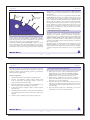
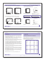
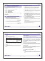
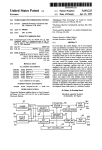
Cytokine Secretion Assays
Cytokine Secretion Assays
Cytokine
Secretion Assay
Miltenyi Biotec S.r.l. (Italy)
Miltenyi Biotec GmbH
Friedrich-Ebert-Str. 68
51429 Bergisch Gladbach, Germany
Phone: +49 2204 83060
Fax:
+49 2204 85197
[email protected]
Miltenyi Biotec Inc.
2303 Lindbergh Street,
Auburn CA 95602, USA
Phone: 800 FOR MACS, +1 530 888 8871
Fax:
+1 530 888 8925
[email protected]
Miltenyi Biotec Pty. Ltd. (Australia)
Phone: +61 2 8877 7400
Fax: +61 2 9889 5044
[email protected]
Miltenyi Biotec Shanghai Office
Phone: +86 21 62351005
Fax: +86 21 62350953
[email protected]
Miltenyi Biotec (France)
Phone: +33 1 56 98 16 16
Fax:
+33 1 56 98 16 17
[email protected]
IFN-γ Secretion Assay
Cell Enrichment and Detection Kit (PE)
IFN-γ
human Secretion Assay
Phone: +39 51 64 60 411
Fax:
+ 39 51 64 60 499
[email protected]
Miltenyi Biotec K.K. (Japan)
Phone: +81 3 56 46 8910
Fax:
+81 3 56 46 89 11
[email protected]
Cell Enrichment
and Detection Kit (PE*) human
7
Miltenyi Biotec Asia Pacific Pte. Ltd.
(Singapore)
Phone: +65 6238 8183
Fax:
+65 6238 0302
[email protected]
For 50 tests with 10 cells
Order No. 130-054-201
For 50 tests with 107 cells
Order No. 130-054-101
Miltenyi Biotec S.L. (Spain)
Phone: +34 91 512 12 90
Fax:
+34 91 512 12 91
[email protected]
Miltenyi Biotec Ltd. (UK)
Phone: +44 1483 799 800
Fax:
+44 1483 799 811
[email protected]
For further information refer to our website www.miltenyibiotec.com
140-000-274.07
For technical questions, please contact your local distributor or our
Technical Support Team in Germany:
e-mail: [email protected]
phone: +49-2204-830 6 830.
This MACS® product is for in vitro research use only and not for diagnostic or therapeutic procedures.
1. Description
Index
Index
1.
2.
3.
4.
5.
6.
7.
2
1. Description
Description
1.1 Principle of the IFN-γ Secretion Assay
1.2 Background and product applications
1.3 Reagent and instrument requirements
Protocol overview
Experimental set-up
3.1 Controls
3.2 Kinetics of restimulation and proposed time schedule
3.3 Counterstaining of cytokine secreting cells
3.4 Two color cytokine analysis
3.5 Combination with peptide-MHC tetramer staining
3.6 Detection without prior enrichment
Protocol for the IFN-γ Secretion Assay
4.1 Cell preparation
4.2 (Antigen-specific) In vitro stimulation
4.3 Cytokine Secretion Assay
4.4 Magnetic labeling
4.5 Magnetic separation
Detection and analysis of IFN-γ secreting antigen-specific T cells
References
Appendix
A: Flask and dish sizes for stimulation
B: Detection and enrichment of cytokine secreting cells from
whole blood
140-000-274.07
Components
1 mL IFN-γ Catch Reagent: anti-IFN-γ
monoclonal antibody (mouse IgG1) conjugated
to cell surface specific monoclonal antibody
(mouse IgG2a).
1 mL IFN-γ Detection Antibody: anti-IFN-γ
monoclonal antibody (mouse IgG1) conjugated
to PE (R-phycoerythrin).
1 mL Anti-PE MicroBeads: colloidal
superparamagnetic MicroBeads conjugated
to monoclonal mouse anti-PE antibody
(mouse IgG1).
Size
For 50 tests with 107 cells
Product format
All components are supplied as a suspension
containing stabilizer and 0.05% sodium azide.
Storage
Store protected from light at 4–8 °C. Do not freeze.
The expiration dates are indicated on the vial labels.
140-000-274.07
3
1. Description
1. Description
labeled with a second IFN-γ-specific antibody, the IFN-γ Detection
Antibody conjugated to R-phycoerythrin (PE) for sensitive detection by
flow cytometry.
Anti-PE
MicroBeads
IFN-γ Catch
Reagent
IFN-γ Detection
Antibody (PE)
The IFN-γ-secreting cells can now be magnetically labeled with AntiPE MicroBeads and enriched over a MACS® Column which is placed
in the magnetic field of a MACS Separator. The magnetically labeled
cells are retained in the MACS Column while the unlabeled cells run
through. After the column has been removed from the magnetic field,
the magnetically retained cells can be eluted as positively selected cell
fraction, enriched for cytokine secreting cells. The cells can now be used
for cell culture or analysis. Since viable cells are analyzed, non-specific
background can be minimized by dead cell exclusion. This provides
highest sensitivity of analysis.
IFN-γ secreting cell
1.2 Background and product applications
1.1 Principle of the IFN-γ Secretion Assay
Antigen-specific T cells are analyzed and isolated using the IFN-γ Secretion
Assay starting from whole blood, PBMC or other leukocyte containing
single-cell preparations. The cells are restimulated for a short period of
time with specific peptide, protein or other antigen preparations.
The IFN-γ Secretion Assay - Cell Enrichment and Detection Kit is
designed for the detection, isolation and analysis of viable IFN-γ
secreting leukocytes. It is specially developed for the detection and
isolation of antigen-specific T cells. After restimulation with specific
antigen in vitro secretion of IFN-γ is induced. IFN-γ is predominantly
secreted by activated CD4+ and CD8+ memory and effector T cells and by
NK cells upon activation.
Subsequently, an IFN-γ-specific Catch Reagent is attached to the cell
surface of all leukocytes. The cells are then incubated for a short time at
37 °C to allow cytokine secretion. The secreted IFN-γ binds to the IFN-γ
Catch Reagent on the positive, secreting cells. These cells are subsequently
Quantitative analysis of antigen-specific T cell populations can provide
important information on the natural course of immune responses.
MACS enrichment of the antigen-specific T cells increases the sensitivity
of analysis, allowing detection of frequencies as low as one in a million
cells.
4
140-000-274.07
140-000-274.07
5
1. Description
1. Description
The MACS enrichment also enables further functional characteri-zation
of the antigen-specific cells and downstream experiments, as well as the
expansion of antigen-specific cells allowing research on potential future
immunotherapeutical applications.
1.3 Reagent and instrument requirements
●
Buffer (degassed): Prepare a solution containing PBS (phosphate
buffered saline) pH 7.2, 0.5% BSA and 2 mM EDTA by diluting
MACS BSA Stock Solution (# 130-091-376) 1:20 with autoMACS™
Rinsing Solution (# 130-091-222). Keep buffer cold (4−8 °C).
Examples of applications
●
Detection and enrichment of viable IFN-γ-secreting leukocytes.
●
●
Detection and enrichment of IFN-γ-secreting, antigen-specific
T cells for enumeration and phenotypic analysis as well as for
expansion and functional characterization.
Culture medium, e.g. RPMI 1640 (# 130-091-440), containing 5%
human serum, like autologous or AB serum (do not use BSA or
FCS because of non-specific stimulation!).
●
Propidium iodide (PI) or 7-AAD for flow cytometric exclusion of
dead cells. For cell fixation and flow cytometric exclusion of dead
cells, the Fixation and Dead Cell Discrimination Kit (# 130-091-163)
is recommended.
●
(Optional) Staining reagents such as CD4-FITC (# 130-080-501)
or CD8-FITC (# 130-080-601) and CD14-PerCPTM.
●
Monitoring and analysis of antigen-specific T cell immunity, e.g. in
infection, autoimmunity, cancer, allergy or alloreactivity.
●
Isolation and expansion of antigen-specific T cells for research in
immunotherapy.
●
Enrichment and analysis of IFN-γ secreting cells for determination of
functional antigens in disease and for T cell receptor (TCR) epitope
mapping.
●
Analysis or cloning of TCR repertoire of antigen-specific T cells.
6
140-000-274.07
140-000-274.07
7
2. Protocol overview
1. Description
●
MACS Columns and MACS Separators:
Column
MS
max. number
of labeled cells
10
7
max. number
of total cells
8
2 × 10
2. Protocol overview
Separator
A. Cell preparation (see 4.1)
Whole blood, PBMC, cell culture or tissue preparation
MiniMACS, OctoMACS;
with Column Adapter:
VarioMACS, SuperMACS
LS
108
2 × 10⁹
MidiMACS;
with Column Adapter:
VarioMACS, SuperMACS
autoMACS
2 × 10⁸
4 × 10⁹
autoMACS
B. (Antigen-specific) In vitro stimulation (see 4.2)
antigen sample
incubation with
antigen
control sample
incubation without
antigen
3–16 hours,
37 °C
C. IFN-γ Secretion Assay (see 4.3)
▲ Note: Column adapters are required to insert certain columns into VarioMACS™
Separator or SuperMACS™ Separator. For details, see MACS Separator data sheets.
• Labeling with
IFN-γ Catch Reagent
(5 minutes on ice)
●
Refrigerated centrifuge (4−8 °C).
●
Rotation device for tubes: MACSmixTM tube rotator (# 130-090-753).
• IFN-γ secretion period
(45 minutes, 37 °C)
●
(Optional) Pre-Separation Filters (# 130-041-407) to remove cell
clumps.
• Labeling with
IFN-γ Detection
Antibody
(10 minutes on ice)
• Magnetic labeling with
Anti-PE MicroBeads (see 4.4)
(15 minutes, 4–8 °C)
D. Magnetic Separation (see 4.5)
over 2 MS or LS Columns
or with the
autoMACS
E. Detection, analysis (see 5.), cell culture or
subsequent experiment
8
140-000-274.07
9
140-000-274.07
3. Experimental set-up
3. Experimental set-up
3.2 Kinetics of restimulation and proposed time schedule
3. Experimental set-up
3.1 Controls
Negative control
For accurate detection of IFN-γ-secreting antigen-specific cells, a negative
control sample should always be included. This will provide information
about IFN-γ secretion unrelated to the specific antigen-stimulation, but
e.g. due to ongoing in vivo immune responses. The control sample should
be treated exactly the same as the antigen-stimulated sample except for
the addition of antigen, or by using a control antigen.
Positive control
When setting up a new experiment, it is recommended to include
a positive control. As a positive control, a sample stimulated with
the superantigen Staphylococcal Enterotoxin B (Sigma) 1 μg/mL for
3−16 hours, may be included in the experiment.
▲ Note: Mitogens like PHA or PMA/Ionomycin are not recommended for stimulation
of a positive control, as the resulting high frequencies of IFN-γ secreting cells do not
allow conclusions on the performance (e.g. sensitivity) of the IFN-γ Secretion Assay.
Peptides
Upon stimulation with peptide, the cells can be analyzed for IFN-γ secretion
3−6 hours later.
It is possible to prepare the cells first and take them into culture
overnight, but without adding the antigen (see 4.2 step 2.).
Peptide is then added the next morning for 3 hours of stimulation, directly
followed by the IFN-γ Secretion Assay.
Proteins
Upon stimulation with protein, the cells can be analyzed for IFN-γ secretion
6−16 hours later.
It is possible to start the stimulation of the cells late in the afternoon, and
to perform the IFN-γ Secretion Assay the following morning.
Costimulation
The addition of costimulatory agents like CD28 or CD49d antibody may
enhance the response to the antigen. If costimulatory agents are added to
the antigen sample, they also have to be included in the control sample.
3.3 Counterstaining of cytokine secreting cells
The IFN-γ secreting cells are stained with PE-conjugated IFN-γ
Detection Antibodies. To identify cells of interest, counterstaining
for T cells with e.g. CD4-FITC (# 130-080-501) or CD8-FITC
(# 130-080-601) is important.
10
140-000-274.07
140-000-274.07
11
3. Experimental set-up
3. Experimental set-up
▲ Do not use tandem conjugates of phycoerythrin, like Cy-Chrome®
(PharMingen), PE-Cy5 (Serotec), ECD, PC5 (Coulter-Immunotech) etc.,
they may also be recognized by the Anti-PE MicroBeads.
▲ Upon activation of T cells, TCR and some associated molecules, like
CD3, might be down-regulated.
▲ The samples should be stained with propidium iodide (PI) or
7-AAD prior to acquisition, to exclude dead cells from analysis. This will
reduce non-specific background staining and increase sensitivity.
▲ For optimal sensitivity, we recommend labeling of undesired non-T
cells such as monocytes with antibodies conjugated to PerCP™, e.g. CD14PerCP™. These cells can then be excluded together with PI stained dead
cells by gating.
3.4 Two color cytokine analysis
IFN-γ-secreting-cells can be analyzed simultaneously for IL-2 or
IL-10 production by two color cytokine analysis combining the IFN-γ
Secretion Assay with the IL-2 Secretion Assay - Detection Kit (APC)
(# 130-090-763), or the IL-10 Secretion Assay - Detection Kit (APC)
(# 130-090-761). Detailed protocols are included in the data sheets of the
Cytokine Secretion Assay - Detection Kits (APC) and are available from
our website www.miltenyibiotec.com.
12
140-000-274.07
3.5 Combination with peptide-MHC tetramer staining
IFN-γ-secreting cells can be analyzed simultaneously for peptide-MHC
tetramers by combining the IFN-γ Secretion Assay (PE) with APCconjugated peptide-MHC tetramers. For combination with PE-conjugated
peptide-MHC tetramers the IFN-γ Secretion Assay - Detection Kit
(APC) (# 130-090-762) and the IFN-γ Secretion Assay - Detection Kit
(FITC) (# 130-090-433) are available. Detailed recommendations for the
experimental setup and the procedure are included in the data sheets of
the Cytokine Secretion Assay - Detection Kits (APC) and are available
from our website www.miltenyibiotec.com.
3.6 Detection without prior enrichment
(Optional) If the sample contains more than 0.01−0.1% of IFN-γsecreting cells, you may be able to analyze IFN-γ-secreting cells without
prior enrichment (see also: IFN-γ Secretion Assay - Detection Kit (PE),
# 130-054-202). The assay can also be performed directly starting from
whole blood. A detailed protocol is included in the data sheet of the IFN-γ
Secretion Assay - Detection Kit (PE) and is available from our website
www.miltenyibiotec.com.
13
140-000-274.07
4. Protocol for the IFN-γ Secretion Assay
4. Protocol for the IFN-γ Secretion Assay
4. Protocol for the IFN-γ Secretion Assay
Protocol for in vitro stimulation
4.1 Cell preparation
For the detection and isolation of cytokine secreting cells, best results
are achieved by starting the assay with fresh PBMC, or other leukocyte
containing single-cell preparations from tissues or cell lines. Alternatively,
frozen cell preparations can be used.
▲ Note: PBMC may be stored over night. The cells should be resuspended and
incubated in culture medium as described in 4.2 step 2., but without addition of antigen.
The antigen is then added to the culture on the next day.
1.
Wash cells by adding medium, centrifuge at 300×g for
10 minutes.
2.
Resuspend cells in culture medium, containing 5% human serum,
adjust to 107 cells/mL and 5×10⁶ cells/cm2 (see 7. Appendix A: Flask
and dish sizes for stimulation).
3.
Add antigen or control reagent:
peptide: 3−6 hours at 37 °C, 5−7% CO2, e.g. 1−10 μg/mL
protein: 6−16 hours at 37 °C, 5−7% CO2, e.g. 10 μg/mL
SEB:
3−16 hours at 37 °C, 5−7% CO2, e.g. 1 μg/mL
▲ Note: Remove platelets after density gradient separation. Resuspend cell pellet, fill
tube with buffer and mix. Centrifuge at 200xg for 10–15 minutes at 20°C. Carefully
remove supernatant.
Special protocols for whole blood: You can start the IFN-γ Secretion
Assay directly from whole blood. For details on the procedure, see
7. Appendix B: Detection and enrichment of cytokine secreting cells
from human whole blood. This special protocol is also available from our
website www. miltenyibiotec.com.
For comparison of different experiments, the stimulation time should
always be the same (see 3.2).
4.
Collect cells carefully by using a cell scraper, or by pipetting up and
down when working with smaller volumes. Rinse the dish with cold
buffer. Check microscopically for any remaining cells, if necessary,
rinse the dish again.
4.3 Cytokine Secretion Assay
4.2 (Antigen-specific) In vitro stimulation
▲ Always include a negative control in the experiment. A positive
control may also be included (see 3.1).
▲ Do not use media containing any non-human proteins, like BSA or
FCS because of non-specific stimulation.
14
140-000-274.07
General considerations
▲ The assay is optimized for cell samples containing < 5% of total
IFN-γ-secreting cells. If ≥ 5% of IFN-γ-secreting cells are expected,
it is necessary to dilute the cells further during the cytokine
secretion period, and therefore a larger test tube will be needed
140-000-274.07
15
4. Protocol for the IFN-γ Secretion Assay
4. Protocol for the IFN-γ Secretion Assay
Labeling cells with IFN-γ Catch Reagent
(see table below). The dilution prevents non-specific staining of cells not
secreting IFN-γ during this period.
▲ For each test with 107 total cells, prepare:
100 mL of cold buffer (4−8 °C)
100 μL of cold medium (4−8 °C)
10 mL (or 100 mL; see table below) of warm medium (37 °C).
1.
▲ Note: For larger cell numbers, scale up all volumes accordingly. For fewer than 107
cells, use same volumes.
2.
▲ Work fast, keep the cells cold, use pre-cooled solutions which will prevent
capping of antibodies on the cell surface and a non-specific cell labeling
(exception: warm medium during secretion period).
▲ Volumes shown below are for 107 total cells. When working with
fewer than 107 cells, use the same volumes as indicated. When working
with higher cell numbers, scale up all reagent volumes and total volumes,
accordingly (e.g. for 2×10⁷ total cells, use twice the volume of all indicated
reagent volumes and total volumes).
▲ Do not remove supernatant by decanting. This will lead to cell loss and
incorrect incubation volumes. Pipette off or aspirate supernatant.
▲ Dead cells may bind non-specifically to MACS® MicroBeads or
antibodies. Therefore, when working with cell preparations containing
large amounts of dead cells, they should be removed before starting the
IFN-γ Secretion Assay, e.g. by density gradient centrifugation or by using
the Dead Cell Removal Kit (# 130-090-101).
Use 107 total cells in a 15 mL closable tube per sample.
Wash cells by adding 10 mL of cold buffer, centrifuge at 300×g for 10
minutes at 4−8 °C, pipette off supernatant completely.
▲ Note: Do not remove supernatant by decanting. This will lead to cell loss and incorrect
incubation volumes.
3.
Resuspend cell pellet in 80 μL of cold medium per 107 total cells.
4.
Add 20 μL of IFN-γ Catch Reagent per 107 total cells, mix well and
incubate for 5 minutes on ice.
IFN-γ secretion period
1.
Add warm (37 °C) medium to dilute the cells according to the
following table:
Expected number of Dilution
IFN-γ secreting cells
106 cells/mL
< 5%
≥≥
≥ 5%
5
≤ 10 cells/mL
Amount of medium to
add per 107 total cells
10 mL
100 mL
▲ Note: For frequencies of cytokine secreting cells >> 20% the cells need to be further
diluted, e.g. by a factor of 5.
16
140-000-274.07
17
140-000-274.07
4. Protocol for the IFN-γ Secretion Assay
4. Protocol for the IFN-γ Secretion Assay
2.
Incubate cells in closed tube for 45 minutes at 37 °C under slow
continuous rotation using the MACSmixTM tube rotator (# 130-090-753),
or turn tube every 5 minutes to resuspend settled cells.
4.4 Magnetic labeling
Magnetic labeling with Anti-PE MicroBeads
▲ Note: During this step it is crucial to prevent contact of cells to avoid cross
contamination with cytokines.
Labeling cells with IFN-γ Detection Antibody
1.
Resuspend cell pellet in 80 μL of cold buffer per 107 total cells.
2.
Add 20 μL of Anti-PE MicroBeads per 107 total cells, mix well and
incubate for 15 minutes at 4−8 °C.
▲ Note: Incubate in refrigerator at 4−8 °C, do not work on ice during this step.
1.
Put the tube on ice.
2.
Wash the cells by filling up the tube with cold buffer, and centrifuge at
300×g for 10 minutes at 4−8 °C. Pipette off supernatant completely.
▲ Note: If the volume of the cell suspension was higher than the volume of added buffer,
repeat wash step.
3.
Resuspend cell pellet in 80 μL of cold buffer per 107 total cells.
4.
Add 20 μL of IFN-γ Detection Antibody (PE) per 107 total cells.
5.
(Optional) Add additional staining reagents, e.g. 10 μL of CD4-FITC
(# 130-080-501) or 10 μL of CD8-FITC (# 130-080-601) and CD14PerCPTM.
6.
Mix well and incubate for 10 minutes on ice.
7.
Wash cells by adding 10 mL of cold buffer, centrifuge at 300×g for 10
minutes at 4−8 °C, pipette off supernatant.
3.
Wash cells by adding 10 mL of cold buffer, centrifuge at 300×g for 10
minutes at 4−8 °C. Pipette off supernatant.
4.
Resuspend cell pellet in 500 μL of cold buffer. For higher cell
numbers than 5×10⁷use a dilution of 108 cells/mL.
5.
(Optional) Take an aliquot for flow cytometric analysis and cell
count of the fraction before enrichment.
6.
Proceed to magnetic separation (see 4.5).
4.5 Magnetic separation
Magnetic separation using MS or LS Columns
▲ Choose an appropriate MACS® Column and MACS Separator
according to the number of total cells (see table in 1.3).
18
140-000-274.07
140-000-274.07
19
4. Protocol for the IFN-γ Secretion Assay
4. Protocol for the IFN-γ Secretion Assay
▲ When enriching antigen-specific T cells, always perform two
consecutive column runs to achieve best results.
1.
Prepare two columns per sample by rinsing with cold buffer:
MS: 500 μL
LS Column: 3 mL
and discard effluent.
2.
Place the first column into the magnetic field of a MACS Separator
(use column adapter with VarioMACS or SuperMACS Separator).
3.
(Optional) Pass the cells through
(# 130-041-407) to remove clumps.
4.
Apply cell suspension onto the column.
5.
Collect unlabeled cells which pass through and wash with appropriate
amount of cold buffer. Perform washing steps by adding buffer
successively once the column reservoir is empty.
MS: 3×500 μL
LS: 3×3 mL
Pre-Separation
Filters
8.
Collect unlabeled cells that pass through and wash with appropriate
amount of cold buffer. Perform washing steps by adding buffer
successively once the column reservoir is empty.
MS: 3×500 μL
LS: 3×3 mL
9.
Remove the second column from separator, place the column on a
suitable collection tube.
10. Pipette appropriate amount of cold buffer onto the column.
Immediately flush out the fraction with the magnetically labeled cells
by firmly applying the plunger, supplied with the column.
MS: 500 μL
LS: 5 mL
▲ Note: For subsequent cell culture, the cells can also be eluted with medium. If part of
the cells are analyzed by flow cytometry, the medium should not contain phenol red.
11. Proceed to analysis (see section 5.), cell culture or other subsequent
experiment.
Collect total effluent. This is the unlabeled cell fraction.
6.
Remove the first column from separator, place the second column
into the separator, and put the first column on top of the second
one.
7.
Pipette appropriate amount of cold buffer onto the first column.
Immediately flush out the fraction with the magnetically labeled cells
by firmly applying the plunger, supplied with the column. directly
onto the second column.
MS: 1 mL
LS: 5 mL
20
140-000-274.07
Magnetic separation using the autoMACS™ Separator
▲ Refer to the autoMACS™ User Manual for instructions on how to use
the autoMACS Separator.
1.
Prepare and prime autoMACS Separator.
2.
(Optional) Pass cells through Pre-Separation Filters (# 130-041-407) to
remove clumps.
140-000-274.07
5. Detection and analysis of IFN-γ-secreting T cells
5. Detection and analysis of IFN-γ-secreting T cells
1.
3.
Place tube containing magnetically labeled cells in autoMACS
Separator. Choose separation program “Posseld”. Collect the
separated fractions from outlet port “pos2”.
2.
4.
Proceed to analysis (see section 5.), cell culture or other subsequent
experiment.
3.
5. Detection and analysis of IFN-γ-secreting T cells
▲ Add propidium iodide (PI) or 7-AAD to a final concentration of
0.5 μg/mL just prior to acquisition for exclusion of dead cells from flow
cytometric analysis. Incubating with PI for longer periods will affect the
viability of the cells.
Do not fix the cells when using PI or 7-AAD.
▲ For optimized sensitivity, an appropriate number of viable cells has
to be acquired from the antigen stimulated sample as well as from the
control sample.
4.
5.
6.
7.
8.
5
-
Acquire 2×10 viable cells from the fraction before enrichment (see
4.4 step 5.).
-
For enumeration of low frequent IFN-γ-secreting cells, acquire all of
the positive fraction. For preparative purposes, acquire an aliquot
of the positive fraction to determine the performance of the cell
enrichment.
To illustrate the analysis, we describe the detection of IFN-γ-secreting
T cells using the IFN-γ Secretion Assay. The detailed description,
including how to set gates, should serve as a model for the analysis of
your own sample.
22
21
140-000-274.07
-
-
9.
107 human PBMC of a CMV+ donor have been restimulated for 16
hours with and without CMV lysate (5 μg/mL; Biowhittaker).
The IFN-γ Secretion Assay was performed on the stimulated and the
unstimulated sample.
Counterstaining of T cells was performed using CD4-FITC.
Monocytes were stained with CD14-PerCP™.
Dead cells were stained with propidium iodide (PI), which was
added just prior to flow cytometric analysis to a final concentration
of 0.5 μg/mL.
200,000 viable cells of the fractions before enrichment and the
complete enriched fractions were acquired by flow cytometry, from
the stimulated and the unstimulated samples.
A lymphocyte gate based on forward and side scatter (FSC/SSC)
properties was activated prior to further gating to exclude monocytes
and debris (see A.).
Dead cells and monocytes were excluded according to PI- and CD14PerCP™-staining in a fluorescence 2 (PE) versus fluorescence 3 plot
(PerCP) (see B.).
The dead cell exclusion is crucial for the analysis of rare antigenspecific T cells, as dead cells may bind non-specifically to antibodies or
MicroBeads. This could lead to false positive events.
The sensitivity of detection is further enhanced by exclusion of
undesired non-T cells, like monocytes which may cause non-specific
background staining.
Analysis of secreted IFN-γ (PE) versus CD4-FITC staining by viable
lymphocytes is displayed (see C.).
140-000-274.07
23
5. Detection and analysis of IFN-γ-secreting T cells
5. Detection and analysis of IFN-γ-secreting T cells
C. Antigen-specific CD4+ T cells stained for secreted IFN-γ
A. Lymphocyte gate in the forward versus side scatter plot
Sample stimulated with CMV lysate
after enrichment
before enrichment
after enrichment
CD4-FITC
side scatter
side scatter
CD4-FITC
before enrichment
anti-IFN-γ-PE
anti-IFN-γ-PE
0.257% of the total CD4+ T cell population
secrete IFN-γ (see formula below).
forward scatter
forward scatter
% IFN-γ+ cells among CD4+
B. Dead cell and monocyte exclusion in FL-2 versus FL-3
before enrichment
=
% IFN-γ+ cells among CD4+
# of IFN-γ+CD4+ cells in the analyzed sample
× 100
# of total CD4+ cells in the analyzed sample
after enrichment
The IFN-γ secreting CD4+ T cells have been
enriched to 70.8%.
1292 IFN-γ+CD4+ T cells were enriched from 106
CD4+ cells (= 0.129%; see formula below).
+
abs. # of total CD4 cells before enrichment
after enrichment
propidium iodide
CD14PerCPTM
CD4-FTIC
propidium iodide
CD14PerCPTM
CD4-FITC
anti-IFN-γ-PE
anti-IFN-γ-PE
24
0.006% of the total CD4+ T cell population
secrete IFN-γ.
anti-IFN-γ-PE
140-000-274.07
≤ 1 IFN-γ+CD4+ T cells were enriched from 106
CD4+ cells (≤ 0.0001%).
25
140-000-274.07
7. Appendix
6. References
6. References
7. Appendix:
1. Manz, R; Assenmacher, M; Pflüger, E; Miltenyi, S; Radbruch, A (1995) Analysis and Sorting of
Live cells According to Secreted Molecules Relocated to a Cell-Surface Affinity Matrix. Proc.
Natl.Acad.Sci. USA 92: 1921–1925. [139]
2. Assenmacher, M; Löhning, M; Scheffold, A; Manz, RA; Schmitz, J; Radbruch, A. (1998)
Sequential production of IL-2, IFN-γ and IL-10 by individual staphylococcal enterotoxin Bactivated T helper lymphocytes. Eur. J. Immunol. 28: 1534–1543. [483]
3. Brosterhus, H; Brings, S; Leyendeckers, H; Manz, RA; Miltenyi, S; Radbruch, A; Assenmacher,
M; Schmitz, J (1999) Enrichment and detection of live antigen-specific CD4+ and CD8+ T cells
based on cytokine secretion. Eur. J. Immunol. 29: 4053–4059. [573]
A: Flask and dish sizes for stimulation
For in vitro stimulation (see 4.2 step 2.) the cells should be resuspended in culture medium,
containing 5% of human serum, at 107 cells/mL and 5×10⁶ cells/cm2. Both the dilution and the
cell density are important to assure optimum stimulation.
The following table lists culture plate, dish and flask sizes suitable for different cell numbers. It
also indicates the appropriate amount of medium to add.
total cell
number
0.15 × 10⁷
medium volume
to add
0.15 mL
culture
plate
96 well
well
diameter
0.64 cm
0.5 × 10⁷
0.5 mL
48 well
1.13 cm
5. Oelke, M; Kurokawa,T; Hentrich, I.; Behringer, D; Cerundolo, V; Lindemann, A; Mackensen,
A (2000) Functional Characterization of CD8+ Antigen-Specific Cytotoxic T Lymphocytes
after Enrichment Based on Cytokine Secretion: Compa-rison with the MHC-Tetramer
Technology. Scand. J. Immunol. 52. 544–549. [970]
1 × 10⁷
1 mL
24 well
1.6 cm
2 × 10⁷
2 mL
12 well
2.26 cm
5 × 10⁷
5 mL
6 well
3.5 cm
6. Bickham, K; Münz, C; Tsang, ML; Larsson, M; Fonteneau, J-F; Bhardwaj, N; Steinmann, R
(2001) EBNA1-specific CD4+T cells in healthy carriers of Epstein-Barr virus are primarily Th1
in function. J. Clin. Invest. 107: 121–130. [1035]
total cell
number
4.5 × 10⁷
medium volume
to add
4.5 mL
culture
dish
small
10 × 107
10 mL
medium
25 × 107
25 mL
large
50 × 107
50 mL
extra large
total cell
number
12 × 107
medium volume
to add
12 mL
culture
flask
50 mL
40 × 107
40 mL
250 mL
75 cm2
80 × 107
80 mL
720 mL
162 cm2
120 × 107
120 mL
900 mL
225 cm2
4. Oelke, M; Moehrle, U; Chen, JL; Behringer, D; Cerundolo, V; Lindemann, A; Mackensen, A
(2000) Generation and purification of CD8+ Melan-A-Specific Cytotoxic T Lymphocytes for
Adoptive Transfer in Tumor Immunotherapy. Clin. Cancer Res. 6: 1997–2005. [663]
7. Pittet, MJ; Zippelius, A; Speiser, DE; Assenmacher, M; Guillaume, P; Valmori, D; Lienard,
D; Lejeune, F; Cerottini, JC; Romero, P (2001) Ex vivo IFN-γ secretion by circulating CD8 T
lymphocytes: Implications of a novel approach for T cell monitoring in infectious malignant
diseases. J. Immunol. 166: 7634–7640. [1037]
8 . Becker, C; Pohla, H; Frankenberger, F; Schüler, T; Assenmacher, M; Schendel, DJ; Blankenstein,
T (2001) Adoptive tumor therapy with T lymphocytes enriched through an IFN-γ capture
assay. Nature Medicine 7, 10: 1159–1162. [1207]
For further information visit our website www.miltenyibiotec.com.
26
× 100
Unstimulated control sample
before enrichment
anti-IFN-γ-PE
=
abs. # of IFN-γ+CD4+ cells in the enriched fraction
140-000-274.07
140-000-274.07
dish
diameter
3.5 cm
6 cm
10 cm
15 cm
growth
area
25 cm2
27
7. Appendix
7. Appendix
B:
Detection and enrichment of cytokine
secreting cells from whole blood
●
Anticoagulant: sodium heparin
●
Buffer (degassed): Prepare a solution containing PBS (phosphate
buffered saline) pH 7.2, 0.5% BSA and 2 mM EDTA by diluting
MACS BSA Stock Solution (# 130-091-376) 1:20 with autoMACS™
Rinsing Solution (# 130-091-222). Keep buffer cold (4−8 °C).
●
Culture medium, e.g. RPMI 1640 (# 130-091-440) containing 20%
of human serum, like autologous serum or AB serum (do not use
BSA or FCS because of non-specific stimulation).
●
Erythrocyte lysing solution (1x):
prepare freshly from 10× stock solution.
10× stock solution: 41.4 g NH4Cl (1.55 M), 5 g KHCO3 (100 mM), 1
mL 0.5 M EDTA (1 mM), adjust pH to 7.3, fill up to 500 mL with dd
H2O.
B1. Reagent and instrument requirements
B2. Protocol
B 2.1
(Antigen-specific) in vitro stimulation
B 2.2
Cytokine Secretion Assay
B 2.3
Magnetic labeling
B 2.4
Magnetic separation
The following special protocol can be used in combination with one
of the Cytokine Secretion Assay - Cell Enrichment and Detection Kits
for human cells.
B 1.
●
-
▲ Note: Do not use FACS Lysing solution™.
Reagent and instrument requirements
●
Cytokine Secretion Assay Kit, for example:
(Optional) Staining reagents: CD4-FITC (# 130-080-501) or CD8FITC (# 130-080-601) and CD14-PerCPTM.
IFN-γ Secretion Assay - Cell Enrichment and Detection Kit (PE)
(# 130-054-201)
▲ Note:: Do not use tandem conjugates of phycoerythrin, like Cy-Chrome®
(PharMingen), PE-Cy5 (Serotec), ECD, PC5 (Coulter-Immunotech) etc., they may also
be recognized by the Anti-PE MicroBeads.
▲ Note: Upon activation of T cells, TCR and some associated molecules, like CD3,
might be down-regulated.
IL-2 Secretion Assay - Cell Enrichment and Detection Kit (PE)
(# 130-090-488)
▲ Note: For optimal sensitivity, we recommend labeling of undesired non-T cells such as
monocytes with antibodies conjugated to PerCPTM, e.g. CD14-PerCPTM. These cells can then
be excluded together with PI stained dead cells by gating.
IL-4 Secretion Assay - Cell Enrichment and Detection Kit (PE)
(# 130-054-101)
IL-10 Secretion Assay - Cell Enrichment and Detection Kit (PE)
(# 130-090-435)
28
140-000-274.07
29
140-000-274.07
7. Appendix
7. Appendix
●
●
Propidium iodide (PI) or 7-AAD to exclude dead cells from
analysis.
B 2.
MACS Columns and MACS Separators:
Protocol
B 2.1 (Antigen-specific) in vitro stimulation
Column
max. number
of labeled cells
max. number
of total cells
Separator
MS
107
2 × 10⁸
MiniMACS, OctoMACS;
with Column Adapter:
VarioMACS, SuperMACS
autoMACS
2 × 10⁸
4 × 10⁹
autoMACS
▲ Note: Column adapters are required to insert certain columns into VarioMACS™
Separator or SuperMACS™ Separator. For details, see MACS Separator data sheets.
●
(Optional) Rotation device for tubes: MACSmix™ tube rotator (# 130090-753)
●
(Optional) Pre-Separation Filters (# 130-041-407) to remove cell
clumps.
▲ The peripheral blood should not be older than 20 hours and
should be supplemented with anticoagulant sodium heparin.
Do not use EDTA, or ACD. Lymphocyte activation and secretion of
cytokines requires calcium, and is consequently inhibited by chelating
anticoagulants.
▲ Note: Whole blood may be stored overnight at room temperature.
▲ Always include a negative control sample in the experiment. A positive
control with e.g. Staphylococcal Enterotoxin B (SEB) may be included in
the experiment (see also detailed protocol provided with the Cytokine
Secretion Assay Kits).
▲ Do not use media containing any non-human proteins, like BSA or
FCS because of non-specific stimulation.
Protocol for in vitro stimulation
1.
30
140-000-274.07
Start with 5 mL of fresh, sodium heparinized, human blood
(containing about 107 lymphocytes) in a 50 mL conical polypropylene
tube.
140-000-274.07
31
7. Appendix
7. Appendix
2.
Add the antigen or, as a positive control, 1 μg/mL SEB for 3–16 hours
at 37 °C, 5−7% CO2 (for details on the kinetics of cytokine secretion
and on concentrations of antigen to add, refer to Cytokine Secretion
Assay data sheet, 3.1–3.2).
▲ Work fast, keep the cells cold, use pre-cooled solutions which will prevent
capping of antibodies on the cell surface and a non-specific cell labeling
(exception: warm medium during secretion period and room temperature
during lysing step).
3.
A negative control sample, treated exactly the same as the antigenstimulated sample but without addition of antigen, should always be
included in the experiment.
▲ Do not remove supernatant by decanting. This will lead to cell loss and
incorrect incubation volumes. Pipette off or aspirate supernatant.
4.
(Optional) Costimulatory agents like CD28 and CD49d antibodies
may be added.
B 2.2 Cytokine Secretion Assay
▲ This protocol is optimized for cell samples containing < 5% of total cytokine
secreting cells. If ≥ 5 % of cytokine secreting cells are expected, it is necessary
to dilute the cells further during the cytokine secretion period, and therefore
a larger test tube will be needed. The dilution avoids non-specific staining of
cells not secreting cytokines during this period.
▲ For each sample with 5 mL whole blood prepare:
100 mL of cold buffer (4−8 °C)
200 μL of cold medium (4−8 °C)
7 mL of warm medium (37 °C)
45 mL of erythrocyte lysing solution (room temperature).
32
140-000-274.07
▲ Dead cells may bind non-specifically to MACS® MicroBeads
or antibodies. Therefore, when working with cell preparations
containing large amounts of dead cells, they should be removed
before starting the Cytokine Secretion Assay, e.g. by density gradient
centrifugation or by using the Dead Cell Removal Kit (# 130-090-101).
▲ Higher temperatures and longer incubation times for staining should
be avoided. This will lead to non-specific cell labeling.
Lysis of erythrocytes
1.
After stimulation add 45 mL of erythrocyte lysing solution to 5 mL
whole blood sample.
2.
Mix gently and incubate for 10 minutes at room temperature.
Rotate tube continuously using the MACSmixTM tube rotator
(# 130-090-753), or turn tube several times during incubation.
3.
Centrifuge cells at 300×g for 10 minutes at room temperature,
remove supernatant completely.
33
140-000-274.07
7. Appendix
7. Appendix
Labeling cells with Cytokine Detection Antibody
Labeling cells with Cytokine Catch Reagent
1.
Put the tube on ice.
1.
Resuspend cell pellet in 15 mL of cold buffer, and transfer into a
15 mL conical propylene tube.
2.
Wash cells by adding 8 mL of cold buffer, centrifuge at 300×g for
10 minutes at 4−8 °C. Pipette off supernatant completely.
2.
Centrifuge at 300×g for 10 minutes at 4−8 °C. Pipette off supernatant
completely.
3.
Resuspend cell pellet in 160 μL of cold buffer.
3.
Resuspend pellet in 160 μL of cold medium.
4.
Add 40 μL of Cytokine Detection Antibody (PE).
4.
Add 40 μL of Cytokine Catch Reagent, mix well and incubate for
5 minutes on ice.
5.
(Optional) Add additional staining reagents, e.g. 20 μL of CD4-FITC
(# 130-080-501) or CD8-FITC (# 130-080-601) and CD14-PerCP™.
Cytokine secretion period
1.
6.
Mix well and incubate for 10 minutes on ice.
7.
Wash cells by adding 10 mL of cold buffer, centrifuge at 300×g for
10 minutes at 4−8 °C. Pipette off supernatant completely.
Add 7 mL of warm medium (37 °C) to dilute the cells.
B 2.3 Magnetic labeling
▲ Note: For frequencies of cytokine secreting cells ≥ 5 % the cells need to be further
diluted, e.g. by a factor of 5.
2.
Incubate cells in a closed tube for 45 minutes at 37 °C under slow
continuous rotation using the MACSmix tube rotator, or turn tube
every 5 minutes to resuspend settled cells.
▲ Note: During this step it is crucial to prevent contact of cells to avoid cross
contamination with cytokines.
Magnetic labeling with Anti-PE MicroBeads
1.
Resuspend cell pellet in 160 μL of cold buffer.
2.
Add 40 μL of Anti–PE MicroBeads, mix well and incubate for
15 minutes at 4−8 °C.
▲ Note: Incubate in refrigerator at 4−8 °C; do not work on ice during this step.
34
140-000-274.07
140-000-274.07
35
7. Appendix
7. Appendix
3.
Wash cells by adding 10 mL of cold buffer, centrifuge at 300×g for 10
minutes at 4−8 °C. Pipette off supernatant completely.
4.
Resuspend cell pellet in 500 μL of cold buffer.
5.
(Optional) Take an aliquot for flow cytometric analysis and cell
count of the fraction before enrichment.
6.
Proceed to magnetic separation.
5.
Collect unlabeled cells which pass through and wash with
3×500 μL of cold buffer. Perform washing steps by adding buffer
successively once the column reservoir is empty. Collect total effluent.
This is the unlabeled cell fraction.
6.
Remove first column from separator, place second column into the
separator, and put the first column on top of the second one.
7.
Pipette 1 mL of cold buffer on top of the first column. Immediately
flush out the fraction with the magnetically labeled cells by firmly
applying the plunger, supplied with the column, directly onto the
second column.
8.
Collect unlabeled cells that pass through and wash with
3×500 μL of cold buffer. Perform washing steps by adding buffer
successsively once the column reservoir is empty.
9.
Remove second column from separator, place column on a suitable
collection tube.
B 2.4 Magnetic separation
Magnetic separation using MS Columns
▲ When enriching antigen-specific T cells, always perform two
consecutive MS Columns to achieve best results.
1.
Prepare two MS Columns per sample by rinsing with 500 μL cold
buffer, discard effluent.
2.
Place first column into the magnetic field of a MACS® Separator (use
column adapter with VarioMACS or SuperMACS Separator).
3.
(Optional) Pass cells through Pre-Separation Filters (# 130-041-407) to
remove clumps.
4.
Apply cell suspension onto the column.
36
140-000-274.07
10. Pipette 500 μL of cold buffer on top of the column. Immediately flush
out the fraction with the magnetically labeled cells by firmly applying
the plunger, supplied with the column.
▲ Note: For subsequent cell culture, the cells can also be eluted with medium. If part of
the cells are analysed by flow cytometry, the medium should not contain phenol red.
11. Proceed to flow cytometric analysis (see detailed protocol), cell
culture or other subsequent experiment.
140-000-274.07
37
7. Appendix
7. Appendix
Warnings
Magnetic separation using the autoMACS™ Separator
▲Refer to the autoMACS™ User Manual for instructions on how to use
the autoMACS Separator.
Reagents contain sodium azide. Under acidic conditions sodium azide yields hydrazoic acid, which
is extremely toxic. Azide compounds should be diluted with running water before discarding.
These precautions are recommended to avoid deposits in plumbing where explosive conditions
may develop.
Warranty
1.
Prepare and prime autoMACS Separator.
2.
(Optional) Pass cells through Pre-Separation Filters (# 130-041-407) to
remove clumps.
3.
Place tube containing magnetically labeled cells in autoMACS
Separator. Choose separation program “Posseld”. Collect the
separated fractions from outlet port “pos2”.
The products sold hereunder are warranted only to be free from defects in workmanship
and material at the time of delivery to the customer. Miltenyi Biotec GmbH
makes no warranty or representation, either expressed or implied, with respect to
the fitness of a product for a particular purpose. There are no warranties, expressed
or implied, which extend beyond the technical specifications of the products.
Miltenyi Biotec GmbH’s liability is limited to either replacement of the products or refund of the
purchase price. Miltenyi Biotec GmbH is not liable for any property damage, personal injury or
economic loss caused by the product.
4.
Proceed to flow cytometric analysis (see detailed protocol), cell
culture or other subsequent experiment.
Cy-Chrome® is a trademark of PharMingen.
Peridin Chlorophyll Protein (PerCP™) is a trademark of Becton Dickinson.
MACS® is a registered trademark of Miltenyi Biotec GmbH.
38
140-000-274.07
140-000-274.07
39