1
Order No. 130-052-501
Magnetic separation with the autoMACS™ Separator
▲ Refer to the "autoMACS™ User Manual" for instructions on how to use
the autoMACS Separator.
1.
Prepare and prime autoMACS Separator.
2. Place tube containing the magnetically labeled cells in the autoMACS
Separator. For a standard separation, choose following separation
programs:
Positive selection: "Possel"
Depletion: "Depletes"
▲ Note: Program choice depends on the isolation strategy, the strength of magnetic
labeling and the frequency of magnetically labeled cells. For details see autoMACS
User Manual: "autoMACS Cell Separation Programs".
3.
When using the program "Possel", collect positive fraction (outlet
port "pos1"). This is the purified CD49b+ cell fraction.
When using the program "Depletes", collect unlabeled fraction (outlet
port "neg1"). This is the CD49b- cell fraction.
Warnings
Reagents contain sodium azide. Under acidic conditions sodium azide yields hydrazoic
acid, which is extremely toxic. Azide compounds should be diluted with running water
before discarding. These precautions are recommended to avoid deposits in plumbing
where explosive conditions may develop.
Warranty
The products sold hereunder are warranted only to be free from defects in workmanship
and material at the time of delivery to the customer. MILTENYI BIOTEC GmbH makes
no warranty or representation, either expressed or implied, with respect to the fitness
of a product for a particular purpose. There are no warranties, expressed or implied,
which extend beyond the technical specifications of the products. MILTENYI BIOTEC
GmbH’s liability is limited to either replacement of the products or refund of the
purchase price. MILTENYI BIOTEC GmbH is not liable for any property damage,
personal injury or economic loss caused by the product.
MACS® is a registered trademark of Miltenyi Biotec GmbH.
3. Example of a separation using
CD49b (DX5) MicroBeads
Relative cell number
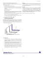
NK cells were isolated from a mouse spleen cell suspension using CD49b
(DX5) MicroBeads, a MiniMACS™ Separator and an MS Column. The
cells are fluorescently stained with CD49b (DX5)-FITC (# 130-091-814).
Cell debris and dead cells were excluded from the analysis based on
scatter signals and PI fluorescence.
CD49b (DX5)+ cells
CD49b (DX5)- cells
Spleen cells before separation
CD49b (DX5)-FITC
4. References
1.
Tomasello, E; Desmoulins, P-O; Chemin, K; Guia, S; Cremer, H; Ortaldo, J; Love,
P; Kaiserlian, D; Vivier, E (2000) Combined Natural Killer Cell and Dendritic
Cell Functional Deficiency in KARAP/DAP12 Loss-of-Function Mutant Mice.
Immunity 13: 335–364. [945]
2.
Shi, FD; Takeda, K; Akira, S; Sarvetnick, N; Ljunggren, HG (2000) IL-18 Directs
Autoreactive T Cells and Promotes Autodestruction in the Central Nervous System
Via Induction of IFN-γ by NK Cells. J. Immunol. 165: 3099–3104. [1021]
3.
Mühlen KA, Schümann J, Wittke F, Stenger S, van Rooijen N, van Kaer L, Tiegs
G (2004) NK cells but not NKT cells, are involved in Pseudomonas aeruginosa
exotoxin a-induced hepatotoxicity in mice. J. Immunol. 172: 3034–3041. [4274]
4.
Hsieh GC, Loukas A, Wahl AM, Bhatia M, Wang Y, et al. (2004) A secreted protein
from the human hookworm necator americanus binds selectively to NK cells and
induces IFN-γ production. J. Immunol. 173: 2699–2704. [4275]
140-000-103.03
This MACS® product is for in vitro research use only
and not for diagnostic or therapeutic procedures.
page 3/3