1
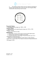
 Cochise Regional Hospital
Hematology and Urinalysis Policies and Procedures
Quality Control
POLICY
It is the policy of Southeast Arizona Medical Center Laboratory to follow all current
federal, state and manufacturer's requirements for quality control for all lab testing.
PROCEDURE
Below is a quality control program for each clinical lab area. Quality Control data in each area
is printed and reviewed at least monthly by the supervisor.
Chemistry (Architect i1000)
1.
Two levels of chemistry control are run each day of use. Standards or
calibrations are run as outlined by each procedure. Assays are calibrated as needed
by the manufacturer's recommendations, each new lot number change, or as needed
per the QC Troubleshooting procedure.
2.
Participation in a JCAHO/CLIA/CAP approved proficiency program.
lmmunochemistry (Architect i1000)
1.
Two levels of chemistry control are run each day of use.
2.
Calibrations are run as outlined by each procedure. Assays are calibrated as
needed by the manufacturer's recommendations, each new lot number
change, or as needed per the QC Troubleshooting procedure.
3.
Participation in a JCAHO/CLIA/CAP approved proficiency program.
Hematology (Ruby Cell Dyn)
1.
Two levels of Ruby Cell Dyn are run each 8 hour shift in use.
2.
If a manu44ifprential is performed it is evaluated by the technologist for
agreement with the Ruby results.
3.
Participation in a JCAHO/CLIA/CAP approved proficiency program.
Coagulation
1.
Two levels of Dade ci-Trol Coagulation controls are run each 8 hour shift in use.
2.
Participation in a JCAHOICLIA/CAP approved proficiency program.
Sedimentation Rates
1.
Two levels of Polymedco Sed Chek Controls are run each day in use.
2.
Participation in Polymedco Quality Assurance Program.
3.
Participation in a JCAHO/CLIA/CAP approved proficiency program.
Reviewed: 11/14
Revised: 11/14
-
"•
Cochise Regional Hospital
Hematology and Urinalysis Policies and Procedures
Urinalysis
1.
2.
Two levels of Kova-Trol are run each day of use.
Participation in a JCAHO/CLIA/CAP approved proficiency program.
Serology
1.
2.
Each kit has manufacturer supplied controls that are run as per
manufacturer's recommendations.
Participation in a JCAHO/CLIAJCAP approved proficiency program.
Blood Bank
1.
2.
Ortho Confidence system is performed each day of use.
Participation in a JCAHO/CLIAJCAP approved proficiency program.
Microbiology
1.
2.
3.
Gram Stains are done on slides with known organisms.
Rapid Strep kits are Qc'd with each lot number change or on the first of
each month. Internal control results are documented on each test
performed.
Participation in a JCAHO/CLIA/CAP approved proficiency program.
Reviewed: 11/14
Revised: 11/14
Quality Control
Troubleshooting Protocol for QC Out of Control
Quality Control programs exist in all laboratory areas. When Quality Control values fall
outside the established limits, the following steps shall be followed:
1.
Do not report patient results.
2.
Review procedures and analytic system for identifiable errors.
3.
Pour or reconstitute fresh control(s). Run both prior control(s) and fresh
control(s).
a.
If fresh control(s) is/are within limits, repeat the patient run and report.
Include a fresh control with the repeat patient run.
b.
If fresh control(s) and repeat control(s) remain unacceptable, make or
change reagent.
4.
Rerun control(s) using new reagent.
a.
If control(s) is/are now within limits, repeat patient run (including a
control) and report results.
b.
If control(s) remain unacceptable, troubleshoot procedure or
instrument in conjunction with the primary operator, hotline
personnel or lab manager.
5.
Discard any control material/reagents which yield unacceptable results.
6.
Do not report any patient results until you have an acceptable control run.
7.
Document all action taken in the work/troubleshooting log. Plot all control values
including those out of range.
Reviewed: 11/14
Revised: 11/14
CALIBRATIONS
Principle
Calibrations provide a standard of analysis using preset determinations in
order to establish continuity of quality control.
Policy
It is the policy of Southeast Arizona Medical Center Laboratory to perform
calibrations on the following instruments:
Ruby Cell Dyn
1. Calibrations are to be run: every 6 months.
2. Calibrations are to be run: when any instrumentation has been replaced.
3. Calibrations are to be run: when drifts and trends in quality control/correlations appear.
CA-500
1. Calibrations are to be run: every 6 months.
2. Calibrations are to be run: with each new lot number of control for DDIMER
3. Calibrations are to be run: with-each-new-lot-number of PT Recombinant Reagent:
4. Calibrations are to be run: when drifts and trends in quality control/correlations appear.
Architect C400
1. Calibrations are to be run: according to manufacturer's instructions.
2. Calibrations are to be run: for Basic Metabolic profile every 8 hours.
3. Calibrations are to be run: for each new lot number of reagent.
4. Calibrations are to be run: for Drugs of Abuse once a week
5. Calibrations are to be run: when drifts or trends appear in quality control/correlations.
Architect i1000
1. Calibrations are to be run: According to manufacturer's instructions.
2. Calibrations are to be run: for each new lot number of reagent.
3. Calibrations are to be run: when drifts or trends appear in quality control.
Reviewed: 11/14
Revised: 11/14
Calibration Verification
Principle
Calibration verifications are performed in order to validate calibrations and quality control.
Policy
It is the policy of Cochise Regional Hospital Laboratory to run calibration verification testing
every six months.
Materials
Chemistry - Maine Standards Validate Kits.
Glucometers - RNA Medical Glucose Calibration Verification Control
Hematology/Streak CVA for cell-Dyn
Procedure
1.
2.
3.
4.
Run linearity kits for each analyte according to manufacturer's instructions.
Fill out appropriate paperwork per kit.
Include instrument printouts and give results to Lab Manager.
Notify Lab Manager of any problems.
Reviewed: 11/14
Revised: 11/14
Operation of the Cell-dyn Ruby Hematology Analyzer
PRINCIPLE
The cell-dyn Ruby is a multi-parameter automated hematology analyzer for in vitro
diagnostic use in counting and characterizing blood cells, with the capability of a five
part leukocyte differential by flow cytometry which uses a laser beam to intersect
cells, light is scatter at different angles that are measure to give the data about cells.
Ruby is capable of running in both modes open and closed; with the purpose of
identify patient results with normal system generating parameters and to flag
patient's data that require additional studies. Ruby also has an automated
reticulocyte mode.
START UP
At the beginning of the day the system shall be on standby, but if the system is idle
for 4 hours it automatically goes into standby, then F12-prime key must be pressed
to run a background count which is used to confirm that the system's baseline
performance meets stated criteria; if results are within acceptable ranges proceed to
run QC.
DAILY QUALITY CONTROL
1- Two controls shall be run every 8 hours.
2- Controls shall be run following a background count (Prime).
OPERATION- CBC
1- From the main screen press F11 to select the mode desirable to be run.
2- If closed mode is to be run load the specimen with the barcode on the rack and
place it on the loader and press F12. Ruby has a cap piercing system that will allow
running closed tubes.
3- If open mode is to be run, input the patient's information in the next open tube
entry either manually or using the hand-held bar code reader.
4- Mix the specimen according to the manufacturer's recommendation.
5- Place the specimen under the aspiration probe, raise the tube until the end
of the probe is deeply immersed in the sample.
6- Press the touch plate behind the probe to start the run cycle, and take
out the specimen while the rinsing probe pushes the specimen down.
7- Results are displayed on the run view screen and printed automatically if auto
print is on.
Reviewed: 11/14
Revised: 11/14
QUALITY CONTROL OUT OF CONTROL
Department: ___________________ Month/Year
Date
Reviewed: 11/14
Revised: 11/14
Problem
Rey.By/Date: ____________________
Corrective Action
Tech
Correlation of Back Up Instrumentation with Primary Systems
Principle
Correlation of patient samples between analyzers is performed to check the
accuracy and precision of each analyzer.
Policy
It is the policy of Southeast Arizona Medical Center Laboratory to run
correlations between each primary analyzer and its corresponding back-up
system twice a year. The following is a list of those analyzers:
Primary
HMX
ACL 8000
DXC600
Back-Up
ACT-5
ACL 1000
CX3
Polyme deo Sedimat 15
Excyte 10
Procedure
1.
2.
3.
4.
5.
6.
7.
Choose 5-7 patient samples covering the ranges of low, normal and high.
Run samples on HMX, ACL 8000 and DXC600,
Run samples on ACT-5, ACL 1000 and CX3.
Compare results for any deviations. Document any troubleshooting action.
Notify Lab Manager of any problems.
Results should be given to Lab Manager for evaluation.
LV % for each analyte should be 90% acceptance.
Reviewed: 11/14
Revised: 11/14
Criteria for Manual Blood Smear Review
A manual differential should be performed if any of the following conditions exist: and remits
entered into the opos system using the test code manditt, results are also recorded in the
instrument result form.
Leukocyte count is
less than 2,500/cmm
greater than 15, 000/cmm
Granulocyte % is greater than 85%
Monocyte % is greater than 12%
Eosinophil % is greater than 15%
Reverse Differential (lymphs>gans) except pediatric patients
Vote out on any cell line
Review Ruby Cell-Dyn flag messages and perform a manual slide for morphology if any of the
Following conditions exist and remits entered into the Opos system using the last code
mandiff.
Abnormal RBC distribution
RDW greater than 16
MCH less than 27
MCV greater than 100
MCV less than 75
Platelet Count <50,000 or > 600,000
Abnormal platelet distribution
Flag for nucleated RBC
Reviewed: 11/14
Revised: 11/14
Manual Blood Smear
Policy
It is the policy of Cochise Regional Hospital Laboratory to reflex any manual blood smear that exceeds the
criteria for manual blood smear review. According to Medicare reimbursement clinical reflexing will not be
reimbursed without an order from a doctor. It will be the policy of this laboratory to notify the doctor that a
blood smear is indicated. Lab results will then be entered upon receipt of the order for blood smear.
Results will also be recorded on the instrument printout.
Principle
The examination of the blood smear is an important part of the hematologic evaluation.
Procedure
Preparation of smear:
I. Place a drop of blood 2 to 3 mm in diameter about 1 cm from the end of a clean dust free
slide. Place the slide on a flat surface.
2. With the thumb and forefinger of the right hand hold the end of a second (spreader)
slide against the surface of the first at an angle of 30 to 45 degrees and draw it back
against the drop of blood until contact is established. Allow the blood to run completely
accross the end filling the angle between the two slides.
3. Push the "spreader" slide at a moderate speed forward so that the blood spreads evenly
on the other side behind the edge of the spread keeping contact between the two until all
the blood has been spread into a moderately thin film.
4. Air dry the smear.
Staining the smear:
1. Dip slide in stain for 10 seconds.
2. Dip slide in distilled water (ph 6-7) for 20 seconds or more for darker staining.
3. Air dry.
Performance of Differential
Reviewed: 11/14
Revised: 11/14
I.
Examine slide under low power. Observe distribution of white cells and choose the area
near the thin end where red cells touch but do not overlap.
2. Shift to oil immersion objective and classify each WBC until 100 cells have been counted.
Note presence of nucleated RBC's and correct WBC count if necessary.
Corrected WBC count = WBC x 100
100 + # of Nrbc's
3. Note RBC morphology, estimate of platelet count (adequate, decreased,
increased, etc.). When the platelet count is normal 8-20 platelets are noted per
high power field.
Procedural Notes and Limitations
A good differential depends on a good slide and proper staining technique. Slides should
not be counted if:
•
•
•
there are ridges, lines or holes
poor margination
bluish cast after staining
All abnormalities should be brought to the attention of a supervisor or should be
repeated for Pathologist review.
Result Reporting
In order to result a manual differential the technologist must contact the primary or attending
physician of the need for the review. Upon receipt of the order the results may be entered into
the-114MS -WsreaP S using the test code. While reflexing the blood smear will still be policy
reporting the results will not occur until the order has been received. Manual differentials should
not be made on blood tubes older than 24 hours old.
References
1. Clinical Diagnosis and Management by Laboratory Methods, 16th edition, Todd,
Sanford and Davidson.
2. Wright-Geimsa Stain label.
Reviewed: 11/14
Revised: 11/14
Criteria for Referral of Blood Smear to Pathologist
1
Evidence of hemoglobinopathy.
2.
Sickle cells or hemoglobin C crystals.
3.
Evidence of metabolic disease, systemic disease or deficiency
state.
a.
Teardrops
b.
Howell Jolly bodies (unless patient has had splenectomy)
c.
Cabot rings
d.
Greater than moderate burr cells
4.
Differential count abnormalities
a.
Atypical lymphocytes->20%
b.
Metamyelocytes and myelocytes->10%
c.
Lymphocytes>60 if >10 years; >80 if < 10 years
d.
Monocytes->20%
e.
Eosinophil->15%
f.
Basophil->8%
g.
Promyelocyte and blasts- any
5.
Plasma cells in peripheral blood.
6.
Any unidentified cells.
7.
Any physician request for pathologist evaluation.
Send 2-3 unstained slides with a copy of the CBC report to the
pathologist.
Reviewed: 11/14
Revised: 11/14
LABORATORY
Title: Autozero Westergren Erythrocyte Sedimentation Rate (ESR) by Sediplast®
Date: August 20, 2013
Reviewed by _____________________________________ Date: _____________
_____________________________________________________________________
Principle:
A pipette (tube) is filled with 1.0 mL of anticoagulated (EDTA) blood and allowed
to stand for a period of one hour. After one hour, a reading is taken at the plasma
meniscus. This reading represents the distance in mm from the zero mark on the
tube and is equal to the Erythrocyte Sedimentation Rate.
Materials
1. Fixed bore pipettes, with self-zeroing cap.
2. Vials
3. Timer
Test Procedure
1. Assemble and check for integrity of supplies. Remove the pink stopper on
the prefilled vial (0.2 ml of 3.8% Sodium Citrate is used as diluent).Using a
transfer pipette, fill the vial to the bottom of the indicated fill line, with 0.8
ml of blood to make the required dilution ratio.
2. Replace pink pierceable stopper and gently invert several times to obtain
homogeneous mixing.
3. Place vial in its rack on a level counter surface free from vibration.
4. The diaphragm of the pink stopper is calibrated to break under the light
pressure made by inserting the pipette.
5. Carefully insert the pipette through the pierceable stopper and gently push
downwards until the pipette comes into contact with the blood sample.
Next, while holding the middle of the pipette, gently twist the pipette and
push downwards until the pipette rests on the bottom of the vial.
6. The pipette will autozero the blood and any reasonable excess will flow
into the reservoir compartment.
7. To ensure proper results, it si essential that the pipette makes firm contact
with the bottom of the vial.
Reviewed: 11/14
Revised: 11/14
8. Let sample stand for exactly one hour and then read the numerical results
of the erythrocyte sedimentation rate in millimeters.
9. Dispose of properly after use.
Reviewed: 11/14
Revised: 11/14
Interpretation of Results
MALE: (under 50) 0-15 mm/hr -(over 50) 0-20 mm/hr
FEMALE: (under 50) 0-20 mm/hr —(over 50) 0- 30 mm/hr
Quality Control
Normal and Abnormal reacting quality control material is available in the
laboratory. Controls are tested once each day of use and patient results are not
reported until quality control results are within published acceptable limits.
References Polymedco Sediplast® user manual. Cortland Manor, New York.
Reviewed: 11/14
Revised: 11/14
Cell Count
Principle
Normal CSF is crystal clear with viscosity comparable to water. There are many conditions which
cause the CSF to change in turbidity and viscosity. Cell counts are performed to provide the
physician with the necessary information to diagnose these conditions.
Reagents and Equipment
Hemocytometer
Pasteur pipette
saline
Procedure
1st tube – Chemistry
2nd tube – Hematology
3rd tube – Microbiology
4h tube – Serology/Cytology
1.
2.
3.
4.
Mix the CSF in tube 3 well.
Fill both sides of the hemocytometer with undiluted CSF.
Allow the hemocytometer to settle for 10 minutes.
Count all red and white blood cells in the 9 large squares on each side of the chambers.
Procedural Notes and Limitations
If there are too many cells present to count dilute the CSF 1:1 with saline before filling chamber.
(Additional serial dilutions may be needed). If greater than 5 WBC/cu mm are seen a
differential must be done.
Calculations
number of cells counted x dilution = cells/cu mm
.9
OR
number of cells counted x dilution x depth (10) = cells/cu mm
number of squares counted (9)
Reviewed: 11/14
Revised: 11/14
Expected Values
Adults — 0-5 cells/cu mm
Neonates — 0- 20 cells/cu mm
Result Reporting
In the HMS system the technologist will enter the results for both white and red blood cells
using the test code CSFCELLS.
References
Clinical Diagnosis and Management by Laboratory Methods 17th Edition, p. 463-464Spinal
Fluid Procedure - http://www.pathguy.com/lectures/csf.htm.
Reviewed: 11/14
Revised: 11/14
Operation of the SYSMEX CA-500 Coagulation Analyzer.
The Sysmex CA-500 is an automated Blood Coagulation analyzer fully
automated capable of 5-parameter random analysis for in vitro analysis. This
instrument has the capability to analyze 10 regular samples, and also one Stat
giving us the opportunity of running everything at the same time. Moreover, it
allows analyzed data to be display and printed out together with reaction curves,
thus making it possible to obtain highly reliable results.
MAINTENENCE
Daily : Daily maintenance must be started at the beginning of every day, before
quality control and patient analysis. It must be done on the following order:
1- Turn off and on the Sysmex CA-500.
2- Empty tube trash box located at the right side. Also empty the waste container.
3- Replenish water container with Deionized water.
4- Check all supplies and refill everything with fresh reagents; CA-CLEAN
must be changed every 24 hrs., Buffer every 8 hour shift.
5- Perform probe rinse. If visible residue, wipe probe with 70% alcohol.
6- Wipe condensation from reagent cooler.
7- Check the trap chamber and drain, if needed.
8- Check temperatures.
Monthly Maintenance:
1- Replace reagent Bottles.
Quarterly Maintenance:
1- Clean diH2O Rinse bottle with alcohol.
2- Perform LED calibration.
Yearly Maintenance:
1- Replace Rinse Filter/Rinse and prepare
As needed Maintenance:
1234-
Replace fuse
Perform "Rinse and Prepare"
Perform maintenance rinse.
Clean instrument surface.
Reviewed: 11/14
Revised: 11/14
DAILY QUALITY CONTROLS
Two levels of ci-Trol Should be run every 8 hours shift for PT and aPTT tests.
Two levels of Innovance D-dimer Should be run every 8 hours when
there are specimens to be run.
OPERATION OF SYSMEX CA-500
1- Go to the main menu.
2- Place sample in the rack with the barcode facing outward if the specimen
does not have barcode input the specimen id by pressing the ID No. Entry icon
which gives the numeric keyboard, then select the test desire to be run and
press START, then press continue to start running the specimen.
3- When the analyzer finishes the run automatically the result is printed.
Reviewed: 11/14
Revised: 11/14
Urinalysis Quality Control
A.
General
KOVA-trol Normal and Abnormal urine controls should be used
each day for the urine dipsticks. Controls need to be run for
confirmatory tests when the tests are performed. Expected values
are posted in the laboratory.
B.
Out of Control QC
1.
Check expiration date of controls and reagents.
2.
Repeat test with new control.
3.
If still not expected results, repeat with new dipstick or reagent.
4.
Record corrective action on worksheet.
Reviewed: 11/14
Revised: 11/14
Clintek Status
Principle:
Clinitek status Urine chemistry analyzer is a portable instrument for reading traditional Bayer
Reagent Strips for Urinalysis. Refer to the bottle label or carton for the tests that are
included with each product. The analyzer can also report the color of the urine sample. No
special training is needed to use the instrument.
Materials :
1. Multistix Pro
2. Paper Towel (for blotting) .
Specimen :
Random Urine
Quality control :
Possitive and Negative control solutions should be tested on a regular basis . This
provides a check to ensure that the test strips are reacting properly and the instrument is
reading strips properly. Testing controls also helps detect errors caused by incorrect user
techinique.Prepare the control solutions as instructed in the package insert that comes
with control product. Then test the control solution using the same procedure as you use
when testing patient urines.
Record the controls results . If any results are not within the expected range, notify the lab
supervisor or physician. Refer to the bottle label and / or package insert for storage
information and expiration date.
You should test controls:
1. At the start of the day:
2. When you open a new bottle of test strips.
3. Whenever test results are in doubt:
4. When training instrument operators.
Procedure :
Routine Urines . The urine specimen should be fresh , well- mixed, and uncentrifuged. If the
urine depth is less than about 3 inches pour the specimen into a narrow tube,such as a
URIN-TEK Specimen tube .
1. DIP A BAYER REAGENT STRIP into the urine . Be sure ALL the test pads are wet
2. Immediately Remove the reagent strip from the urine, dragging the edge of the strip
against the side of the container as you remove the strip, At The SameTime , press either
green (Start) key . You now have 10 seconds to complete Steps 3 and 4 .
3. Blot the strip to remove excess urine by Touching the edge to a paper towel. Do not
drag the strip across the towel : touch the edge only .
4. Place the Reagent Strip . With the test pads facing up, into the middle trough of the
test strip table until it touches the end of the trough.
5. The table is automatically pulled into the instrument for reading . Results are available in
one minute. Be sure not to move or bump the table .
Reviewed: 11/14
Revised: 11/14
Complete Urinalysis
Principle:
The examination of urine is performed in three steps. The first step involves
evaluation of the physiochemical characteristics of the urine sample. The second step
involves the chemical examination of the urine specimen and the third step is the microscopic
examination of the urine sediment by trained laboratory personnel.
Materials:
1. Specimen collection device as supplied in the laboratory.
2. Urine centrifuge tubes as supplied in the laboratory.
3. Bayer 10 SG Urine Dipsticks as supplied in the laboratory.
4. Microscope as supplied in the laboratory.
5. Slides and cover slips as supplied in the laboratory.
6. Centrifuge as supplied in the laboratory.
Specimen:
The specimen of choice for this procedure is a fresh (mid-stream or clean
catch) specimen, early morning samples are recommended, collected into a clean, dry
container. Samples may be stored for 1 hour before testing at room temperature then must
be refrigerated at 2-8° C for up to 24 hours.
Quality Control:
Quality control testing for this procedure is outlined in the "UA dipstick"
procedure for biochemical dipstick testing found elsewhere in this manual. Proficiency
testing supplied to prove competency for identification of formed elements found in a urine
specimen are supplied on a quarterly basis.
Remedial Action:
In the event that a member of the laboratory staff cannot prove his
competency with proficiency testing, that staff member will take part in an educational in
service meant to increase his/her skills in identifying the formed elements of the urine
specimen.
Procedure:
1. Obtain a properly labeled urine specimen.
2. Pour off 12 ml of urine into a separate centrifuge tube.
3. Perform a dipstick examination as described in the "UA Dipstick" procedure found
elsewhere in this manual.
4. If blood, leukocyte esterase, protein (greater than a trace) or nitrite are positive, a
microscopic examination is required. If the listed tests are negative, even though
other tests are positive, a microscopic examination need not be performed or
performed upon physician request.
5. Centrifuge the 12 ml sample of urine for two (2) minutes in the urine centrifuge.
6. Decant all but 1 ml of the sample.
7. Mix the 1 ml sample well and place one (1) drop of the concentrated urine onto a
clean dry slide. Cover with a cover slip as supplied in the laboratory.
8. Perform a microscopic examination of the urine sample.
Reviewed: 11/14
Revised: 11/14
Interpretation of Results:
The slide is first examined under low power magnification to locate casts and
elements that are present in only a few fields. Casts tend to congregate near the edges of
the cover slip. After the entire slide has been scanned, a further examination is made
under high power magnification in order to identify specific types of cells, crystals,
elements and objects present in the urine and to delineate the various types of casts. A
minimum of 10 to 15 high power fields should be scanned for this examination. Good
technique requires varying the intensity of the light source of the microscope in order to
correctly identify the various components of the sediment. Some elements are more
easily recognized in subdued light, others in brighter light.
Red blood cells, leukocytes and epithelial cells are conventionally reported in terms of
cells per high power field; casts are counted per low power field. For each determination, the
number of elements seen in at least 10 fields should be counted and the average number
used for the reported value. Other elements, such as bacteria, parasites, crystals and
spermatozoa are usually reported as well.
In this laboratory, results are reported as one of the following for each category:
Appearance:
Color
Formed Elements
CLEAR
STRAW
TRACE
SLT HAZY
LT YELLOW
1+
HAZY
YELLOW
2+
TURBID
DK YELLOW
3+
CLOUDY
ORANGE
4+
RED
RARE
BROWN
FEW
2-4
5-10
10-20
20-50
>50
NEGATIVE
POSITIVE
EXPECTED RESULTS:
• Red Blood Cells: The finding of more than one or two red blood cells per
high power field is an abnormal condition. It can indicate a variety of renal
and systemic diseases, including trauma to the kidney. It may also be found
following violent exercise. It may follow traumatic catheterization, the
passage of stones or contamination from menstrual blood. Hematuria
occurs with pyelonephritis, tuberculosis of the genitourinary tract, cystitis,
prostatitis, renal calculi, renal tumors and other malignancies of the urinary
tract, and hemorrhagic diseases such hemophila, etc. Red blood cells will
tend to lyse or dissolve if allowed to stand in urine which is alkaline or dilute.
• White Blood Cells: The presence of large numbers of white blood cells
(leukocytes) usually indicates bacterial infection in the urinary tract.
Reviewed: 11/14
Revised: 11/14
•
Pyuria may also be seen in acute glomerulonephritis. The cells are
segmented neutrophils or "polys". Large amounts of mononuclear cells
"Lymphs", in a patient with a kidney transplant may indicate early tissue
rejection phenomena. Epithelial Cells: Large numbers of renal epithelial
cells may indicate active tubular degeneration. These cells are noted in
the urine of patients with acute tubular necrosis and necrotizing papillitis.
Squamous epithelial cells appear frequently in normal urine.
Crystals: The type and quantity of crystalline precipitate vary with the pH of the
urine. Amorphous material is of little importance. Crystals in normal urine are
formed as the specimen cool. Crystals of abnormal urine cystine, leucine and
tyrosine and cholesterin. The following table lists some crystals found in urine
sediment and the physical-chemical characteristics associated with them.
Reviewed: 11/14
Revised: 11/14
ACID URINE
INORGANIC SEDIMENT:
SOLUBILITY
NAME
COLOR
--ALK ACID ALCOHOL "C OTHER
SHAPE
Amorphous Brick-Red
urates
80°C =—
Granules
Acetic
Acid
Uric Acid
Yellow-Brown Polymorphous—whetstones,
rosettes of prisms, rhombohedral prisms, hexagonal
Sodium
Colorless to
Yellow
plate
urate
Cystine
(rare)
Fan of slender prisms
+
80°C
−
1) Colorless
Flat hexagonal plates with
2) Highly retractile
well-defined edges
Singly or in clusters
+
+
-
-
Cholesterol 1) Colorless
(rare)
"Broken window panes" with
notched corners
Leucine
1) Yellow or
Brown
2) Highly retractile
Spheroids with striations
Pure form hexagonal
Tyrosine
(rare)
Colorless or
Yellow
Fine silky needles in sheaves
or rosettes
+
+
Bilirubin
Brown
Reddish
Cubes
Rhombic plates
Amorphous needles
+
+
Calcium
Colorless
Ether
Chloroform
-
Flat plates
(rare)
oxalate
Hippuric
4-
Slightly
In hot
H2 O
-
−
Acetone
80°C Chloroform
ACID, NEUTRAL, OR SLIGHTLY ALKALINE URINE
Octahedral
Dumbells
- +
Often small—use h.p.
Dilute
HCI
- -
Colorless
Hot I-1,0
acid
ALKALINE, NEUTRAL, OR SLIGHTLY ACID URINE
Triple
phosphate
Colorless
"Coffin lids"
3-6 sided prism
__,....
Dilute
-
Acetic
Acid
-
Occ. fern-leaf
ALKALINE URINE
Calcium
carbonate
Colorless
Needles
- - - - Acetic
Spheres
Dumbells
Ammonium Yellow
Writes
Opaque
Brown
Acid
"Thorn apple" spheres
Rhombic plates
Four sided prisms
60°C
Dumbells
with
Sheaves of needles
Acid
Calcium
phosphate
Colorless
Prisms
Plates
Needles
Reviewed: 11/14
Revised: 11/14
Dilute
- - - - Acetic
Acid
Acetic
• Bacteria: Normal urine contains no bacteria. If proper and careful technique was
used to obtain the sample and if the specimen was protected from contaminants
before examination, the presence of bacteria in significant numbers may indicate
urinary tract infection. The presence of leukocytes helps to differentiate between
"contamination" and a "true Infection".
• Yeast and Parasites: Yeast cells (Candida albicans) may be indicative of urinary
moniliasis, especially in patients with diabetes mellitus. Frequently, yeast appears
as a contaminant in the urine of female patients with vaginal moniliasis. The
majority of parasites observed in urine are contaminants from fecal or vaginal
material. A urinary tract parasite infestation may be associated with the presence
of red blood cells, as in Schistosoma haematobium.
• Spermatazioa: Spermatazoa are frequently seen in urine following nocturnal
emissions or sexual intercourse.
• Casts: Cast formation occurs usually in the distal convoluted tubule of the nephron.
Casts may also occur in the ascending loop of Henle or the collecting duct.
Requirements for cast formation are an acid condition, high salt concentration,
reduced urine flow and protein. Casts are named according to the matrix of the
inclusions contained in them, e.g., red blood cell cast, white blood cell cast, etc.
• Red Blood Cell Casts indicate the presence of an acute inflammatory or
vascula disorder in the glomerulus causing renal hematuria. They should
always be regarded as pathological and may be the only manifestation of
acute glomerulonephritis, renal infarction, collagen disease, or kidney
involvement in subacute bacterial endocarditis.
• White Blood Cell Casts may be found in the urine from patients with acute
glomerulonephritis, nephrotic syndrome or pyelonephritis. Since
pyelonephritis may remain completely asymptomatic even though it is
progressively destroying renal tissue, careful examination of the urinary
sediment for leukocyte casts is important. In some cases, it may be the only
significant laboratory finding in an asymptomatic situation.
• Epithelial Cell Casts are formed by fused desquamated tubular cells. Since
the tubule is a living membrane, it is always replacing itself. Thus, the finding
of an occasional renal epithelial cell or clump is not remarkable. However, in
any disease producing damage to tubular epithelium, the appearance of
many casts may indicate excessive desquamation such as may occur in
nephrosis, eclampsia, amyloidosis and in the presence of poisoning with
heavy metals and a variety of other toxins.
• Hyaline Casts are formed of the gel of Tamm-Horsfall protein. The presence of
hyaline casts imply damage to the glomerular capillary membrane, permitting
leakage of proteins through the glomerular filter. Such damage may be
permanent or transient as a result of fever or the effects of posture (orthostatic,
iordotic), emotional stress or strenuous exercise.
• Granular Casts: The terms "coarsely granular" and "finely granular" are
merely descriptive, indicating the degree of degeneration that has occurred in
the cellular inclusion-the cells having been broken down into coarse or finer
particles. While an occasional granular cast may be found in normal
individuals, their presence beyond "occasional cast" may indicate
pyelonephritis. Granular casts are also found in chronic lead intoxication.
• Waxy and Fatty Casts are associated with tubular inflammation and
degeneration. The broad, waxy cast is formed in the collecting tubules when the
urine flow them is reduced. Both waxy and fatty casts are found in chronic renal
disease.
Reviewed: 11/14
Revised: 11/14
Reporting of Patient Results:
Patient results are entered into the LIS using the above table for reporting
of results.
Inoperable Test System:
In the event that complete urinalysis testing cannot be performed, samples be sent to the
Copper Queen Hospital.
Clinical Significance:
The benefit of performing complete urinalysis examination is well known as
a means to assess kidney function.
References:
Modern Urine Chemistry. 1982, Ames Company, Elkhardt, Indiana.
Reviewed: 11/14
Revised: 11/14
Urinalysis (MuliStiz 10 SG)
Principle:
The MultiStix SG Reagent strips for Urinalysis are firm plastic strips to which
are affixed several separate reagent areas. The reagent test areas on the Reagent Strips
are ready to use upon removal from the bottle and the entire reagent strip is disposable.
The following are chemical principles of the reagent ares.
1. Glucose: This test is based on a double sequential enzyme reaction. One
enzyme, glucose oxidase, catalyzes the formation of gluconic acid and
hydrogen peroxide from the oxidation of glucose. A second enzyme,
peroxidase, catalyzes the reaction of hydrogen peroxide with a potassium
iodide chromogen to oxidize the chromogen to colors ranging from green to
brown
2. Bilirubin: This test is based on the coupling of bilirubin with diazotized
dichloroaniline in a strongly acid medium. The color ranges through various
shades of tan.
3. Ketone: This test is based on the development of colors ranging from buff-pink,
for a negative reading, to purple when aceroacetic acid reacts with
nitroprusside.
4. Specific Gravity: This test is based on the apparent pKa change of certain
pretreated polyelectrolytes in relation to ionic concentration. In the presence of
an indicator, colors range from deep blue-green in urine of low ionic
concentration through green and yellow-green in urines of increasing ionic
concentration.
5. Blood: this test is based on the peroxidase-like activity of hemoglobin, which
catalyzes the reaction of diisopropylbenzene dihydroperoxide and 3,3",5,5"tetramethylbenzidine. The resulting color ranges from orange through green;
very high levels of blood may cause color development to continue to blue.
6. pH: The test is based on the double indicator principle that gives a broad range of
colors covering the entire urinary pH range. Colors range from orange through
yellow and green to blue.
7. Protein: This test is based on the protein-error-of -indicators principle. At a
constant pH, the development of any green color is due to the presence of
protein. Colors range from yellow for "Negative" through yellow-green and
green to green-blue for "Positive" reactions.
8. Urobilinogen: This test is based on a modified Ehrlich reaction, in which pdiethylaminobenzaldehyde in conjunction with a color enhancer reacts with
urobilinogen in a strongly acid medium to produce a pink-red color.
9. Nitrite: This test depends upon the conversion of nitrate (derived from the diet)
to nitrite by the action of Gram negative bacteria in the urine. At the acid pH of
the reagent area, nitrite in the urine reacts with p-arsanilic acid to form a
diazonium compound. This diazonium compound in turns couples with 1,2,3,4tetrahydrobenz(h)quinolin-3-ol to produce a pink color.
10.Leukocytes: Granulocytic leukocytes contain esterases that catalyze the
hydrolysis of the derivatized pyrrole amino acid ester to liberate 3-hydroxy-5phenyl pyrrole. This pyrrole then reacts with a diazonium salt to produce a
purple product.
Reviewed: 11/14
Revised: 11/14
Materials:
1. MultiStix 10 SG as supplied in the laboratory. Product is stable until the
expiration date printed on the container label when stored at room temperature
between 15-30° C. Do not store the container in direct sunlight. All unused
strips must remain in the original container after opening. Do not remove the
desiccant(s) from the container. Do not remove the strip from the container until
immediately before it is to be used for testing. Replace the cap immediately and
tightly after removing the reagent strip. Do not touch the test areas of the
reagent strip. Work areas and specimen containers should be free of detergents
and other contaminating substances.
2. Specimen collection device as supplied in the laboratory.
3. Cleansing wipes as supplied in the laboratory for performing a "Clean Catch"
collection.
Specimen:
The specimen of choice for this procedure is urine collected in a clean
container and tested as soon as possible. Do not centrifuge. The use of urine
preservatives is not recommended. If testing cannot be performed within an hour after
voiding, the specimen may be refrigerated for up to 24 hours before testing. Allow the
specimen to warm to room temperature if refrigerated.
Nitrite test results are optimized by using a first morning specimen or one
that has incubated in the bladder for four (4) hours or more. It is especially important to use
fresh urine to obtain optimal results with tests for bilirubin or urobilinogen, as these
compounds are very unstable when exposed to room temperature and light.
Prolonged exposure of urine to room temperature may result in microbial
proliferation with resultant changes in pH. A shift to alkaline pH may cause false positive
results with the protein with the protein test area. Urine containing glucose may decrease
in pH as organisms metabolize the glucose. Bacterial growth from contaminating organism
may cause false positive blood reactions from the peroxidases produced. In random urine
urine specimens from females, a positive result for leukocytes may be due to a source
external to the urinary tract.
Contamination of the urine specimen with skin cleansers containing
chlorhexidine may affect protein (and to a lesser extent specific gravity and bilirubin) test
results. The user should determine whether the use pf such skin cleansers is warranted.
Quality Control:
Known positive and negative specimens are supplied in the laboratory for
quality control testing. Known positive and negative specimens are tested once each day of
use in the same manner as patient samples are tested. Water should not be used as a
negative control. Patient results are never reported until acceptable results are obtained form
quality control testing.
Remedial Action:
In the event that quality control testing does not produce acceptable results,
the following procedure may be used to correct any deficiencies.
1. Repeat the testing being careful to follow incubation times exactly.
2. Reconstitute new control material and repeat testing.
3. Obtain a new vial of test strips and repeat testing.
4. Contact the Laboratory Manager.
Reviewed: 11/14
Revised: 11/14
Procedure:
1. Collect a fresh urine specimen in a clean, dry container. Mix well immediately
before testing.
2. Remove one strip from the container and replace cap.
3. Completely immerse the reagent areas of the strip in the urine sample and remove
immediately to avoid dissolving out reagents.
Note: An alternate method of applying the sample is to use a clean disposable
pipet to dispense the urine specimen onto the reagent pads, one drop to a pad.
4. While removing the strip from the specimen, run the edge of the entire length of
the strip against the rim of the urine container to remove excess urine.
5. Hold the strip in a horizontal position to prevent possible mixing of chemicals from
adjacent reagent areas and/or contaminating the hands with urine and touch one
side to a clean paper towel to remove excess urine.
Note: If the sample has been applied with a pipet, turn the strip on one side against a
clean paper towel to remove excess urine.
6. Compare the reagent areas to the corresponding Color Chart on the container at the
times specified. Hold the strip close to the color blocks and match carefully. Avoid
laying the strip directly on the Color Chart, as this will result in the urine soiling the
chart.
Proper read time is critical for optimal results. Read the glucose and
bilirubin tests at 30 seconds after dipping. Read the ketone test at 40 seconds; the
specific gravity test at 45 seconds; pH, protein, urobilinogen, blood and nitrite at 60
seconds; and leukocytes at 2 minutes. The pH and protein areas may also be read
immediately or at any time up to 2 minutes after dipping.
After dipping the strip, check the pH area. If the color on the pad is not
uniform, read the reagent area immediately, comparing the darkest color to the
appropriate Color Chart. All reagent areas except leukocyte may be read between 1 and
2 minutes for identifying negative specimens and for determination of the pH and SG. A
positive reaction (Small or greater) at less than 2 minutes on the leukocyte test may be
regarded as a positive indication of leukocytes in urine. Color changes that occur after 2
minutes are of no diagnostic value.
Interpretation of Results:
Results with Bayer Reagent strips are obtained in clinically meaningful
units directly from the Color Chart comparison.
Limitations:
1. As with all laboratory tests, definitive diagnostic or therapeutic decisions should not
be based on any single result or method.
2. Substances that cause abnormal urine color, such as drugs containing azo dyes
(e.g., Pyridium, Azo Gantrisin, Azo Gantanol), nitrofurantoin (Macrodantin,
Furadantin), and riboflavin, may affect the readability of the reagent areas on
urinalysis reagent strips. The color development on the reagent pad may be
masked, or a color reaction may be produced on the pad that could be interpreted
visually as a false positive.
3. Glucose: Ascorbic acid concentrations of 50 mg/dL or greater may cause false
negatives for specimens containing small amounts of glucose (75-125 mg/dL).
Ketone bodies reduce the sensitivity of the test; moderately high ketone levels
(40 mg/dL) may cause false negatives for specimens containing small amounts
of glucose (75-125 mg/dL) but the combination of such ketone levels and low
glucose levels is metabolically improbable in screening. The reactivity of the
glucose test decreases as the SG of the urine increases. Reactivity may also
vary with temperature. This test is specific for glucose; no substance excreted in
urine other than glucose is known to give a positive test. The reagent area does
not react with lactose, galactose, fructose nor reducing metabolites of drugs
(e.g., salicylates and nalidixic acid).
Reviewed: 11/14
Revised: 11/14
4. Bilirubin: Indican (indoxyl sulfate) can produce a yellow-orange to red color
response that may interfere with the interpretation of a negative or a positive
bilirubin reading. Metabolites of Lodine (etodolac) may cause false positive or
atypical results; ascorbic acid concentrations of 25 mg/dL or greater may cause
false negatives. Since very small amounts of bilirubin may be found in the
earliest stages of liver disease, the user must consider whether the sensitivity of
the reagent strip to bilirubin is sufficient for the intended use. When very small
amounts of bilirubin in urine are sought (e.g., earliest phase of hepatitis),
ICTOTEST reagent tablets should be the method of choice
5. Ketone: False positive results (Trace or less) may occur with highly pigmented
urine specimens or those containing large amounts of levodopa metabolites.
Compounds such as mesna (2-mercaptoethane sulfonic acid) that contain
sulfhydryl groups may cause false positive results or an atypical color reaction.
This test reacts with acetoacetic acid in urine, it does not react with acetone or 8hydroxbutyric acid.
6. Specific Gravity: The chemical nature of the reagent test strips may cause
slightly different results from those obtained with other specific gravity methods
when elevated amounts of certain urine constituents are present. Highly
buffered alkaline urines may cause low readings relative to other methods.
Elevated specific gravity readings may be obtained in the presence of moderate
quantities of protein (100-750 mg/dL).
7. Blood: Elevated specific gravity may reduce the reactivity of the blood test.
Capoten may also cause decreased reactivity. Certain oxidizing contaminants,
such as hypochlorite, may produce false positive results. Microbial peroxidase
associated with urinary tract infections may cause false positive reactions. Levels
of ascorbic acid normally found in urine do not interfere with this test.
8. pH: If proper procedure is not followed and excess urine remains on the strip, a
phenomenon known as "runover" may occur, in which the acid buffer from the
protein reagent will run onto the pH area, causing a false lowering of the pH
result.
9. Protein: False positive results may be obtained with highly buffered or alkaline
urines. Contamination or the urine specimen with quaternary ammonium
compounds (e.g., from some antiseptics and detergents) or with skin cleansers
containing chlorhexidine may also produce false positive results. The reagent
area is more sensitive to albumin than to globulins, hemoglobin, Bence-Jones
proteins and mucoprotein; a negative result does not rule out these other
proteins.
10.Urobilinogen: The reagent area may react with interfering substances known to
react with Ehrlich's reagent, such as p-amino-salicylic acid and sulfonamides.
Atypical color reactions may be obtained in the presence of high concentrations
of p-amino-benzoic acid. Strip reactivity increases with temperature; the
optimum temperature is 22-26° C. The test is not a reliable method for the
detection of porphobilinogen. The absence of urobilinogen cannot be
determined with this test.
11.Nitrite: Pink spots or pink edges should not be interpreted as a positive result.
Any degree of uniform pink color development should be interpreted as a
positive result suggesting the presence of 100,000 or more organisms per mL,
but color development is not proportional to the number of organisms present. A
negative result does not in itself prove that there is no significant bacteiuria.
Negative results may occur when urinary tract infections are caused by
organisms that do not contain reductase to convert nitrate to nitrite; when urine
has not been retained in the bladder long enough (four hours or more) for
reduction of nitrate to nitrite to occur; or when dietary nitrate is absent, even if
organisms containing reductase are present and bladder incubation is ample.
Sensitivity of the nitrite test is reduced for urines with high specific gravity.
Ascorbic acid concentrations of 25 mg/dL or greater may cause false negative
results with specimens containing small amounts of nitrite ion (0.06 mg/dL or
less).
Reviewed: 11/14
Revised: 11/14
12.Leukocytes: Elevated glucose concentrations (> 3 g/dL) or high specific gravity
may cause decreased test results. The presence of cephalexin (Keflex,
cephalothin (Keflin), or high concentrations of oxalic acid may also cause
decreased test results. Tetracycline may cause decreased reactivity, and high
levels of the drug may cause false negative reaction.
Expected Values:
Expected values for the typical "normal" healthy population and the
abnormal population are listed below for each reagent.
1. Glucose: Small amounts of glucose are normally excreted by the kidney. These
amounts are usually below the sensitivity of this test. Results at the first positive
level may be significantly abnormal if found consistently.
2. Bilirubin: Normally no bilirubin is detectable in urine by even the most sensitive
of methods. Even trace amounts of bilirubin are sufficiently abnormal to require
further investigation. Atypical colors (colors unlike the negative or positive color
blocks shown on the Color Chart) may indicate that bilirubin-derived bile
pigments are present in the urine sample and may be masking the bilirubin
reaction. These colors may indicate bile pigment abnormalities and the urine
specimen must be tested further with a confirmative test such as the
ICTOTEST.
3. Ketone: Normal urine specimens ordinarily yield negative results with this
reagent. Detectable levels of ketone may occur in urine during physiological
stress conditions such as fasting, pregnancy and frequent strenuous exercise. In
ketoacidosis, starvation or with other abnormalities of carbohydrate or lipid
metabolism, ketones may appear in urine in large amounts before serum ketone
concentrations are elevated.
4. Specific Gravity: Random urines may vary in specific gravity from 1.001 —1.035.
Twenty-four hour urines from normal adults with normal diets and normal fluid
intake will have specific gravity of 1.016 — 1.022.
5. Blood: The significance of the Trace reaction may vary among patients, and
clinical judgment is required for assessment in an individual case. Development
of green spots (intact erythrocytes) or green color (free hemoglobin/myoglobin)
on the reagent area within 60 seconds indicates the need for further
investigation. Blood is often, but not always, found in urine of menstruating
females. The test is highly sensitive to hemoglobin and thus complements the
microscopic examination.
6. pH: Both the normal and abnormal urinary pH range is from 5 to 9.
7. Protein: Normally no protein is detectable in urine, although a minute amount is
excreted by the normal kidney. A color matching any block greater than Trace
indicated significant proteinuria. For urine of high specific gravity, the test area
may most closely match the Trace color block even though only normal
concentrations of protein are present. Clinical judgment is needed to evaluate
the significance of Trace results.
8. Urobilinogen: The normal urobilinogen range obtained with this test is 0.2-1.0
mg/dL (1 mg is approximately equal to 1 Ehrlich Unit/dL). A result of 2.0 mg/dL
represents the transition from normal to abnormal, and the patient and/or urine
specimen should be evaluated further.
9. Nitrite: Normally no nitrite is detectable in urine. The proportion of positive nitrite
tests in cases of significant infection depends on how long the urine specimens
Reviewed: 11/14
Revised: 11/14
were retained in the bladder prior to collection. Identification of known positive
cases with the nitrite test ranges from as low as 40%, when little bladder incubation
occurred, to as high as approximately 80%, when a minimum of four hours of
bladder incubation occurred.
Leukocytes: Normal urine specimens generally yield negative results; positive results
(Small or greater) are clinically significant. Individually observed Trace results may
be of questionable clinical significance; however, Trace results observed repeatedly
may be clinically significant. Positive and repeated Trace results indicate the need
for further testing of the patient and/or urine specimen, according to medically
accepted procedures for pyuria. Positive results may occasionally be found with
random specimens from females due to contamination of the specimen by vaginal
discharge.
Reporting of Patient Results:
Dipstick urine results are reported by means of the LIS. Reporting is
performed by the input of the levels obtained from the reagent strip.
Inoperable Test Conditions:
In the event that Reagent Strips are unavailable for testing, the sample will be
spun down and a microscopic examination will be made. Clinitest tablets, IctoTest, AceTest
and a Refractometer are available for use as backup biochemical testing.
Clinical Significance:
The benefit of reagent strip testing as aid to provide information regarding the
status of carbohydrate metabolism, kidney and liver function, acid-base balance and urinary
tract infection is well documented.
References:
Bayer Multisick 10 SG package insert, 4/99, Bayer Corporation, Elkhardt,
IN.
Reviewed: 11/14
Revised: 11/14
Sulfosalicylic Acid Confirmatory Test for Protein
Principle
Sulfosalicylic acid tests for both albumin and globulin. It is a simple precipitation
test that in the presence of protein turns a clear urine specimen to turbid. Changes in
urinary solute concentrate effect the Chemstrip results but not the sulfosalicylic acid
method.
Quality Control
Normal and Abnormal KOVA - trol should be performed when confirmatory
testing is needed.
Procedure
A.
Using a 12 x 75 test tube, add one part sulfosalicylic acid to one part
centrifuged urine and observe for turbidity holding the tube against a white
background with black print.
B.
Grade and report as follows:
1.
2.
3.
4.
5.
6.
Neg: no turbidity
trace: slight, distinct turbidity
1+: definite turbidity, print readable
2+: light cloud, print readable
3+: moderate cloud, slight precipitate
4+: heavy cloud, flocculent
Expected Values
Negative for normal urine
Result Reporting
Log on urinalysis worksheet.
References
Fundamentals of Clinical Chemistry, 2nd Edition
package insert
Reviewed: 11/14
Revised: 11/14
Clinitest is not specific for glucose and will react with urine that contains
sufficient quantities of other reducing substances. An example would be galactose. It
is important that the physician be notified when the Clinitest is positive, but the urine
dipstick is negative.
Result Reporting
Log on urinalysis worksheet
Reference
Clinitest Reagent tablet insert, September 1995
Reviewed: 11/14
Revised: 11/14
Ictotest Confirmatory Test for Bilirubin
Principle
Ictotest reagent tablets are used to test for the presence of bilirubin in urine. The
presence of bilirubin is an important finding in the evaluation of liver function.
Reagents and Equipment
absorbent test mat
Ictotest reagent tablet
transfer pipet
DI water
Quality Control
Normal and Abnormal KOVA-trol should be performed when confirmatory testing is
needed.
Procedure
1.
2.
3.
Place a square of the absorbent test mat supplied onto a paper towel. Place 10 drops
of urine onto the center of the test mat.
Shake one ICTOTEST Reagent Tablet into the bottle cap and transfer the tablet to
the center of the moistened mat. Do not handle tablet with the fingers. Recap the
bottle promptly.
Place one drop of water onto the tablet. Wait 5 seconds, then place a second drop
of water onto the tablet so that the water runs off tablet onto the mat.
Expected Values
Negative for normal urine
Procedural Notes and Limitations
The presence of a blue or purple color on the mat indicates that bilirubin is present.
A slight pink or red color should be ignored.
Metabolites of Phridium give bright red-orange colors which may mask the reaction of
small amounts of bilirubin. Elevated concentrations of urobilinogen do not mask the reaction
of small amounts of bilirubin, but atypical orange colors are produced. Chlorpromazine in
large amounts may give a false positive result, and metabolites of Lodine (etodolac) may
cause false positive or atypical results.
Reviewed: 11/14
Revised: 11/14
Result Reporting
Log on urinalysis log.
Referenced:
Icotest package insert, September 2009
Reviewed: 11/14
Revised: 11/14
Acetest Test for Ketone or Acetone
(Urine or Serum)
Principle
This test is based on the nitroprusside reaction with Ketone bodies to give a
purple color. The presence of Ketone bodies is important in the evaluation of
carbohydrate metabolism.
Reagents and Equipment
white paper
Acetest tablet
Quality Control
Normal and Abnormal KOVA-trol should be performed when confirmatory
testing is needed.
Procedure
1.
2.
3.
4.
Remove tablet from bottle and recap promptly. Place tablet on clean, dry, white
paper.
Put one drop of urine or serum directly on top of tablet.
For urine testing- compare color tablet to charts 30 seconds after application of
specimen
For serum testing- compare color of tablet to color chart two minutes after
application of serum.
Expected Values
Ketones are not found in blood or urine in normal patients or normal
carbohydrate metabolism.
Result Reporting
Log on urinalysis log
Serum Acetone should be reported as "positive" or "negative" in the Opos
computer system.
References
Acetest Package insert, August 2010
Reviewed: 11/14
Revised: 11/14
Sure-Vue Serum/Urine Pregnancy Test
Principle:
The Sure-Vue Serum/Urine Pregnancy Test utilizes a combination of
monoclonal and polyclonal antibody reagents to selectively detect elevated levels of hCG
in serum or urine. The assay is conducted by the addition of a serum or urine specimen
into the test device sample well and observing for the formation of colored lines. The
specimen migrates via capillary action along the membrane and reacts with the colored
conjugate. A positive specimen reacts with the hCG-specific antibody colored conjugate
and forms a colored line in the T (test) window. Absence of this colored line suggests a
negative result. To serve as a control for the procedure, a colored line in the C (control)
window will always appear regardless of the presence or absence of hCG.
Materials:
1. Sure-Vue Serum Pregnancy test device as supplied in the laboratory. The device is
stable until the expiration date printed on the package label when stored at room
temperature (15-30° C). The test device must remain sealed in the protective pouch
until ready for use.
2. Timer
3. Specimen collection container.
Specimen:
Urine:
The specimen of choice for this procedure is a fresh urine specimen
collected into a clean, dry container, either plastic or glass. Specimens collected at
random may be used, however, the first morning urine generally contains the highest
concentration of hormone. A urine sample exhibiting visible precipitates should be
centrifuged or allowed to settle before testing. Gross hematuria may prevent an accurate
reading of test results by masking the positive line.
Serum:
The specimen of choice for this procedure is blood collected by approved
laboratory methods into a sterile tube without any anticoagulants and processed to
produce a clear serum specimen. If the serum specimen is cloudy or highly viscous,
dilute it with equal parts of saline before testing. Hemolyzed specimens may require a
new sample for accurate results. Lipemic specimens may be centrifuged for a short
period of time.
Specimen Storage:
Specimens, urine or serum, may be stored at 2-8° C for up toP241 ours prior
to testing. If specimens are refrigerated, they must be equilibrated to room temperature
before testing. Serum specimens can be frozen at -20° C for 3 months before testing. Frozen
samples must be thawed and mixed before testing.
Quality Control:
Internal Controls:
The appearance of a Control Line in the C region of the device is a positive
procedural control. Correct procedural technique, specimen flow and device
performance is confirmed when a colored line appears in the C (control) area of the
membrane. If the colored line fails to appear in the C (control) area, the test result is
invalid.
A clear background is an internal negative procedural control. The
Reviewed: 11/14
Revised: 11/14
background color should be white to light pink and should not interfere with the reading of
the membrane. If a more intensely red background color appears, it may interfere with the
ability to read the test result, therefore the test should be repeated.
External Controls:
Urine/Serum positive and negative quality control material is supplied in the laboratory.
External quality controls are tested with each new lot or shipment of kits received, every 30
days and as a means of troubleshooting procedural problems.
Remedial Action:
In the event that unacceptable quality control or patient results are obtained, the following
procedure is suggested:
1. Obtain a new testing device and repeat the testing carefully following procedural
steps.
2. Repeat the testing including quality control material.
3. Obtain a new patient specimen and repeat the testing.
4. Obtain a new package or lot number of testing devices and repeat the testing.
5. Contact the Laboratory Manager.
Patient results are not reported until acceptable quality control results are obtained.
Procedure:
The pouch must be at room temperature before opening to avoid condensation of moisture
on the membrane. Allow specimen and/or controls to reach room temperature prior to testing.
1. Remove enough test devices from their protective pouches to perform the
necessary testing. Place the devices on a flat surface.
2. Label the devices as necessary with patient or control identification.
3. Dispense 4 drops (approximately 0.15 mL) of specimen into the round sample well.
4. Wait for the colored lines to appear.
5. Read results at 4 minutes. Positive results may be observed in as short as 30
seconds depending on the concentration of hCG. The presence of the Control
Line is not indicative of the test being completed. Wait the entire 4 minutes for
completion of the test. Read under direct light to avoid interference of shadows
in the T and C windows.
Interpretation of Results:
Negative Results:
The test result is negative if a colored line appears only in the C (control)
window.
Positive Results:
The test result is positive if one colored line appears in the T (test) window
and one colored line appears in the C (control) window. Any colored line in the T (test )
window should be considered positive. Colored lines may be lighter or darker than each
other. Specimens with hCG levels near the threshold of the test may develop color (faint
lines) overtime after the 4 minutes reading. In such cases another test should be
performed with a new specimen in 48-72 hours. A line that appears after 15 minutes
should be ignored.
Invalid Results:
The test is invalid if no colored line appears in the C (control) window even if a
colored line appears in the T (test) window.
Serum:
If no colored line appears in the C (control) window or the migration of the
specimen is too slow or incomplete, add 1 to 2 additional drops of serum or add 1 to 2 drops
of deionized water or saline into the sample well and wait an additional 4 minutes. If a
colored line still does not appear in the C (control) window, the serum could be too
Reviewed: 11/14
Revised: 11/14
viscous. Dilute the serum 1:1 with saline or deionized water and repeat the test using
another device. Dilution with saline or deionized water to specimens with low levels of
hCG will dilute sample lines. Care must be used to read the assay correctly.
Urine:
If no colored line appears in the C (control) window, add 1 to 2 additional
drops of urine and wait an additional 4 minutes. If a colored line still does not appear in the
C (control) window, the test is invalid and should be repeated using another device.
Expected Values:
Negative results are expected in healthy non-pregnant women and healthy
men. Healthy pregnant women have hCG present. The amount will vary with gestational age
and between patients. Sure-Vue Serum/Urine hCG can detect hCG levels as low as 25
mIU/mL.
Limitations:
1. False negative results may occur when levels of hCG are below the sensitivity
level of the test. When pregnancy is still suspected, a fresh serum or a first
morning urine specimen should be collected 48 hours later and tested.
2. Elevated levels of hCG may be found in trophoblastic disease, choriocarcinoma and
embryonal cell carcinoma. Islet cell tumors may also produce hCG as well as other
carcinomas.
3. Detectable levels of hCG may remain several weeks following normal pregnancy,
delivery by cesarean section, or spontaneous or therapeutic abortion.
4. Ectopic pregnancies may produce very low levels of hCG. A negative test,
therefore, does not exclude ectopic pregnancy. If this condition is suspected,
further testing using a quantitative test may be desirable. Abnormally high levels of
hCG may be seen in molar pregnancies. Samples form abnormal pregnancies are
beyond the intended use for qualitative hCG tests.
5. Approximately one third of all conceptions end in natural termination. This may
produce positive results when testing early in the pregnancy, followed by negative
results after the natural termination. Low positive results may be confirmed by
retesting with a fresh serum or first morning urine specimen collected 48 hours
later.
6. This test provides a presumptive diagnosis for pregnancy. Physicians should
evaluate all clinical and laboratory findings before making a definitive diagnosis.
7. A viscous specimen (high specific gravity) may exhibit a slower flow rate, therefore
requiring more time for the test to be completed.
8. A high dose "hook effect" may occur where the intensity of sample line decreases as
the concentration of hCG increases. If a "hook effect" is suspected, dilution of
specimens may increase color intensity of the sample line.
9. This test is designed to be a qualitative test only and does not correlate directly to
quantitative hCG tests. The intensity of color in a positive line should not be evaluated
as "quantitative or semi-quantitative".
10.Sensitive immunoassays may demonstrate false positive results with specimens
containing heterophile antibodies. Assays may also exhibit false positive or false
negative results with specimens containing human anti mouse antibodies. These
specimens may come from patients receiving preparations of mouse monoclonal
antibodies for diagnosis or therapy or have been exposed to mice. If the qualitative
interpretation is inconsistent with the clinical evaluation, results should be confirmed by
an alternate hCG method.
Reporting of Patient Results:
Patient results are reported as either "Positive" or "Negative" in the LIS.
Inoperable Test System:
In the event that the Sure-Vue Serum/Urine hCG test kit is unavailable,
Quantitative BhCG may be performed using the Access Chemistry Instrument supplied in
the laboratory.
Reviewed: 11/14
Revised: 11/14
Clinical Significance:
The detection of hCG (human chorionic gonadotropin) in serum and urine
has proven valuable in the presumptive diagnosis of pregnancy.
References:
Sure-Vue Urine/Serum hCG Test Kit insert, 3/03, Fisher Scientific Co.,
Houston, Texas.
Reviewed: 11/14
Revised: 11/14
Refractometer
Principle
The Reichert Refractometer is designed for simple and rapid microanalysis of body
fluids. The three scale reticle provides read out for urine specific gravity while the
remaining scale is calibrated for protein calibration of plasma or serum. The
instrument is temperature compensated for temperatures between 60 degrees F (16C)
and 100 degrees F (38C). There is no need to adjust reading for either temperature of
sample or ambient temperatures.
Materials
A.
B.
C.
D.
Plastic disposable pipette
distilled water
refractometer
sample
Procedure
A. Hold instrument horizontally and swing cover plate up to expose the
measuring prism. Clean the prism and bottom surface of cover plate if necessary.
B. Place a drop or two of sample on the prism. Close cover plate immediately.
**NOTE: Do not use glass or metal applicators as they may scratch prism surface.
-orClose cover plate and place drop on exposed portion of prism. Drop will be
drawn into space between cover plate and prism. Avoid lifting.
C. Hold instrument by placing your finger on the cover plate and press gently,
but firmly. This will spread the specimen in a thin layer over prism.
D. Point instrument towards a light source. Tilt until the contrast between light and
dark is obtained.
Reviewed: 11/14
Revised: 11/14
The reading should be taken at the point where the dividing line
between light and dark fields crosses the scale. (See diagram below).
Values can be taken directly from the scale.
E.
Expected Values
Urine specific gravity ranges are 1.005 to 1.030.
Normal Values
Normal values for urine specific gravity ranges are 1.010 to 1.025.
Quality Control
Quality control will be run daily.
Maintenance
A. Clean with soft cloth or tissue paper moistened with distilled water.
B. Wipe the prism and bottom surface of cover plate.
C. Dry with a soft cloth or tissue paper. Erroneous results may occur if
surfaces are not cleaned properly.
**NOTE: Do not immerse eyepiece in water. Never use gritty cleaning
compounds or very hot water. Do not expose instrument to temperatures above
150 degrees F (60C).
Reviewed: 11/14
Revised: 11/14
Calibration
The Reichert Refractometer rarely needs adjustment according to
manufacturer documentation, however if readjustment is warranted take
the following steps:
A. Temperature of instrument should be between 70 degrees F
(21C) and 85 degrees F (29C).
B. Using distilled water apply sample to prism.
C. Take reading. If reading departs from 1.000 an adjustment may be
made by adjusting the screw at the bottom of instrument. Turning screw first
counterclockwise will move the dividing line below 1.000. Turn clockwise to
bring back to 1.000. Final adjustment is always clockwise. Do not remove
screw from instrument.
Reviewed: 11/14
Revised: 11/14
Semi-quantitative Confirmatory Testing for Albumin in Urine
Using Cargille Kingsbury-Clark Standards and Sulfosalicylic
Acid Method
PRINCIPLE
The Kingsbury-Clark standards consist of eight (8) standards which are turbidity
suspensions in a gel to represent 5, 10, 20, 30, 40, 50, 75 and 100 mg of albumin
as part of a total protein precipitation in 100 ml or urine.
Albumin and other proteins precipitate from urine with a 3% solution of
sulfosalicylic acid in a graduated albumin tube. The solution is mixed and
compared to the standards after 10 minutes.
The Cargille Kingsbury-Clark standards were developed in order to provide a set of
stable standards for visual comparison with urine treated with sulfosalicylic acid.
The standards have stepped degrees of turbidity which are compared with the
turbidity of the specimen which is caused by the precipitation of protein by the
sulfosalicylic acid.
SUMMARY
The Kingsbury-Clark standards rack holds eight (8) standards alternately placed in
order to accommodate the urine albumin tube. The black background shows the
presence of faint traces of turbidity under opaque back lighting. Critical comparisons
are made using the black and white comparison strip.
PROCEDURE (Recommended)
1.
2.
3.
4.
5.
Fill a 6 ml albumin comparison tube to the lower graduated mark (4.5m1)
with distilled water.
Add specimen to the 6 ml mark.
Add one (1) 135mg sulfosalicylic acid tablet and invert until dissolved.
Let stand for 10 minutes after addition of the tablet, then shake to
suspend precipitate.
Compare tube to standard tube to determine semi-quantitative value for
albumin.
Reviewed: 11/14
Revised: 11/14
Alternate Procedure:
Fill 6 ml albumin comparison tube to the lower graduated mark (4.5m1) with 3%
sulfosalicylic acid solution.*
Add specimen to the 6 ml mark.
Invert the tube several times to mix thoroughly.
Let stand 10 minutes, then shake to suspend precipitate.
Compare tube to standard tube to determine quantitative albumin.
1.
2.
3.
4.
5.
•
To make 3% solution, make 30gm of sulfosalicylic acid up to 1 liter with distilled
water. A red or blue color indicates the presence of iron; distilled water is best.
STORAGE
Standards should be stored at ambient temperatures, 4-40C, away from heat and sunlight.
Cracking or crazed gelatin is a symptom of freezing.
RESULTS
Compare turbidity of treated urine sample with the turbidity of the standard to match
concentration. This procedure will be used to confirm positive proteins on dipstick
urinalysis. If confirmatory testing matches the dipstick report out dipstick value. If
confirmatory testing is negative report out as negative.
Reviewed: 11/14 Revised: 11/14


![Corel Office Document [PFP#241512617]](http://vs1.manualzilla.com/store/data/005699212_1-655f6a875c479857ca50d39f97eeaf8f-150x150.png)