1
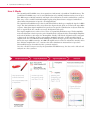
User Manual QuantiGene® FlowRNA Assay in situ gene expression assay for flow cytometry P/N 19221 Rev.A ii QuantiGene®Flow User Manual For research use only. Not for use in diagnostic procedures. Trademarks Affymetrix® and QuantiGene® are registered trademarks of Affymetrix, Inc is a registered trademark exclusively licensed to Affymetrix. All other trademarks are the property of their respective owners. Limited License Subject to the Affymetrix terms and conditions that govern your use of Affymetrix products, Affymetrix grants you a non-exclusive, nontransferable, non-sublicensable license to use this Affymetrix product only in accordance with the manual and written instructions provided by Affymetrix. You understand and agree that, except as expressly set forth in the Affymetrix terms and conditions, no right or license to any patent or other intellectual property owned or licensable by Affymetrix is conveyed or implied by this Affymetrix product. In particular, no right or license is conveyed or implied to use this Affymetrix product in combination with a product not provided, licensed, or specifically recommended by Affymetrix for such use. Citing QuantiGene® FlowRNA Assay in Publications When describing a procedure for publication using this product, please refer to it as the QuantiGene® FlowRNA Assay from Affymetrix. Disclaimer Affymetrix, Inc. reserves the right to change its products and services at any time to incorporate technological developments. This manual is subject to change without notice. Although this manual has been prepared with every precaution to ensure accuracy, Affymetrix, Inc. assumes no liability for any errors or omissions, nor for any damages resulting from the application or use of this information. Copyright © 2012 Affymetrix Inc. All rights reserved. Contents iii Contents About This User Manual . . . . . . . . . . . . . . . . . . . . . . . . . . . . . . . . . . . . . . . . . . . . . . . . . . . .1 Contacting Technical Support . . . . . . . . . . . . . . . . . . . . . . . . . . . . . . . . . . . . . . . . . . . . . . . .1 About the QuantiGene FlowRNA Assay Kits . . . . . . . . . . . . . . . . . . . . . . . . . . . . . . . . . . . . .1 How it Works . . . . . . . . . . . . . . . . . . . . . . . . . . . . . . . . . . . . . . . . . . . . . . . . . . . . . . . . . . . .2 QuantiGene FlowRNA Reagent System Contents and Storage Conditions . . . . . . . . . . . . . . .3 Required Equipment and Materials Not Supplied . . . . . . . . . . . . . . . . . . . . . . . . . . . . . . . . . .4 Experimental Guidelines . . . . . . . . . . . . . . . . . . . . . . . . . . . . . . . . . . . . . . . . . . . . . . . . . . . .4 Assay Procedure I. Fixation and Permeabilization . . . . . . . . . . . . . . . . . . . . . . . . . . . . . . . . . . . . . . . . . . . . . . .6 II. Target Probe Hybridization . . . . . . . . . . . . . . . . . . . . . . . . . . . . . . . . . . . . . . . . . . . . . . . .7 III. Signal Amplification and Detection . . . . . . . . . . . . . . . . . . . . . . . . . . . . . . . . . . . . . . . . . .9 Appendix A1: Troubleshooting . . . . . . . . . . . . . . . . . . . . . . . . . . . . . . . . . . . . . . . . . . . . . . . . . . . . . .10 A2: Lab setup guide . . . . . . . . . . . . . . . . . . . . . . . . . . . . . . . . . . . . . . . . . . . . . . . . . . . . . .13 A3: Cytometer Setup . . . . . . . . . . . . . . . . . . . . . . . . . . . . . . . . . . . . . . . . . . . . . . . . . . . . .14 A4: Incorporating antibody staining with the QG FlowRNA Assay . . . . . . . . . . . . . . . . . . . .17 A5: Expected results . . . . . . . . . . . . . . . . . . . . . . . . . . . . . . . . . . . . . . . . . . . . . . . . . . . . . .19 A6: Validated cell lines . . . . . . . . . . . . . . . . . . . . . . . . . . . . . . . . . . . . . . . . . . . . . . . . . . . .20 A7: Temperature Validation Procedure for Incubator . . . . . . . . . . . . . . . . . . . . . . . . . . . . . .21 iv QuantiGene® FlowRNA Assay User Manual 1 About This User Manual This manual is for anyone who has purchased a QuantiGene FlowRNA Assay Kit for the following sample types: Cultured mammalian suspension cells Peripheral Blood Mononuclear Cells (PBMC) from cryopreserved or freshly collected whole blood and purified by Ficoll®. Suspension cells sorted by a flow cytometry sorting instrument, e.g. BD FACSAria®, or purified by antibody-coated magnetic beads Contacting Technical Support NOTE: For the most current version of user documentation, go to our website at www.ebioscience.com For technical support, contact the appropriate resource provided below based on your geographical location. For an updated list of FAQs and product support literature, visit our website at www.ebioscience.com. Location Contact Information North America 1-888-810-6168, [email protected] Europe [email protected] About the QuantiGene FlowRNA Assay Kits The QuantiGene FlowRNA RNA Assay System consists of 3 modules (each sold separately). QuantiGene FlowRNA Fixation and Permeabilization Kit. Contains reagents and buffers to ensure that the cell membranes are properly permeabilized and the RNA is fixed within the cell. QuantiGene FlowRNA Assay Kit. Contains the reagents for running the assay. QuantiGene ViewRNA Probe Set. Contains gene specific probe sets for specified RNA targets. Sample Type Suspension Cells including PBMC, bone marrow cells and suspension cell lines. Species mammalian Plex level Up to 3 RNA targets Assay format 1.5-mL microfuge tube Detection fluorescent Instrumentation for detection Standard flow cytometer equipped with blue (488 nm) and red (633 nm or similar) lasers, and filter sets for FITC, APC and APC-Cy7 2 QuantiGene® FlowRNA Assay User Manual How it Works The QuantiGene® FlowRNA Assay is an expansion of Affymetrix’s QuantiGene ViewRNA Assay. The QuantiGene FlowRNA assay is an in situ hybridization assay enabling simultaneous detection of up to three RNA targets with high sensitivity and single-cell resolution for use with a standard flow cytometer. This assay can be combined with immunophenotyping using fluorochrome-conjugated antibodies to allow further discrimination of specific cell subpopulations. The QuantiGene FlowRNA Assay is based on branched DNA technology to amplify the signal of a RNA target. The first hybridization of the assay allows the oligonucleotide probe set to bind to the target RNA sequence. Subsequent signal amplification is predicated on specific hybridization of adjacent Probe Set pairs. A typical Probe Set contains on average 20 oligonucleotide pairs. Next signal amplification is achieved via a series of sequential hybridization steps. The PreAmplifier molecules hybridize to their respective pair of bound Probe Set oligonucleotides, then multiple Amplifier molecules hybridize to their respective PreAmplifier. Finally, Label Probe oligonucleotides conjugated to fluorescent dye hybridize to their corresponding Amplifier molecules. A fully assembled signal amplification "tree" has 400 Label Probe binding sites. When all target-specific oligos in the Probe Set bind to the target mRNA transcript, an 8,000 fold amplification is achieved. Affymetrix currently offers three different amplification “tree” structures that allow simultaneous measurement of up to three different RNA targets for multicolor flow cytometric analysis. Once the cells have been processed by the QuantiGene FlowRNA Assay, the data can be collected and analyzed on a flow cytometer. 3 QuantiGene FlowRNA Reagent System Contents and Storage Conditions The components of the QuantiGene FlowRNA Assay Kit and their recommended storage conditions are listed below. Refer to the package insert for quantities of individual components supplied. Kit components have a shelf life of six months from the date of receipt. Each QuantiGene FlowRNA Assay Kit is supplied in three separate parts, based on storage temperature. Box 1: Shipped on dry ice and store at -20°C Component Description Storage 100x Label Probe Mix Fluorescent dye-labeled oligonucleotides in aqueous buffered –20°C solution containing Alexa 647-LP1 (uses APC channel), Alexa 488LP4 (uses FITC channel) and Alexa 750-LP6 (uses APC-Cy7 channel) Box 2: Shipped on blue ice and store at 2-8°C Component Description Storage Target Probe Diluent Aqueous solution containing formamide and detergent 2-8°C PreAmp Mix DNA (PreAmp1, PreAmp4 and PreAmp6) in aqueous solution containing formamide and detergent 2-8°C Amp Mix DNA (Amp1, Amp4 and Amp6) in aqueous solution containing formamide and detergent 2-8°C Label Probe Diluent Aqueous solution containing detergent 2-8°C Wash Buffer Aqueous buffered solution 2-8°C Storage Buffer Aqueous buffered solution 2-8°C 1.5-mL microfuge tube Clear copolymer tube with a flat cap (RNase free) for low cell binding. DO NOT SUBSTITUTE. Room Temp Box 3: Fixation and Permeabilization Kit: Shipped at room temperature and store at 2-8°C Component Description Storage Solution A Aqueous buffered solution 2-8°C Solution B Aqueous buffered solution 2-8°C Solution C Aqueous buffered solution containing detergent 2-8°C Solution D Aqueous buffered solution 2-8°C QuantiGene FlowRNA Probe Sets In addition to the QuantiGene FlowRNA Assay Kit, Probe Set(s) specific to your RNA target(s) of interest must be purchased separately. Refer to the Product Insert for more information. Our current probe set catalog can be found at http://www.ebioscience.com/application/flowrna.htm. Component Description Storage QuantiGene FlowRNA Probe sets RNA-specific oligonucleotides designed against a target of interest and are compatible with TYPE 1, 4 or 6 Signal Amplifiers. –20°C 4 QuantiGene® FlowRNA Assay User Manual WARNING: All chemicals should be considered potentially hazardous. We recommend that this product and its components be handled by those trained in laboratory techniques and be used according to the principles of good laboratory practice. Required Equipment and Materials Not Supplied Required Equipment/Material Source Part Number 16% Paraformaldehyde (PFA) aqueous solution (in 10-mL amber tube) Electron Microscopy Sciences 15710 Methanol (HPLC-grade, 99.9% pure) Fisher Scientific A452-4 QuantiGene ViewRNA Temperature Validation Kit Affymetrix QV0523 Flow cytometer with the following MLS1(e.g. BD Biosciences, Beckman Coulter…) components: - Two lasers, blue (488 nm) and red (633 nm or similar) - Detection channels optimized for FITC, APC, and APC-Cy7 fluorophores Many (e.g. Calibur from BD Biosciences, FC500 from Beckman Coulter…) Refrigerated low-speed centrifuge with swinging bucket for 15-mL tubes with inserts for 1.5-mL microfuge tubes MLS, (e.g. Eppendorf) Eppendorf 5804 with A-4-44 Rotor (swing bucket) Incubator (validated to maintain 40 ± 1 °C) Affymetrix QS0704 (120V) QS0712 (220V) Metal tube rack (heat block) for 1.5mL microfuge tubes (placed inside incubator) MLS (e.g. VWR, Fisher Scientific...) VWR 13259-002 Vacuum aspirator, adjustable to 0.5 mL/ sec MLS, (e.g. Fisher Scientific, VWR...) Fisher Scientific 07200564 Vortexer MLS, (e.g. VWR, Fisher Scientific...) VWR 58816-121 (Optional) 15 mL polypropylene tubes MLS, (e.g. Fisher Scientific, BD...) Fisher Scientific 14-959-70C (Optional) RNasin Promega N2611 1 Multiple Lab Supplier Experimental Guidelines 1. Start with cell culture in good physiological conditions. Cells should be in active growth phase to preserve RNA integrity and minimize cell lysis in processing. If using freshly isolated primary cells, make sure the cells are healthy after purification. 5 2. Selection of probe type and fluorescent channel for optimal RNA detection Type 1 /650 probe set produces the most sensitive detection when compared to Type 4 /488 and Type 6/ 750 probe sets. Type 1/650 channel is about two to five folds more sensitive than the other two channels. We recommend using the following probe type/excitation line for the low, medium and high expression genes, respectively. In addition, background fluorescence in the Type 4/488 channel is higher compared to Type 1/650 and Type 6/750 channels. Gene Expression Level Probe Type Cytometer Laser line Ex/Em Low Type 1 633 650/668 Medium to High Type 4 488 495/519 Medium to High Type 6 633 749/775 3. QuantiGene FlowRNA Assay Controls Below are recommended assay controls that we recommend for first time users to perform. Control Gene Positive Control B2M - high expression in PBMC Human (Type 1) VA1-10611 (Type 4) VA4-13460 (Type 6) VA6-11782 Mouse (Type 1) VB1-11142 (Type 4) VB4-14643 (Type 6) VB6-12836 GAPDH - medium expression in PBMC Human (Type 1) VA1-10119 (Type 4) VA4-10641 (Type 6) VA6-10337 Mouse (Type 1) VB1-10150 (Type 4) VB4-10414 (Type 6) VB6-10574 Negative assay control No Target Probe Bacterial DapB gene Catalog Number for Probe Set Not Applicable (Type 1) VF1-11712 (Type 4) VF4-10408 (Type 6) VF6-10407 6 QuantiGene® FlowRNA Assay User Manual Assay Procedure This protocol contains the following sections. It can be performed in two days with optional stop points. Day 1: I. Fixation and Permeabilization II. Target Probe Hybridization Day 2: III. Signal Amplification and Detection IMPORTANT: This protocol is optimized for the swinging bucket centrifuge. We do not recommend using a fixed-angle centrifuge as severe cell loss may result. I. Fixation and Permeabilization NOTE: The fixation and permeabilization can be performed in bulk if desired. However, polypropylene conical tubes (e.g. Fisher Scientific cat. #14-959-70C) must be used. DO NOT use polystyrene tubes such as FACS tube (e.g. BD Falcon cat. #352052) which would lead to low signal. Step Action Step 1. (Optional) Antibody staining - see Appendix for details. Step 2. Perform the following tasks to prepare enough reagents for 24 samples. a. Pre-warm Target Probe Diluent to 40°C. b. Equilibrate Solutions A, B, C, and D to room temperature (RT). c. Prepare a 4% paraformaldehyde (PFA) in Solution A. NOTE: PFA solution must be freshly prepared before use. Add 30 mL of Solution A to a 50 mL conical tube and keep at RT in the dark. Open an amber tube containing 10 mL of 16% PFA stock solution and add the solution to the conical tube containing Solution A. Mix well and keep at RT in the dark. d. Ensure incubator and heat block are calibrated to 40°C using a traceable digital thermometer (see QuantiGene ViewRNA Temperature Validation Kit for details). e. Pre-chill methanol to -20°C. f. Set the temperature of centrifuge to 4°C. Step 3. Hybridization must be performed in the 1.5-mL microfuge tubes provided. For each reaction, start by transferring 1-3 million cells into each tube. Step 4. Centrifuge the tubes in a swinging bucket at 400 x g at 4°C for 5 min to pellet the cells. Aspirate the supernatant leaving a residual volume of 100 µL. Vortex tubes to resuspend cells evenly. Add 1 mL Solution A, then invert tubes a few times to mix. NOTE: Always leave a residual volume of 100 µL when aspirating in subsequent steps to avoid disturbing the cell pellet. The lowest mark within the graduations on the microfuge tube is 100 µL. Step 5. Centrifuge cells at 400 x g at 4°C for 5 min and aspirate the supernatant leaving a residual volume of 100 µL. Step 6. Vortex tubes to resuspend cells evenly. To each tube, add 1 mL of 4% PFA in Solution A prepared in Step 2c. Briefly invert the tubes to mix and incubate at RT for 60 min in the dark, inverting the tubes 1-2 times during incubation. Step 7. Centrifuge the tubes at 800 x g at 4°C for 5 min and aspirate the supernatant leaving a residual volume of 100 µL. Briefly vortex tubes to resuspend the cell pellets. 7 Step Action Step 8. To each tube, add 1 mL of Solution B and invert the tubes to mix. Centrifuge cells at 800 x g at 4°C for 5 min and aspirate the supernatant leaving a residual volume of 100 µL. Briefly vortex tubes to resuspend the cell pellets. Step 9. To each tube, add 1 mL of ice-cold methanol solution. Briefly vortex the tubes to mix and incubate on ice for 10 min. NOTE: Optional stop point: For storage, transfer cells to -80°C after adding methanol. Samples should be stable for 3 months. However, we do not recommend sample storage in methanol if antibody staining has been performed as in Step 1. Step 10. Perform the following tasks: NOTE: PFA solution must be freshly prepared before use. a. Prepare 4% PFA in Solution C. Add 30 mL of Solution C to a 50-mL conical tube. Keep at RT in the dark. Open an amber tube containing 10 mL of 16% PFA stock solution and add the solution to the conical tube containing Solution C. Mix well and keep at RT in the dark. b. Prepare a 1:1 solution of methanol and 4% PFA in Solution C Add 13 mL of ice-cold methanol to a 50-mL conical tube. Add 13 mL of 4% PFA in Solution C (from Step 10a). Keep on ice in dark. c. Thaw the target probe sets at RT and then place on ice. Step 11. Centrifuge tubes at 800 x g at 4°C for 5 min and aspirate the supernatant leaving a residual volume of 100 µL. Briefly vortex tubes to resuspend the cell pellets. Step 12. To each tube, add 1 mL of ice-cold methanol/ 4% PFA in Solution C (1:1) prepared in Step 10b. Invert the tubes to mix and incubate on ice for 5 min. Step 13. Centrifuge tubes at 800 x g at 4°C for 5 min and aspirate the supernatant leaving a residual volume of 100 µL. Briefly vortex tubes to resuspend cell pellet. Step 14. To each tube, add 1 mL of freshly prepared 4% PFA solution in Solution C prepared in Step 10a. Invert the tubes to mix and incubate at RT for 30 min. Invert tubes once during incubation. Step 15. Centrifuge tubes at 800 x g at RT for 5 min and aspirate the supernatant leaving a residual volume of 100 µL. Briefly vortex tubes to resuspend the cell pellets. Step 16. To each tube, add 1 mL of Solution D. Invert tubes to mix and centrifuge tubes at 800 x g at RT for 5 min, then aspirate the supernatant leaving a residual volume of 100 µL. Briefly vortex tubes to resuspend the cell pellets. Step 17. Repeat wash with Solution D and leave a residual volume of 100 µL. II. Target Probe Hybridization Step Action Step 1. Dilute the target probes 1/20 with Target Probe Diluent. Add 100 µL of diluted target probes to each tube from Step 17. Step 2. Briefly vortex tubes to mix. Place tubes in the pre-warmed heat block inside the incubator. Incubate at 40°C for 2 hours. Invert tubes to mix after 1 hour of incubation. Step 3. To each tube, add 1 mL of Wash Buffer and invert tubes to mix. Centrifuge the tubes at 800 x g at RT for 5 min to pellet the cells, then aspirate leaving a residual volume of 100 µL. Briefly vortex tubes to resuspend the cell pellets. 8 QuantiGene® FlowRNA Assay User Manual Step Step 4. Action Repeat wash by adding 1 mL of Wash Buffer (Optional: add 1 µL/mL of RNasin if used as stop point) and invert tubes to mix. Centrifuge the tubes at 800 x g at RT for 5 min to pellet the cells, then aspirate leaving a residual volume of 100 µL. Briefly vortex tubes to resuspend the cell pellets. NOTE: Optional stop point: store samples overnight at 4°C in the dark. 9 III. Signal Amplification and Detection Step Step 1. Perform the following tasks: a. Pre-warm PreAmp Mix, Amp Mix and Label Probe Diluent to 40°C. b. Equilibrate Wash Buffer, Storage Buffer and the samples to RT. Step 2. Add 100 µL of pre-warmed PreAmp Mix (with green cap) to each tube and briefly vortex tubes to mix. Place tubes in the pre-warmed heat block inside the incubator. Incubate at 40°C for 1.5 hours. Step 3. Remove tubes from incubator and add 1 mL of Wash Buffer to each tube. Invert tubes to mix. Centrifuge the tubes at 800 x g at RT for 5 min to pellet the cells, then aspirate leaving a residual volume of 100 µL. Briefly vortex tubes to resuspend the cell pellets. Step 4. Repeat wash. Step 5. Add 100 µL of pre-warmed Amp Mix to each tube and briefly vortex tubes to mix. Place tubes in the prewarmed heat block inside the incubator. Incubate at 40°C for 1.5 hours. Step 6. During Amp Mix incubation, thaw Label Probe Mix on ice. Step 7. Dilute Label Probe Mix 1/100 with Label Probe Diluent. Store in the 40°C incubator and protect from light until ready to use. Reagent 1 Tube 10 Tubes 24 Tubes 1 Label Probe 1.2 µL 12 µL 28.8 µL 2 Label Probe Diluent 118.8 µL 1188 µL 2851.2 µL 120 µL 1200 µL 2880 µL Total Step 8. Remove tubes from incubator and add 1 mL of Wash Buffer to each tube. Invert tubes to mix. Centrifuge the tubes at 800 x g at RT for 5 min to pellet the cells then aspirate the supernatant leaving a residual volume of 100 µL. Briefly vortex tubes to resuspend the cell pellets. Step 9. Repeat wash. Step 10. Add 100 µL of Working Label Probe Solution prepared in Step 7 to each tube and briefly vortex tube to mix. Place tubes in the pre-warmed heat block inside the incubator. Incubate at 40°C for 1 hour. Step 11. Turn on the flow cytometer to allow the lasers to warm up. Step 12. Remove tubes from incubator and add 1 mL of Wash Buffer to each tube. Invert tubes to mix. Centrifuge the tubes at 800 x g at RT for 5 min to pellet the cells, then aspirate leaving a residual volume of 100 µL. Briefly vortex tubes to resuspend the cell pellets. Step 13. Repeat wash. Step 14. Add 1 mL of Storage Buffer to each tube and invert tubes to mix. Centrifuge the tubes at 800 x g at RT for 5 min to pellet the cells, then aspirate supernatant leaving 100 µL residual volume. Briefly vortex tubes to resuspend the cell pellets. Protect samples from light. NOTE: Optional stop point: store samples at 4°C in the dark. Step 15. To analyze a sample, pipette to mix. Then transfer 50-100 µL sample to a reading tube containing 250 µL of 1X PBS and analyze on a flow cytometer. For storage, keep samples at 4°C in the dark. Samples are stable for 1 week when stored at 4°C. 10 QuantiGene® FlowRNA Assay User Manual Appendix A1: Troubleshooting Observation Probable Cause Recommended Solution Poor Cell Recovery / Low Cell Count on Cytometer Cell lysis due to cells in poor physiological condition during processing Improper centrifuge and tube adaptor Improper centrifuge settings If using frozen cells, thaw cells carefully following proper cell culture procedures. Check cell viability using trypan blue or other viability dyes before assay. Use swinging bucket for centrifugation. Do not use fixedangled centrifuges. Use correct tube adaptor for proper g-force. Confirm settings are for RCF (xg), not RPM. Set centrifuge to the speed specified in the protocol. Improper tubes used Only use the 1.5-mL microfuge tubes provided with the kit. Tubes from other vendors may result in significant cell loss. High aspiration rate by pipetting or vacuum aspirator when removing supernatant Ensure vacuum setting for aspirator is low. Aspirate no faster than 0.5 mL/ sec. Place a 1-10 µL pipette tip on the tip of the aspirator to help reduce aspiration rate. Follow the meniscus while aspirating down to the 100 µL mark on side of tube. Rough handling Avoid excessive vortexing to prevent cell damage. Vortex in pulses. When pipetting to mix, do so gently. Clog Ensure cytometer is not clogging due to clumps of aggregated cells. Try using gentle vortexing and pipette mixing to break up aggregates, or use a 70 µL filter. Incorrect cell count by flow cytometer Verify the flow rate of cytometer using beads of known concentration such as the Flow Cytometry Absolute Count Standard (Bangs Laboratories, cat. # 580). False Positive Population Compensation not set up correctly See Appendix section on how to set up compensation. Low / No Signal Cytometer not set up properly Check that your instrument is properly set up according to the manufacturer's recommendations. Old 4% PFA solution was used in fixation. Prepare fresh 4% PFA solution before use. 11 Observation Probable Cause Recommended Solution Polystyrene tube (e.g. FACS reading tube) was used in fixation and permeabilization. Use polypropylene tube (e.g. Fisher Scientific 14-959-70C) for fixation and permeabilization. Insufficient number of cells recorded on cytometer Determine the number of total events to collect to ensure at least 100-1000 positive cells are recorded, for example: Total number of cells to count = number of positives desired / frequency of positives (%) To count 1,000 positive cells, Frequency of Positives Total # of cells to count 10% 10,000 5% 20,000 1% 100,000 Incorrect preparation of signal amplification reagents Make sure PreAmp, Amp and Label Probe are used in the specified order. Incorrect incubator temperature Incubator must be able to hold temperature at 40±1°C. Use 1.5-mL heat block for hybridization. Minimize traffic to incubator. Temperature tolerance is 1-2°C. Ensure incubator has been stable at 40°C for at least 12 hours before starting assay as temperature may drift during initial incubator setup. Measure temperature using QG ViewRNA Temperature Validation Kit from Affymetrix (PN QV0523). Incorrect Wash Buffer Do not substitute Wash Buffer with Storage Buffer. Incorrect diluents used Ensure Target Probe Diluent is used with Target Probe, and Label Probe Diluent used with Label Probe. Incorrect dilution of Target Probe/ Label Probe Ensure Target Probe was added to Target Probe Diluent at a 1/20 dilution. Ensure Label Probe Mix was added to Label Probe Diluent at a 1/100 dilution. 12 QuantiGene® FlowRNA Assay User Manual Observation Probable Cause Recommended Solution Gene expressed at very low level High Background Incorrect incubator temperature Insufficient washing Check biological model and confirm gene is expressed in sample by running QuantiGene 2.0 Assay for lysate. Keep in mind that protein expression might not correlate with RNA expression in some cases. Incubator must be able to hold temperature at 40±1°C. Minimize traffic to incubator. Temperature tolerance is 1-2°C. Ensure incubator has been stable at 40°C for at least 12 hours before starting assay as temperature may drift during initial incubator setup. Measure temperature using QG ViewRNA Temperature Validation Kit from Affymetrix (cat. #QV0523). Ensure the Wash Buffer is used at room temperature. Follow instructions of washing steps in the protocol. Ensure uniform cell resuspension by vortexing. Cytometer not set up properly Reduce voltage settings of your cytometer. Note that this assay may require lower settings than typical antibody staining. Over-fixation Follow the protocol for proper fixation time. Excessive Target Probe used Dilute Target Probe at a 1/20 dilution. 13 A2. Lab Setup Guide This guide illustrates the setup of typical equipment and their specifications for the QuantiGene FlowRNA Assay. Consult your equipment manufacturers to make sure the equipment meets the specifications. The major equipment include: incubator, swinging bucket centrifuge, flow cytometer, aspiration system for washing, and temperature validation kit. Equipment Specifications Incubator with heat block inside Refrigerator swinging bucket centrifuge Flow cytometer Aspiration system for washing QuantiGene ViewRNA Temperature Validation Kit Validated to maintain 40±1°C. Metal block for 1.5-mL microfuge tube. Example shown: Incubator (Affymetrix QS0704) Metal heat block (VWR 13259-002) Swinging bucket with adaptors for 15-mL conical tubes and 1.5-mL microfuge tubes. With refrigeration to 4°C. Examples shown: Centrifuge (Eppendorf 5810R) Rotor (Eppendorf A-4-44) Adaptor for 15-mL conical tube (Eppendorf 5804 755.006) Adaptor for 1.5-mL microfuge tube (Eppendorf 5804 750.004) NOTE: Fixed-angle centrifuge is NOT recommended. Two lasers - blue (488 nm) and red (633 nm or similar) Detection optics optimized for FITC, APC, and APC-Cy7 fluorophores Example shown: BD LSRFortessa Aspiration rate adjusted to 0.5 mL / sec. Can use in-house vacuum line or vacuum pump. Example shown: Vacuum bottle (Argos Technologies EV432) Aspirator (Argos Technologies EV514) NIST traceable thermometer with temperature probe in 1.5-mL microfuge tube (shown on left). Affymetrix, cat.# QV0523 14 QuantiGene® FlowRNA Assay User Manual A3. Cytometer Setup The QuantiGene FlowRNA Assay Kit utilizes up to three fluorescent channels for detection of RNA on a flow cytometer. To ensure optimal detection of RNA, it is important that the cytometer is set up properly. In a multi-color assay, signal from a fluorophore often spills over into the other detection channels due to overlapping emission spectra. The process of subtracting the spillover signal is called compensation. The procedures below describe how to set proper PMT voltage and compensation for multi-color RNA analysis. Reagents and samples needed Calibration beads Unstained cells Single-color samples: Type 1 probe (Alexa Fluor® 647)-only labeled cells Type 4 probe (Alexa Fluor® 488)-only labeled cells Type 6 probe (Alexa Fluor® 750)-only labeled cells Single-color antibody-labeled cells (needed only if protein staining is performed) 1. Ensure proper alignment following the instrument manufacturer's recommendations. Alternatively, calibration beads, such as Spherotech Rainbow Calibration Particles (8-peak beads, catalog number RCP-30-5A) or Rainbow Fluorescent Particles (1-peak beads, catalog number RFP-30-5A [1 peak]) may be used to check the linearity of the PMT, PMT voltage range, and alignment of the instrument. 2. Setting PMT Voltages A. Create an acquisition template/workspace that includes a forward scatter versus side scatter plot, one single-parameter histogram plot for every fluorescent channel being used, and a twoparameter plot for every pairwise combination of fluorescent channels being used in the experiment. B. Place the unstained cell sample on the cytometer and begin to collect events. 1) Adjust voltages for forward scatter and side scatter so that cells/events of interest are on scale. Set a threshold on forward scatter to eliminate debris from acquisition. 2) Looking at the histogram plots, ensure that the signals are on scale and adjust PMT voltages, if necessary. 3) Stop collecting events and remove sample from the cytometer. C. Place a single-color sample on the cytometer and begin to collect events. 1) Looking at the histogram plot for the fluorochrome being collected, ensure that events are on scale for the fluorescent channel being collected and adjust PMT voltages, if necessary. 2) Stop collecting events and remove sample from the cytometer. 3) Repeat for each of the single-color samples. D. Confirm that the voltage settings are appropriate for the experimental samples. 1) Place an experimental sample on the cytometer and begin collecting events. 2) Looking at each of the histogram plots, confirm that all fluorescent signals are on scale and adjust PMT voltages, if necessary. 3) Stop collecting events and remove sample from the cytometer. 3. Setting compensation A. If an autocompensation feature is available for the cytometer being used, follow the manufacturer's instructions for use. B. If an autocompensation feature is not available or to set compensation manually: 15 1) Place the unstained sample on the cytometer and begin to collect events. 2) Looking at the two-parameter plots, create quadrant regions on each plot such that the cells fall within the lower left quadrant (see Figure 1). 3) For each two-parameter plot, create statistics windows/tables according to your instrument's acquisition software and include the median for the x- and y-parameters. 4) Stop collecting events and remove sample from the cytometer. C. Place a single-color sample on the cytometer and begin to collect events. NOTE: If your single- color sample does not contain any negative events, a small amount of unstained cells may be added to each of the single-color samples just prior to loading the sample onto the cytometer. Alternatively, the MFI of the unstained sample run in Step 3B above must be noted for all parameters. 1) Looking at the two-parameter plots for the fluorochrome being collected, adjust the quadrants so that the positive events are fully contained within either the lower right (if the parameter is on the x-axis; Figure 2A, left) or the upper left (if the parameter is on the y-axis; Figure 2B, left) quadrant. 2) From the statistics window, note the y-medians (if the parameter is on the x-axis; Figure 2A, left) or the x-medians (if the parameter is on the y-axis Figure 2B, left) of the two populations. 3) Adjust each of the compensation values for the fluorochrome being collected (subtract its fluorescence out of each of the other channels being used in the experiment) until the medians are the same (Figure 2A and 2B, right). 4) Stop collecting events and remove sample from the cytometer. 16 QuantiGene® FlowRNA Assay User Manual 5) Repeat for each of the single-color samples. D. Confirm that the compensation settings are appropriate for the experimental samples. 1) Place an experimental sample on the cytometer and begin collecting events. 2) Looking at each of the two-parameter plots, confirm that the compensation is set correctly by looking at the medians. 3) Stop collecting events and remove sample from the cytometer. 17 A4. Incorporating Antibody Staining with the QG FlowRNA Assay Fluorochrome-conjugated antibodies can be used to stain surface proteins for the purpose of immunophenotyping cells that will be further analyzed for RNA levels using the QuantiGene FlowRNA Assay. Due to the nature of the RNA labeling protocol, additional considerations for staining are warranted. The following two options are recommended for staining with antibodies: 1. Cells may be stained with fluorochrome-conjugated antibodies before the Fixation and Permeabilization Procedure only if the fluorochromes are resistant to exposure to methanol. Methanolresistant and methanol-sensitive fluorochromes are listed in the table below. Although some fluorochromes are resistant to methanol, there may still be some loss in signal intensity, therefore, performance of a specific reagent in this assay should be determined empirically. 2. If the antibody is not conjugated to a methanol-resistant fluorochrome, it may be possible to utilize an indirect staining protocol. Cells may be stained with an unconjugated antibody before the Fixation and Permeabilization Procedure. A secondary reagent (fluorochrome-conjugated anti-Ig secondary antibody) can then be used to detect the primary antibody. Staining with the secondary reagent must be done after Signal Amplification and Detection. It is important to note that only one unconjugated antibody from a given host may be used unless the isotypes of the unconjugated antibodies used are different. In this case, isotype-specific secondaries must be used; a pan anti-mouse IgG secondary antibody will bind to all mouse IgG antibodies present on the cells. Methanol-Resistant and Methanol-Sensitive Fluorochromes Methanol Resistant Fluorochromes Methanol Sensitive Fluorochromes FITC PE Alexa Fluor® 488 PE-Cy5 Alexa Fluor® 700 PE-Cy5.5 Alexa Fluor® 647 PE-Cy7 eFluor® PerCP 450 Pacific BlueTM PerCP-Cy5.5 Pacific OrangeTM PerCP-eFluor 710 Brilliant Violet 421TM APC Brilliant Violet 605TM APC-Cy7 APC-H7 APC-Alexa Fluor® 750 APC-eFluor® 780 Qdot® reagents eFluor® Nanocrystal (NC) reagents How to validate an antibody for QuantiGene FlowRNA Assay: Each antibody needs to be validated with the assay protocol to confirm its compatibility. Experimental conditions: 1. Antibody staining alone 18 QuantiGene® FlowRNA Assay User Manual 2. Antibody staining followed by RNA detection procedure without target probe Analyze and compare two samples for: 1. Signal intensity of the protein staining 2. Percentage of positive cells For a qualified antibody: 1. Sufficient separation between the positive and negative signals should be maintained after the RNA detection procedure to allow identification of desired populations 2. The percentage of desired cell populations should be comparable. 19 A5. Expected Results This is an example of typical results of the QuantiGene FlowRNA Assay. Experimental conditions Human PBMC were stimulated with 1 µg/mL of bacterial lipopolysaccharides (LPS) and 2.5 µg/ mL of R-848 for 4 hours. Cells were subjected to analysis by the QuantiGene FlowRNA Assay. Cells were stained with anti-human CD14-FITC to identify monocytes and labeled with probes for the RNA expression of IL-1beta, IL-6, IL-8 and TNF alpha. Cells in the monocyte gate were used for analysis. CD14 staining is shown on the y-axis and RNA labels shown on the x-axis. Result 20 QuantiGene® FlowRNA Assay User Manual A6. Validated Cell Lines The QuantiGene FlowRNA Assay has been validated with the following cell types: Primary cells Human PBMC (cryopreserved and freshly purified by Ficoll) Human T cells (CD3+)* Human B cells (CD19+)* Human monocytes (CD14+)* Mouse bone marrow cells *Cells were purified by antibody-coated magnetic beads. Cultured suspension cell lines NKT, human natural killer cell line Jurkat, human lymphocytic cell line U937, human monocytic cell line K562, human myelocytic cell line M1, mouse myeloid cell line 21 A7. Temperature Validation Procedure for Incubator Temperature control is critical for the success of the QuantiGene FlowRNA Assay. The incubator should be validated before use. Improper hybridization temperature will result in high background and/or weak signal. Required materials Item Description Incubator Capable of maintaining temperature at 40±1°C, e.g. Affymetrix cat. # QS0704 or QS0712 QuantiGene View Temperature Validation Kit (Affymetrix cat. # QV0523) NIST calibrated digital thermometer with Type-K beaded probe 1.5-mL microfuge tube 1.5-mL microfuge tube used for assay (included in the QuantiGene FlowRNA Assay Kit) Metal heat block for 1.5-mL microfuge tube Metal block to hold the microfuge tube, e.g. Fisher Scientific 11718-9Q Parafilm Major Laboratory Supplier 22 QuantiGene® FlowRNA Assay User Manual Procedure Step Action Step 1 Prepare the incubator A. Turn on the incubator. B. Set the temperature to 40°C. C. Put the metal heat block into the incubator near the center of the middle incubator. D. Allow the incubator and heat block to equilibrate for overnight. Step 2 Assemble unit A. Insert the battery to activate the digital thermometer. B. Use a pointed object, e.g. a ballpoint pen to drill a hole at the lid of the 1.5-mL microfuge tube, along the circular markings. C. Add 0.2 mL deionized water to the tube, and then close the lid. D. Insert the Type-K beaded probe into the digital thermometer, and the other end into the pre-drilled hole of the provided 1.5-mL tube containing 0.2 mL of deionized water. E. Wrap parafilm around the top of the 1.5-mL tube and probe to form a seal. Avoid an excessive amount of parafilm around the sides of the tube; otherwise it may not fit properly into the heat block. F. Turn on the digital thermometer device 23 Step Action Step 3 Measure and adjust incubator temperature A. Insert the 1.5-mL tube with probe and deionized water into the heat block that has been pre-warmed in the incubator in Step 1. B. Close the door making sure there is sufficient slack in the wiring. C. Wait 15 min for temperature to equilibrate. D. Record the temperature. E. If necessary, adjust the dry incubator temperature settings so that the digital thermometer reads 40°C. After adjustment, allow the incubator and heat block to equilibrate. Then recheck the temperature. F. Repeat Step E as needed. IMPORTANT: We recommend calibrating the incubator at least once a month to ensure accuracy. Step 4 Assess incubator temperature uniformity Repeat step 3 to measure the temperature at multiple positions in the incubator to determine temperature uniformity. IMPORTANT: The temperature for all positions should be 40±1°C. Step 5 Assess ramp-up time A. Open the incubator door for 1 min then close the door. Measure the time needed for the temperature to return to 40°C and monitor the temperature profile during recovery. B. Repeat the Step 5A two more times. IMPORTANT: Do not use the incubator for the assay if it takes more than 5 minutes to return to 40°C or if it overshoots by more than 2°C during recovery. 24 QuantiGene® FlowRNA Assay User Manual