1
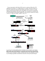
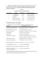
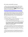
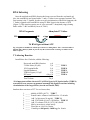
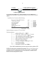
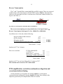
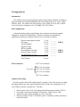
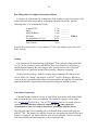
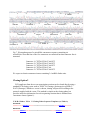
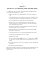
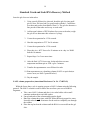
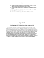
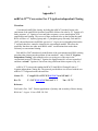
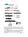
INTEGRATED DNA TECHNOLOGIES miRCat miRCat-33 TM TM microRNA Cloning Kit Technical Manual THE CUSTOM BIOLOGY COMPANY WWW.IDTDNA.COM Contents Overview and Kit Contents 3 RNA Isolation and Size Fractionation 7 RNA Linkering 8 11 12 17 19 Appendix 2: PCR Purification from Agarose Gels 21 22 Appendix 4: Additional Products for Small RNA Cloning 27 Reverse Transcription PCR Amplification, Concatemerization, and Cloning References Appendix 1: RNA Recovery from PAGE Appendix 3: miRCat‐33TM Conversion 3 miRCat™ Small RNA Cloning Kit Technical Manual Overview MicroRNAs (miRNAs) are small non-coding RNAs that are involved in posttranscriptional gene regulation (cf., Bartel, 2004). First identified in C. elegans just over a decade ago (Lee et al., 1993), miRNAs have been identified in virtually every metazoan and plant species examined. Experimental evidence is rapidly accumulating that shows miRNAs play key roles in process such as cellular differentiation, cell death, and cell metabolism. MiRNA biogenesis has been mapped to at least a first-order level. An outline of miRNA development is presented in Bartel (2004). In general, an miRNA is composed of a highly conserved core sequence of 21-23 nucleotides (the mature miRNA) contained within a less well conserved precursor sequence (pre-miRNA) ranging in size from 60 nucleotides to more than 120 nucleotides. This pre-miRNA sequence is part of a larger primary transcript that may contain a single pre-miRNA or two or more premiRNAs arranged as paired or polycistronic transcripts. Following transcription, premiRNAs form a characteristic stem-loop structure that is processed by the RNase III enzyme DROSHA (Lee et al., 2003) in concert with accessory proteins such as PASHA and DGCR8 (Gregory et al., 2004; Denli et al., 2004). The pre-miRNA is thus released to be exported from the nucleus whereupon it is further processed by the DICER/RISC complex releasing the mature miRNA to carry out its regulatory function. A number of investigators have reported on methods for cloning miRNAs from primary RNA sources (Berezikov et al., 2006; Cummins et al., 2006; Elbashir et al., 2001; Lau et al., 2001; Pfeffer et al., 2003; Sunkar and Zhu, 2004). Here, we provide a User’s Guide for cloning miRNAs and other small RNAs from primary RNA sources using IDT’s miRCat™ Small RNA Cloning Kit. A schematic representation of the cloning process is shown in Figure 1. There are three distinct experimental phases involved in cloning microRNAs and other small RNA species using the miRCatTM Kit. RNA Isolation and Enrichment Phase RNA species in the 18 to 26 nucleotide size range are purified from total RNA. Best results are obtained if 50-100 ug of total RNA is used, however cloning can be performed with less mass if RNA is scarce. This size range contains mature microRNA sequences. There are several options to perform purification, including denaturing PAGE, the miRVanaTM kit (Ambion®), or the flashPAGETM fractionator (Ambion®). Cloning Linker Attachment Phase The 3’ and 5’ cloning linkers are ligated to purified small RNA species in preparation for cDNA synthesis and amplification. Amplification and Cloning Phase 4 Reverse transcription of the linkered RNA species is carried out followed by PCR amplification and cloning. Two cloning options are available. The preferred option is a SAGE-like method where the small RNA cloning units (miRNA + linkers) are serially ligated (concatemerized) and then cloned. This method is more efficient for sequencing using sequencing platforms with long read lengths. The second option is to directly clone the PCR amplicons. In both options cloning can be done using any available PCR cloning vectors (e.g., TOPO-TA Cloning®, pGEM® T-Easy, etc.). TISSUE SOURCE Total RNA + TRIZOL; RNA STAT 60; OR OTHER ORGANIC EXTRACTION REAGENT 3’ Cloning Linker Ligation P04 Internal RNA Control Small RNA Enrichment Pg. 7 P04 Pg. 8 PAGE Purification 5’ M-R-S Linker Ligation P04 Pg. 9 Reverse Transcription Pg. 10 PCR Pg. 11 A A Ban I Digestion Pg. 12 Concatemers Pg. 13 DIRECT CLONING AND SEQUENCING KEY Small RNAs CONCATAMER CLONING AND SEQUENCING miSPIKE RNA 3’ cloning linker 5’ cloning linker Fig. 1. The process of cloning miRNAs and other small RNAs from any total RNA source using the miRCat™ Kit is shown. Desired small RNAs are represented by the yellow bar, the spike-in internal RNA control is shown as the green bar, the 3’ cloning linker is shown as the red bar, and the 5’ M-RS Linker is shown as the blue bar. The Ban I restriction enzyme sites in the cloning linkers are shown by cross hatching. Page numbers in the manual are noted where each step of the protocol begins. 5 Sufficient materials are provided in this kit to generate more than ten small RNA libraries. The two most important aspects of miRNA cloning are the quantity and quality of the starting RNA and the maintenance of relative mass relationships during the Cloning Linker Attachment Phase. Total cellular RNA can be used to clone small RNA species but the absolute mass of small RNAs is very small and larger RNA species will compete for linker molecules. For this reason it is best to prepare a highly enriched and purified small RNA fraction at the outset (see below). Once purified small RNA species are obtained, it is crucial that sufficient linker mass is used to ensure efficient 3’ and 5’ linker attachment. We strongly encourage using the 3’ and 5’ linkers in the amounts and at the concentrations called for in the Cloning Linker Attachment Phase. Reductions in the mass of linker in either of the linkering steps will result in a substantial reduction in linkering efficiencies. Getting Started miRCat™ Kit Contents Tube Number 1 Contents 3’ cloning linker 2 5’ cloning linker 3 miSPIKE™ internal control RNA 4 Forward PCR primer 5 Reverse transcription/PCR primer 6 IDT RNase/DNase/pyrogen-free water 7 IDTE (pH 7.5) 8 10X ligation buffer 9 Ligation enhancer 10 10 mM ATP 11 10 mg/ml Glycogen 12 3 M NaOAc (pH 5.2) 13 T4 RNA Ligase (5 U/µl) 14 T4 DNA Ligase (30 U/µl) Storage Recommendations: Store all kit components at -20oC. (NOTE: Repeated freezing and thawing of ATP is not recommended. It is best to store ATP in smaller aliquots and to thaw only as much as will be needed for each library construction). 6 Cloning linkers, miSPIKETM internal RNA control, and PCR primers are provided dry. Before opening, centrifuge the tubes containing dried material and follow the instructions in Table 1 below. All reagents should be handled with gloves under RNase-free conditions. Table 1 Rehydration of Stock Oligonucleotides Tube Number Contains 1 3’ cloning linker IDTE (Tube 7) 20 µl Final Concentration 50 µM (50 pmole/µl ) 2 5’ cloning linker 20 µl 50 µM (50 pmole/µl ) 3 miSPIKETM 12 µl 8.3 µM (8.3 pmole/µl ) 4 Forward PCR primer 100 µl 10 µM (10 pmole/µl ) 5 RT/Reverse PCR primer 100 µl 10 µM (10 pmole/µl ) Additional Reagents and Supplies In addition to the materials supplied in this kit you will need the following: Reagent Recommended Vendor Ban I restriction endonuclease New England Biolabs (Cat. No. R0118S) GelStar® Nucleic Acid Stain* Lonza BioScience (Cat. No. 50535) Reverse Transcriptase SuperScript™ III (Invitrogen-Cat. No. 18080-093) PCR Reagents No specific recommendation 100% EtOH No specific recommendation PCR amplicon cloning kit pGEM® T-EASY (Promega-Cat. No. A1380) TOPO TA Cloning® (Invitrogen-Cat. No. K4550) DTR desalting columns Edge Biosystems (Cat. No. 42453) NAP-5 desalting columns GE Healthcare (Cat. No. 17-0853-02) Disposable pestles for 1.5 ml tubes Kontes Glass Co. (Cat. No. 749521-1590) UV transilluminator (312nm) No specific recommendation QIAQuick® PCR clean-up columns QIAGEN (Cat. No. 28104) Materials and equipment to run PAGE and Agarose gels No specific recommendation *NOTE: ethidium bromide staining is not appropriate to visual single-stranded nucleic acids on gels. GelStar or other single-strand binding dye should be employed. 7 RNA Isolation and Small RNA Enrichment While every step in a protocol is important and should be followed as closely as possible, the very first step in cloning miRNAs is absolutely crucial to your ultimate success. RNA isolation methods utilizing glass fiber filters (GFF) or silicate adsorption should not be used as these will deplete small RNAs. Organic extraction reagents such as Trizol or RNA STAT 60 are recommended. In addition, it is important to take great care in maintaining an RNase-free environment (see IDT Technical Report: “RNase AlertTM User’s Guide” available on-line at: http://www.idtdna.com/support/technical/TechnicalBulletinPDF/RNaseAlertTM_User_Guide.pdf). Once total RNA is extracted and purified, the next step is to enrich for small RNAs. The mass of RNAs in the miRNA size range of 18 nt to 26 nt is very small relative to total RNA, so removal of as much competing mass as possible is essential. (Note: This is important regardless of whether your total RNA mass is small, such as from cultured cells or micro-dissected tissues, or large, such as from whole organ preps). Recovering the small RNA fraction from a slice of a 12% denaturing (7M Urea) polyacrylamide gel identified by an internal size marker is the conventional method. miSPIKE™ Internal Control Oligonucleotide The miSPIKETM (TUBE 3) is a 21-mer RNA designed specifically to assist in small RNA cloning (Table 1). This oligonucleotide serves three functions: A size control for isolating RNAs in the 21-23 nt size range An internal 3’ ligation control (that subsequently also serves as a size marker for successful 3’ ligated RNAs during the next purification step) A mass carrier/ co-precipitant for small RNAs. Note: This RNA oligonucleotide lacks a 5’ phosphate so it cannot be 5’ linkered and will not participate in subsequent steps. The miSPIKETM sequence does not have any significant homology to any currently known small RNA as determined via BLAST against RNAdb, GenBank and miRBase. Add 10 pmoles (1µl) of miSPIKETM into the total RNA before loading on the PAGE gel. After staining with GelStar® Nucleic Acid Stain, the 21-mer RNA will be clearly visible. To obtain an enriched small RNA fraction, cut the gel 2 mm above and below the control band and recover the RNA from the gel slice (see Figure 3) (Note: Several optional methods for recovering RNAs from acrylamide gel slices are presented in Appendix 1). Ambion® offers two other methods for enriching small RNA species. One of these is the mirVanaTM miRNA Isolation Kit (Cat. No. AM1560) that uses spin columns for selecting RNA less than 200 nt in length. The other is the flashPAGETM fractionator (Cat. No. AM13100) that electrophoretically excludes RNA species greater than 40 nt in length. 8 RNA linkering Once the enriched small RNA fraction has been recovered from the acrylamide gel slice, the small RNAs are ligated with a 3’ and a 5’ linker in two separate reactions. The first reaction is the 3’ ligation. In order to avoid circularization of the RNA fragments, the 3’ linker is ligated to the small RNAs using T4 RNA ligase in the absence of ATP (Figure 2). This reaction requires use of a pre-activated 5’ adenylated (rApp) cloning linker with a 3’ ddC end-block (Lau et al., 2001). RNA Fragments Adenylated 3’ Linker OH PO4 rApp ddC T4 RNA ligase without ATP Fig. 2. Ligation of small RNAs with the pre-activated 3’ cloning linker. The 3’ end of the linker is blocked with a dideoxycytidine to prevent any side reactions from occurring. See table 1 for the linker sequence. 3’ Linkering Reaction In an RNase-free 0.2ml tube, add the following: Recovered small RNA fraction y l 3’ RNA linker (50 M) 1 l 10X Ligation Buffer 2 l Ligation Enhancer 6 l T4 RNA Ligase (1 U/l)* 1 l IDT water (10-y) l Total Volume _____ _____ _____ _____ _____ _____ TUBE 1 TUBE 8 TUBE 9 TUBE 13 TUBE 6 20 l *It is important to dilute the stock 5 U/µl RNA ligase in 1X ligation buffer (TUBE 8) as needed. Excess enzyme can promote unwanted side ligation reactions including circularization of the target RNAs (Aravin and Tuschl, 2005). Incubate these reactions at 22oC for two hours, then, 1. 2. 3. 4. 5. 6. 7. 8. _____ _____ _____ _____ _____ _____ _____ _____ Add 80 l IDTE (pH 7.5) TUBE 7 Transfer entire volume to an RNase-free 1.5 ml tube Add 3 l glycogen (10 mg/ml) TUBE 11 Add 1/10 volume (10 l) 3.0 M NaOAc TUBE 12 Add 2.5 volumes (250 l) -20oC 100% EtOH Mix by inversion or vortex briefly Place tube at –80oC for 30 min. Centrifuge at full speed (~16000 x g) for 10 min. 9 9. _____ Pour off the supernatant 10. _____ Dry the pellet completely 11. _____ Resuspend in 10 l IDT water. TUBE 6 If you cannot continue directly to the next step, store the dry pellet at -20oC. PAGE Purification of 3’-Linkered Species Free 3’-linker competes with the linkered RNAs in the 5’ ligation step and must be removed. Successfully ligated RNAs are 40 nt long while unreacted 3’ linker is 19 nt long. These sizes are easily resolved on a 12% denaturing (7M urea) polyacrylamide gel (Figure 3). 3’ Linkered Material Total RNA 5.8S rRNA 5.0S rRNA tRNAs Gel Slice miSPIKETM Control RNA 40 nt = 3’ Ligated RNAs Unligated 3’ linker Fig. 3. Example of the two PAGE RNA purification gels from Phases 1 & 2. On the left is the acrylamide gel containing total RNA spiked with the 21-mer control RNA (original small RNA enrichment gel from Phase 1). On the right is the second acrylamide gel in which the spiked RNA recovered from the gel slice has been 3’ ligated and re-purified (during Phase 2). Both gels were prepared with 7M Urea, 1x TBE, 12% Polyacrylamide using 1mm thick spacers. Material at the 40nt size is recovered and purified for the subsequent 5’ linkering step. The linkered RNAs are recovered the same way as the enriched small RNA fraction. Stain the gel with GelStar® and cut out a gel slice 2 mm above and below the 40 nt band and recover the RNA (Note: several optional methods for recovering RNAs from acrylamide gel slices are presented in Appendix 1). 5’ Linkering Reaction The 5’ MRS Linker is ligated to the 3’ linkered small RNAs in the presence of 1.0 mM ATP (Figure 4). 10 3’ Linkered RNA Fragments 5’ Linker OH PO4 ddC T4 RNA Ligase withATP Fig. 4. Ligation of the 5’ MRS Linker to 3’ linkered small RNAs. The 22 nt 5’ MRS Linker is composed of 5 DNA nucleotides (5’ end) and 17 RNA nucleotides (3’ end). See Table 1 for the linker sequence. In an RNase-free 0.2 ml tube, add the following: Recovered 3’ linkered RNA fraction 5’ RNA linker (50 M) 10X Ligation Buffer Ligation Enhancer 10 mM ATP T4 RNA Ligase (5 U/l) IDT water Total Volume y l 1 l 2 l 6 l 2 l 1 l (8-y) l _____ _____ _____ _____ _____ _____ _____ TUBE 2 TUBE 8 TUBE 9 TUBE 10 TUBE 13 TUBE 6 20 l Incubate the ligation reactions at 22oC for two hours. Following incubation, 1. _____ 2. _____ 3. _____ 4. _____ 5. _____ 6. _____ 7. _____ 8. _____ 9. _____ 10. _____ 11. _____ Add 80 l IDTE (pH 7.5) TUBE 7 Transfer entire volume to an RNase-free 1.5 ml tube Add 3 l glycogen (10 mg/ml) TUBE 11 Add 1/10 volume (10 l) 3.0 M NaOAc TUBE 12 Add 2.5 volumes (250 l) -20oC 100% EtOH Mix by inversion or vortex briefly Place tube at –80oC for 30 min. Centrifuge at full speed (~16000 x g) for 10 min. Pour off the supernatant Dry the pellet completely Resuspend in 10 l IDT water (TUBE 6). If you cannot continue directly to the next step, store the dry pellet at -20oC. NOTE: It is not necessary to gel purify this reaction. However, if you choose to do so, it should be purified the same as for the small RNA fraction and the 3’ ligation. If you gel purify, be sure to carefully desalt the recovered material as residual salt will interfere with the subsequent RT reaction. 11 Reverse Transcription The 5’ and 3’ ligated RNAs contain both RNA and DNA regions. These are converted to DNA using reverse transcriptase with the RT/REV primer (Tube 5). Note that these cDNA reverse transcripts have Ban I restriction sites at both ends within the linkers (see Figure 5 and Table 1). Ban I Ban I ddC RT/REV Primer Fig. 5. Reverse transcription of the double-linkered small RNAs. The reverse transcription protocol provided below is for SuperScriptTM III Reverse Transcriptase (Invitrogen Cat. Nos. 18080-093 or 18080-044). In an RNase-free 0.2 ml tube, add the following: Recovered linkered RNA fraction dNTPs (10 mM) RT primer (10 M) IDT DNase/RNase/pyrogen-free water y l 1.0 l 1.0 l (11.0-y) l Total Volume _____ _____ _____ TUBE 5 _____ TUBE 6 13.0 l Incubate at 65oC for 5 minutes. Place on ice and add: 5X First Strand Buffer 4 l 0.1 M DTT 1 l 1 l RNase-OUTTM (40 U/l) SuperScriptTM III RT (200 U/l) 1 l Total Volume _____ _____ _____ _____ 20.0 l Incubate at 50oC for one hour followed by 15 minutes at 70oC. This reaction can be stored at -20oC until needed. PCR amplification, restriction endonuclease digestion and concatemerization/cloning At this point there are two options for cloning. Option 1 is serial ligation (concatemerization) followed by cloning. Option 2 is simple direct cloning of the amplicons generated by PCR amplification of the RT reaction. 12 Cloning Option 1 Introduction This miRNA clone concatemerization protocol is based upon elements of SAGE tag protocols and newer methods published by Dr. David Bartel (Lau et al., Science 294: 858-862, 2001), Dr. Andrew Fire (Pak and Fire, Science 315: 241-244, 2007), and Dr. Victor Velculescu (Cummins et al., PNAS 103: 3687-3692, 2006). PCR Amplification Concatemerization requires significantly more amplicon mass than is routinely obtained in a single PCR amplification. Therefore, assemble six parallel PCR amplification reactions in separate nuclease-free 0.2 ml tubes as follows: Reverse transcription reaction IDT water 10X PCR Buffer MgCl2 (25 mM) dNTPs (10 mM) Forward Primer (10 µM) Reverse Primer (10 µM) Taq polymerase (5 U/l) 3.0 l 35.5 l 5.0 l 3.0 l 1.0 l 1.0 l 1.0 l 0.5 l Total Volume 50.0 l ________ ________ TUBE 6 ________ ________ ________ ________ TUBE 4 ________ TUBE 5 ________ 95.0oC for 10 minutes PCR Conditions: 25 cycles 95.0oC for 30 seconds 52.0oC for 30 seconds 72.0oC for 30 seconds 72.0oC for 5 minutes Amplicon Processing Check the quality of the PCR amplification by running 5 l of each reaction on a high percentage agarose gel. The expected amplicon size is 62 bp. The remaining 45 l of each of these reactions is pooled in a 1.5 ml tube: 1. 2. 3. 4. Add an equal volume (270 l) of phenol:chloroform:isoamyl alcohol (25:24:1) Vortex this reaction and centrifuge at full speed (~16000 x g) for 5 min. Transfer the upper (aqueous) phase to a new 1.5 ml tube. Add 1/10 volume (27 l) of 3 M NaOAc (pH 5.2) and three volumes (900 l) of cold 100% EtOH. 13 5. Place the tube at -80oC for 20 minutes. 6. Centrifuge at full speed (~16000 x g) for 10 minutes. 7. Pour off the supernatant and wash the pellet in 900 l of ice cold 70% EtOH. 8. Centrifuge at full speed (~16000 x g) for 10 minutes. 9. Pour off the supernatant and dry the pellet. 10. Add 20 l IDT DNase/RNase/pyrogen-free water (TUBE 6). If you cannot continue directly to the next step, you should store the dry pellet at -20oC. Ban I Digestion of Pooled Amplicons The concentrated amplicon pool is digested with Ban I restriction endonuclease (New England Biolabs R0118S) for 1 hour at 37oC under the following conditions; Amplicon Pool 20 l 10X Ban I Buffer 3 l IDT water 5 l Ban I (20 U/µl) endonuclease 2 l Total Volume _________ _________ _________ TUBE 6 _________ 30l Following digestion: 1. Add 30 l of phenol: chloroform: isoamyl alcohol (25:24:1) 2. Vortex and centrifuge at full speed (~16000 x g) for 3 minutes. 3. Transfer the upper (aqueous) phase to a new tube and add 3 l of 3 M NaOAc (pH 5.2) and 100 l of ice cold 100% EtOH. 4. Place tube at -80oC for 20 minutes 5. Centrifuge at full speed (~16000 x g) for 10 minutes. 6. Pour off the supernatant and wash the pellet in 100l of ice cold 70% EtOH. 7. Centrifuge at full speed (~16000 x g) for 10 minutes. 8. Pour off the supernatant and dry the pellet. 9. Add 17 l IDT DNase/RNase/pyrogen-free water (TUBE 6). If you cannot continue directly to the next step, store the dry pellet at -20oC. Concatemerization The concatemerization reaction is set up as follows: Ban I digested amplicons 10X Ligation Buffer 10 mM ATP T4 DNA Ligase (30 U/l) 15 l ________ 2 l ________ TUBE 8 2 µl ________ TUBE 10 1 l ________ TUBE 14 Total Volume 20 l This reaction is then incubated over night at room temperature. 14 End Filling and Non-templated Adenosine Addition To prepare the concatemers for cloning into a PCR cloning vector it is necessary to fill in the concatemer ends and to add an overhanging adenosine nucleotide. Add the following to the 20 l concatemer reaction: 10 mM dNTPs MgCl2 10X PCR Buffer IDT water Taq polymerase (5 U/l) 1.7 l 2.4 l 3.0 l 2.4 l 0.5 l Total Volume 30 l ________ ________ ________ ________ TUBE 6 ________ Incubate this reaction at 95oC for five minutes, 72oC for ten minutes, then cool to 25oC before cloning. Cloning This reaction can be passed through a QIAQuick® PCR clean up column (QIAGEN Cat. No. 28104) to remove buffers and dNTPs. This is also helpful as it will remove small unligated fragments that will compete in the cloning reaction. The QIAQuick® column removes a significant amount of these smaller, competing fragments. At this point the reaction is ready for cloning using a standard PCR cloning vector such as TOPO TA Cloning® (Invitrogen) or pGEM® T-EASY (Promega). Whichever vector is chosen, cloning should be done as recommended by the supplier. Plasmid DNA preparation and DNA sequencing can be performed using your method of choice. Concatemer Sequencing Concatemerization results in a series of small RNAs separated by well defined linker units in which the Ban I site is reconstituted. The connector sequence will be either CTGTAGGCACCAAGGT (Fig. 7) or ACCTTGGTGCCTACAG (if cloned in reverse orientation). Note that these connector sequences are not always perfectly reconstituted so some care needs to be taken in reading the sequence traces. The following ABI 3130 sequencing trace was obtained from a clone made using the miRCatTM Kit. The identity of 6 miRNAs cloned in a concatemer are shown below their respective sequences. The linker connector units are also indicated. Note the variation seen between different linker/connectors in this concatemer unit. 15 _____________________ 5’ Linker ________________ ________________ Connector 1 miR-26a miR-122a ________________ Connector 3 Connector 5 miR-34a ____________ miR-21 _________ Connector 2 Connector 4 ______ miR-122a ________________ miR-23a 3’ Linker Fig. 7. Electropherogram of a microRNA concatemer sequence containing six microRNAs. Note that four of the five connectors are canonical but that connector four is truncated. Connector 1: CTGTAGGCACCAAGGT Connector 2: CTGTAGGCACCAAGGT Connector 3: CTGTAGGCACCAAGGT Connector 4: CTGTAGGCACCAA Connector 5: CTGTAGGCACCAAGGT We expect to obtain concatemer inserts containing 3-6 miRNA linker units. Cloning Option 2 PCR amplicons from the reverse transcription reaction can be cloned directly using a standard PCR cloning vector such as TOPO TA Cloning® (Invitrogen) or pGEM® TEASY (Promega). Whichever vector is chosen, cloning will proceed according to the protocol supplied with the vector. This method is simpler at the cloning phase but provides much less information for each sequencing reaction than is obtained from concatemer clones (Option 1). 5’ M-R-S Linker/ RNA /3’ Cloning Linker Sequence Templates (see Table 1): Forward: GGAATTCTCGGGCACCAAGGTnnnnnnnnnnnnnnnnnnnnnCTGTAGGCACCATCAATC Reverse: GATTGATGGTGCCTACAGnnnnnnnnnnnnnnnnnnnnnACCTTGGTGCCCGAGAATTCC 16 References Aravin A, and T Tuschl 2005 Identification and characterization of small RNAs involved in RNA silencing. FEBS Letters 579: 5830-5840. Bartel DP, 2004. MicroRNAs: Genomics, biogenesis, mechanism, and function. Cell 116: 281-297. Berezikov E, E Cuppen, and RHA Plasterk 2006 Approaches to microRNA discovery. Nature Genetics 38: S2-S7. Cummins JM, He Y, Leary RJ, Pagliarini R, Diaz LA, Sjoblom T, Barad O, Bentwich Z, Szafranska AE, Labourier E, Raymond CK, Roberts BS, Juhl H, Kinzler KW, Vogelstein B, and VE Velculescu 2006 The colorectal microRNAome. Proceedings of the National Academy of Sciences USA 103: 3687-3692. Datson NA, van der Perk-de Jong J, ven den Berg MP, de Kloet ER, and E Vreugdenhil 1999 MicroSAGE: a modified procedure for serial analysis of gene expression in limited amounts of tissue. Nucleic Acids research 27: 1300-1307. Denli AM, BB Tops, RH Plasterk, RF Ketting, and GJ Hannon 2004 Processing of primary microRNAs by the microprocessor complex. Nature 432: 231-235. Elbashir SM, W Lendeckel, and T Tuschl 2001 RNA interference is mediated by 21and 22- nucleotide RNAs. Genes and Development 15: 188-200. Gregory RI, KP Yan, G Amuthan, T Chendrimada, B Doratoaj, N Cooch, and R Shiekhattar 2004 The microprocessor complex mediates the genesis of microRNAs. Nature 432: 235-240. Lau NC, LP Lim, EG Wienstein, and DP Bartel 2001 An abundant class of tiny RNAs with probable regulatory roles in Caenorhabditis elegans. Science 294: 858-862. Lee RC, RL Feinbaum, and V Ambros, 1993. The C. elegans heterochronic gene lin-4 encodes small RNAs with antisense complementarity to lin-14. Cell 75: 843-854. Pfeffer S, Lagos-Quintana M, and T Tuschl 2003 Cloning of small RNA molecules. Current protocols in Molecular Biology 2003: 26.4.1-26.4.18. Powell J 1998 Enhanced concatamer cloning- a modification to the SAGE(Serial Analysis of Gene Expression) technique. Nucleic Acids Research 26: 3445-3446. Sunkar R, and J-K Zhu 2004 Novel and stress-related microRNAs and other small RNAs from Arabidopsis. The Plant Cell 16: 2001-2019. Ye SQ, LQ Zhang, F Zheng, D Virgil, and PO Kwiterovich 2000 MiniSAGE: Gene expression profiling using serial analysis of Gene expression from 1g total RNA. Analytical Biochemistry 287: 144-152. 17 Table 1. Linker and Primer Sequences miSPIKETM internal control RNA: 5’- rCrUrCrArGrGrArTrGrGrCrGrGrArGrCrGrGrUrCrU-3’ Cloning Linker Sequences: 3’ Linker 5’ Linker 5’-rAppCTGTAGGCACCATCAAT/3ddC/-3’ 5’-TGGAATrUrCrUrCrGrGrGrCrArCrCrArArGrGrU-3’ Primer Sequences: Tm PCR FOR: 5’-TGGAATTCTCGGGCACC -3’ 55.0oC RT/PCR REV: 5’-GATTGATGGTGCCTACAG -3’ 50.2oC# ________________________________________________________________________ # The RT and REV PCR primer is the same sequence. The Ban I restriction endonuclease sites shown as GGCACC . 18 Appendix 1 RNA Recovery from Denaturing PAGE using DTR columns An approach that has works well is a spin column method using Edge Biosystems DTR Gel Filtration Cartridges (Cat. No. 42453). 1. Separate total RNA on a 15% denaturing PAGE (7M Urea) for 90 minutes at 275V. 2. Following manufacturer’s recommendations, stain the gel with GelStar™ nucleic acid stain (Lonza) and place on a medium wavelength (312 nm) UV light box. 3. Select RNA fragment(s) to be purified and excise them from the gel. 4. Place the gel slice in a 1.5 ml tube and crush the gel slice with a glass rod. (NOTE: we have had very good results using the 1.5 ml tubes and disposable pestles from Kontes Glass Company). 5. Add 200 μl sterile, nuclease-free water and continue to crush the gel into a fine slurry. Place the tube at 70oC for 10 minutes. 6. Following manufacturer’s recommendations, prepare a DTR column (EDGE Biosystems) for each gel slice. 7. Vortex the gel slice slurry for 15 to 30 seconds and transfer the entire volume onto the DTR column and spin at 850 x g for 3 minutes. 8. Discard the DTR column. 9. Add 3 μl 10 mg/ml glycogen (TUBE 11), 25 μl of 3 M NaOAc (pH 5.2) (TUBE 12), and 900 μl ice cold 100% EtOH. Mix by inversion and hold at -80oC for 30 minutes. 10. Spin tubes at full speed (~16,000 x g) for 10 minutes. Pour of the supernatant and dry the RNA. 11. Proceed to next procedure/application. This protocol successfully removes the Urea and other salts with substantially less loss of RNA than is seen with conventional crush and soaks methods followed by NAP-5 column desalting. 19 Standard Crush and Soak RNA Recovery Method Once the gel slices are in the tubes: 1. Using a sterile, RNase-free glass rod, break the gel slice into small pieces. Note: We have had very good results with the 1.5 ml RNasefree tubes and pestles from Kontes Glass Co. The gel slice becomes a fine powder in the tubes and is easy to work with. 2. Add an equal volume of IDT Nuclease-free water to the tube (weigh the gel slice to determine this volume at 1 µl/mg). 3. Vortex the suspension for 15-30 seconds. 4. Heat this suspension to 70oC for 10 minutes. 5. Vortex the suspension for 15-30 seconds. 6. Place tube in a –80oC freezer for 30 minutes or in a dry ice/ EtOH bath for 10 minutes. 7. Repeat Steps 3 to 5 two more times. 8. After the final –80oC freeze step, let the tube thaw at room temperature and then spin at 1200 x g for 5 minutes. 9. Transfer the supernatant to a new RNase-free tube. 10. Run supernatant over a desalting column (NAP-5 or equivalent) to remove urea (see NAP-5 protocol below). 11. Dry the sample. NAP-5 column preparation (Amersham Biosciences Cat. No. 17-0853-02) While the freeze-thaw cycle is ongoing, prepare a NAP-5 column using the following protocol. The NAP-5 column is used to remove the urea from your recovered RNA fragments. a. Take a new NAP-5 column and place it in a tube holder with a small container underneath to catch the washes. b. Uncap both ends of the column and decant the liquid. c. Rinse the column three times with IDT DNase/RNase/pyrogen-free water. It usually takes about 7-10 minutes for the wash to completely run through the column. d. Place the cap back on the column until the RNA is recovered from the gel slice. 20 e. Estimate the volume of material recovered from the gel slice at Step 8. Add IDT DNase/RNase/pyrogen-free water to 500 l. f. Load this on the prepared NAP-5 column and let it completely enter the gel bed. g. Place a new RNase-free tube below the column and add 1.0 ml of IDT DNase/RNase/pyrogen-free water. h. Collect the material coming off the column and dry the sample. Appendix 2 Purification of PCR Reactions from Agarose Gels Occasionally, extra bands are observed on RT-PCR gels when using the cloning linkers. These result from two sources. Bands that are smaller than the expected size usually result from linker-linker ligations. Bands that are larger than the expected size are due to promiscuous linkering that sometimes occurs in which the PCR primers participate and concatamers are formed. In both cases, these extra PCR products will interfere with cloning the properly linkered cDNAs. When this occurs, it is recommended that the desired PCR product be purified and reamplified prior to cloning. A number of agarose gel purification products and protocols are available. Among these are the QIAEX® II purification kit (Qiagen), Gene Clean® (Q-BIOgene), Montage™ (Millipore), and Freeze ‘N Squeeze™ columns (BioRad). We have had success with each of these products. 21 Appendix 3 miRCat-33TM Conversion For 5’ Ligation-independent Cloning Overview Conventional small RNA cloning, including the miRCat™ method, begins with enrichment of the small RNA fraction of total RNA followed in order by a 3’ ligation of a linker sequence, a 5’ ligation of a second linker sequence, reverse transcription, PCR amplification and cloning. The success of these methods relies on the fact that the small RNAs will have a 3’ hydroxyl group and a 5’ phosphate group. Recently, Pak and Fire (2007) showed that some small RNA species in C. elegans are tri-phosphorylated on the 5’ end and, therefore, cannot be cloned by the conventional method. This raises the possibility that there are other small RNAs with 5’ modifications that render them refractory to conventional cloning. Pak and Fire (2007) introduced a modification of the conventional small RNA cloning procedure that circumvents the problem of non-standard 5’ ends. Called “5’ Ligationindependent Cloning”, this method involves reversing two of the steps in the conventional protocol. Following 3’ ligation, the ligated material is reverse transcribed and then a second 3’ ligation is carried out using a different linker sequence (Fig. A1). miRCat-33™ converts the standard miRCat™ Small RNA Cloning Kit into a 5’ Ligation-independent Cloning Kit. The protocol provided here is to be used with the two miRCat-33™ oligonucleotides shown below: Linker-33: 5’-rAppTGGAATTCTCGGGTGCCAAGGT/ddC/ -3’ Rev-33: 5’- CCTTGGCACCCGAGAATT- 3’ Tm = 55.3oC Reference Pak J and A Fire 2007 Distinct populations of primary and secondary effectors during RNAi in C. elegans. Science 315: 241-244. 22 Total RNA TISSUE SOURCE TRIZOL; RNA STAT 60; OR OTHER ORGANIC EXTRACTION REAGENT 5’ 3’ PAGE Purification 5’ 3’ Small RNA Enrichment 1st 3’ Cloning Linker Ligation Reverse Transcription PCR 5’ 3’ 5’ 3’ OH Exonuclease I Digestion and 2nd 3’ Cloning Linker Ligation A A Ban I Digestion DIRECT CLONING AND SEQUENCING Concatemers KEY Small RNAs 3’ cloning linker 3’ cloning linker CONCATAMER CLONING AND SEQUENCING Figure A1. The process of cloning miRNAs and other small RNAs from any desired tissue or cell source using miRCat-33™ is shown. Target small RNAs are shown as the yellow bar, the first 3’ cloning linker is the red bar, and the second 3’ cloning linker is the blue bar. The Ban I restriction enzyme sites in the cloning linkers are shown by cross hatching. Note that the miSPIKETM internal control RNA is not used here (see below). Instead of the 5’ Linkering Reaction, Reverse Transcription and PCR Amplification on Pages 10 through 12 of the miRCat™ Technical Manual, substitute the following protocol. Tube Numbers refer to the tubes provided in the miRCat™ Kit. NOTE: if you are using this method you cannot use miSPIKETM as it will be cloned by this method! 23 Reverse Transcription Reaction The 3’ ligated small RNA fragments contain both RNA and DNA regions. This is converted to an all DNA substrate via reverse transcription using the RT/REV primer (Tube 5). The purpose of reverse transcription at this point is that regardless of the composition of the 5’ end, the DNA copy will have a free 3’ hydroxyl group (Figure A2). ? 5’ Ban I 3’ ddC RT/REV Primer 5’ 3’ Ban I OH Fig. A2. Reverse transcription of 3’ linkered small RNAs. The reverse transcription protocol provided below is for SuperScriptTM III Reverse Transcriptase (Invitrogen Cat. Nos. 18080-093 or 18080-044). In an RNase-free 0.2 ml tube, add the following: Recovered linkered RNA fraction dNTPs (10 mM) RT primer (10 M) IDT DNase/RNase/pyrogen-free water Incubate at 65oC for 5 minutes. Total Volume y l 1.0 l 1.0 l (11.0-y) l _____ _____ _____ TUBE 5 _____ TUBE 6 13.0 l Place tubes on ice and add: 5X First Strand Buffer 0.1 M DTT RNase-OUTTM (40U/l) SuperScriptTM III RT (200U/l) Total Volume 4 l 1 l 1 l 1 l _____ _____ _____ _____ 20.0 l Incubate at 50oC for one hour followed by 15 minutes incubation at 70oC. This reaction can be stored at -20oC until needed. 24 Exonuclease Digest At this point an exonuclease digest is carried out to remove the unused deoxynucleotides and the primer. This protocol is for the ExoSAP-IT® (USB Cat. No. 78200) clean up. ExoSAP-IT® contains Exonuclease I and Shrimp Alkaline Phosphatase in a buffer that is compatible with the RT reaction. Thus no buffer exchange or precipitation is required prior to performing the clean up. RT reaction ExoSAP-IT Total volume 20 l 8 l 28 l ________ ________ Incubate at 37oC for 15 minutes, then: 1. _____ 2. _____ 3. _____ 4. _____ 5. _____ 6. _____ 7. _____ 8. _____ 9. _____ 10. _____ 11. _____ Add an equal volume of Phenol:Chloroform:Isoamyl Alcohol (25:24:1) Vortex Centrifuge at full speed (~16000 x g) for 3 min. Transfer the aqueous (upper) phase to a new 0.2 ml tube. Add 2.8 l 3 M NaOAc (TUBE 12) Add 90 l ice cold 100% EtOH Place tube at -80oC for 20 minutes Centrifuge at full speed (~16000 x g) for 10 min. Pour off the supernatant Dry the pellet completely Resuspend in 10 l IDT DNase/RNase/pyrogen-free water. If you cannot continue directly to the next step, store the dry pellet at -20oC. The Second 3’ Ligation In an RNase-free 0.2 ml tube add the following: Resuspended Reverse Transcription Reaction 3’ Linker-33 (50 M) 10X Ligation Buffer Ligation Enhancer T4 RNA Ligase (1 U/l)* Total Volume 10 l 1 l 2 l 6 l 1 l _____ _____ _____ TUBE 8 _____ TUBE 9 _____ TUBE 13 20 l *It is important to dilute the stock 5 U/µl RNA ligase in 1X ligation buffer (TUBE 8) as needed. Excess enzyme has been shown to promote unwanted side ligation reactions including circularization of the target RNAs (Aravin and Tuschl, 2005). 25 Incubate these reactions at 22oC for two hours, then, 1. _____ 2. _____ 3. _____ 4. _____ 5. _____ 6. _____ 7. _____ 8. _____ 9. _____ 10. _____ 11. _____ Add 80 l IDTE (pH 7.5) TUBE 7 Transfer entire volume to an RNase-free 1.5 ml tube Add 3 l glycogen (10 mg/ml) TUBE 11 Add 1/10 volume (10 l) 3.0 M NaOAc TUBE 12 Add 2.5 volumes (250 l) -20oC 100% EtOH Mix by inversion or vortex briefly Place tube at –80oC for 30 min. Centrifuge at full speed (~16000 x g) for 10 min. Pour off the supernatant Dry the pellet completely Resuspend in 10 l IDT DNase/RNase/pyrogen-free water (TUBE 6). If you cannot continue directly to the next step, store the dry pellet at -20oC. PAGE purification Free linker must be removed from this reaction as it will compete for PCR primers in the amplification step. Thus, this reaction is PAGE purified the same way the first 3’ ligation was purified (see miRCat Technical Manual, Pages 9, 18-20). In this purification, the desired RNA species are found at 60 nt. PCR Amplification PAGE purified material IDT DNase/RNase/pyrogen-free water 10x PCR Buffer MgCl2 (25 mM) dNTPs (10 mM) RT Primer (10 pmole) REV-33 Primer (10 pmole) Taq polymerase (5 U/l) Total Volume 3.0 l 35.5 l 5.0 l 3.0 l 1.0 l 1.0 l 1.0 l 0.5 l 50.0 l 95.0oC for 10 minutes PCR Conditions: 25 cycles 95.0oC for 30 seconds 52.0oC for 30 seconds 72.0oC for 30 seconds 72.0oC for 5 minutes ________ ________ TUBE 6 ________ ________ ________ ________ TUBE 5 ________ ________ 26 Continue with miRCat™ Kit protocol at the top of Page 13. miRCat-33TM Sequencing Templates: Forward: GATTGATGGTGCCTACAGnnnnnnnnnnnnnnnnnnnnTGGAATTCTCGGGTGCCAAGG Reverse: CCTTGGCACCCGAGAATTCCAnnnnnnnnnnnnnnnnnnnnCTGTAGGCACCATCAATC Concatemer Connector: Forward: nnnnnnnnnTGGAATTCTCGGGTGCCTACAGnnnnnnnnn Reverse: nnnnnnnnnCTGTAGGCACCCGAGAATTCCAnnnnnnnnn Appendix 4 Additional Products for use with miRCatTM and miRCat-33TM small RNA cloning kits 31-mer piSPIKE™ RNA control oligonucleotide This is a ten base extension of the miSPIKETM internal control RNA also synthesized without a 5’ phosphate. 454 Adapter Primer Sets These are pairs of longer PCR primers (40-mers) that will convert the small RNA libraries made by either version (standard or miRCat-33TM) of the miRCat™ Cloning Kit into PCR libraries that can be directly sequenced on the 454 platform. 3′ miRNA Cloning Linkers IDT offers three different adenylated linker oligos that can be used in miRNA library construction. Linker-1 is the original modban sequence employed by Lau and Bartel in 20011 and contains a Ban-I restriction site. Linker-2 contains Ava-I and Sty-I restriction sites. Linker-3 contains EcoR-I and Msp-I restriction sites and was adapted from Pfeffer and Tuschl2. All three linkers are modified with a 3’-terminal dideoxy-C (ddC) base to prevent self ligation. 5′ M.R.S Cloning Linker The 5’ M.R.S (Multiple Restriction Site) Linker sequence is designed for use with any of the 3’ miRNA Cloning linkers. The sequence has been optimized for linking to the 5’ end of RNAs containing a 5’ phosphate group. STARFIRE® Nucleic Acid Labeling System StarFire® is a proprietary labeling system for generating 32P-radiolabeled oligo probes with 10fold greater specific activity than traditional 5'-end labeling with polynucleotide kinase. It is based on 3'-end labeling with DNA polymerase. This labeling method is particularly useful for probes to identify small regulatory RNA and analyze the expression of miRNA genes.