1
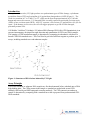
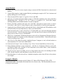
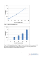
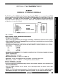
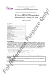
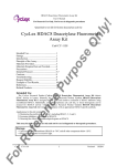
Product Manual OxiSelect™ UV-Induced DNA Damage ELISA Kit (CPD Quantitation), Trial Size Catalog Number STA-322-T 32 assays FOR RESEARCH USE ONLY Not for use in diagnostic procedures Introduction Absorption of ultraviolet (UV) light produces two predominant types of DNA damage, cyclobutane pyrimidine dimers (CPD) and pyrimidine (6-4) pyrimidone photoproducts (6-4PP) (Figure 1). The result is a transition of C to T and CC to TT, which are the most frequent mutations of p53 in both human and mouse skin cancers. UV damaged DNA is usually repaired by nucleotide excision repair (NER) or base excision repair (BER). After UV exposure, cells activate p53 and stall the cell cycle for repair. If the damage is too severe, the cell will trigger apoptosis to get rid of DNA damaged, potentially mutant cells. Cell Biolabs’ OxiSelect™ Oxidative UV-induced DNA Damage ELISA Kit (CPD Quantitation) is an enzyme immunoassay developed for rapid detection and quantitation of CPD in any DNA samples. The quantity of CPD in unknown sample is determined by comparing its absorbance with that of a known CPD-DNA standard curve. This Trial Size kit provides sufficient reagents to perform up to 32 assays, including standard curve and unknown samples. Figure 1: Structures of DNA lesions induced by UV Light Assay Principle CDP-DNA standards or unknown DNA samples are first heat denatured before adsorbed onto a DNA high-binding plate. The CPDs present in the sample or standard are probed with an anti-CPD antibody, followed by an HRP conjugated secondary antibody. The CPD content in an unknown sample is determined by comparing with a standard curve that is prepared from predetermined CPDDNA standards. 2 Related Products 1. STA-320: OxiSelect™ Oxidative DNA Damage ELISA Kit (8-OHdG Quantitation) 2. STA-321: OxiSelect™ DNA Double-Strand Break (DSB) Staining Kit 3. STA-323: OxiSelect™ UV-induced DNA Damage ELISA Kit (6-4PP Quantitation) 4. STA-324: OxiSelect™ Oxidative DNA Damage Quantitation Kit (AP sites) 5. STA-325: OxiSelect™ Oxidative RNA Damage ELISA Kit (8-OHG Quantitation) 6. STA-326: OxiSelect™ Cellular UV-induced DNA Damage ELISA Kit (CPD) 7. STA-327: OxiSelect™ Cellular UV-induced DNA Damage Staining Kit (CPD) 8. STA-328: OxiSelect™ Cellular UV-induced DNA Damage ELISA Kit (6-4PP) 9. STA-329: OxiSelect™ Cellular UV-induced DNA Damage Staining Kit (6-4PP) Kit Components 1. DNA High-Binding Plate (Part No. 232201-T): One strip well plate containing 32 wells (8 x 4), precoated with DNA binding matrix. 2. Anti-CPD Antibody (Part No. 232202-T): One 10 µL vial of anti-CPD. 3. Secondary Antibody, HRP Conjugate (Part No. 10902): One 50 µL vial. 4. Blocking Reagent (100X) (Part No. 232206-T): One 100 μL tube. 5. Assay Diluent (Part No. 310804-T): One 20 mL bottle. 6. 10X Wash Buffer (Part No. 310806-T): One 30 mL bottle. 7. Substrate Solution (Part No. 310807-T): One 4 mL amber bottle. 8. Stop Solution (Part. No. 310808-T): One 4 mL bottle. 9. CPD-DNA Standard (Part No. 232203-T): One 40 µL vial of 25 μg/mL CPD-DNA in 1X TE Buffer. Materials Not Supplied 1. DNA samples such as cell or tissue genomic DNA 2. DNA Extraction Kit 3. Heating Block 4. 5. 6. 7. 8. PBS 10 µL to 1000 µL adjustable single channel micropipettes with disposable tips 50 µL to 300 µL adjustable multichannel micropipette with disposable tips Multichannel micropipette reservoir Microplate reader capable of reading at 450 nm (620 nm as optional reference wave length) 3 Storage Upon receipt, store the CPD-DNA standard at -20ºC and all other components at 4ºC until their expiration dates. Preparation of Reagents • 1X Wash Buffer: Dilute the 10X Wash Buffer to 1X with deionized water. Stir to homogeneity. • 1X Blocking Reagent: Prepare the appropriate volume for the number of samples being tested. Immediately prior to using, dilute the provided 100X Blocking Reagent 1:100 in 1X PBS. Do not store. • Anti-CPD Antibody and Secondary Antibody: Immediately before use dilute the Anti-CPD Antibody 1:1000 and Secondary Antibody 1:1000 with Assay Diluent. Do not store diluted solutions. Preparation of Standard Curve 1. Convert CPD-DNA standard (25 μg/mL) to single-stranded DNA by incubating the DNA sample at 95ºC for 10 minutes and rapidly chilling on ice for 10 minutes. Note: Aliquot and store denatured CPD-DNA standard at -20ºC. Repeat the above denaturation step every time you prepare the CPD-DNA standard. 2. Dilute desired amount of freshly denatured DNA sample 10 fold to 2.5 µg/mL in cold PBS. For example, add 10 µL of the 25 µg/mL CPD-DNA standard to 90 uL of cold PBS. Prepare a dilution series of CPD-DNA standards in the concentration range of 0 ng/mL – 250 ng/mL by diluting the denatured CPD-DNA Standard in cold PBS according to Table 1 below. Standard Tubes 1 2 3 4 5 6 7 8 2.5 µg/mL Denatured CPD-DNA Standard (µL) 80 400 of Tube #1 400 of Tube #2 400 of Tube #3 400 of Tube #4 400 of Tube #5 400 of Tube #6 0 Cold PBS (µL) 720 400 400 400 400 400 400 400 Table 1. Preparation of CPD-DNA Standards 4 CPD-DNA (ng/mL) 250 125 62.5 31.3 15.6 7.8 3.9 0 Assay Protocol 1. Extract DNA from cell or tissue samples using a commercial DNA Extraction kit or other desired method. 2. Convert DNA sample to single-stranded DNA by incubating the sample at 95ºC for 10 minutes and rapidly chilling on ice for 10 minutes. 3. Dilute denatured DNA sample to 2 µg/mL or less in cold PBS. 4. Add 100 µL of unknown denatured DNA sample or CPD-DNA standards to the wells of the DNA High-Binding plate. Incubate at 37ºC for 2 hours or overnight at 4ºC. Each DNA sample including unknown and standard should be assayed in duplicate. 5. Remove the DNA solutions and wash twice with PBS. Blot plate on paper towels to remove excess fluid. Add 150 μL of Assay Diluent to each well and block for 1 hour at room temperature. 6. Aspirate the Assay Diluent from the wells and add 100 µL of the diluted anti-CPD antibody to each well and incubate at room temperature for 1 hour on an orbital shaker. 7. Wash microwell strips 3 times with 250 µL 1X Wash Buffer per well with thorough aspiration between each wash. After the last wash, empty wells and tap microwell strips on absorbent pad or paper towel to remove excess 1X Wash Buffer. 8. Add 150 µL of prediluted 1X Blocking Reagent to each well (see Preparation of Reagents Section). Incubate the plate for 60 minutes at room temperature on an orbital shaker. Wash microwell strips 3 times according to step 7 above. 9. Add 100 µL of the diluted Secondary Antibody-Enzyme Conjugate to each well and incubate at room temperature for 1 hour on an orbital shaker. 10. Wash microwell strips 3 times according to step 7 above. Proceed immediately to the next step. 11. Warm Substrate Solution to room temperature. Add 100 µL of Substrate Solution to each well, including the blank wells. Incubate at room temperature on an orbital shaker. Actual incubation time may vary from 2-30 minutes. 12. Stop the enzyme reaction by adding 100 µL of Stop Solution into each well, including the blank wells. Results should be read immediately (color will fade over time). 13. Read absorbance of each microwell on a standard microplate reader using 450 nm as the primary wave length. Example of Results The following figures demonstrate typical Oxidative UV-induced DNA Damage ELISA (CPD Quantitation) results. One should use the data below for reference only. This data should not be used to interpret actual results. 5 Figure 2: CPD-DNA Standard Curve. Figure 3: DNA Damage Induced by UV Light. 0.2 mg/mL Calf thymus DNA was exposed to UV light inside a cell culture hood for the time indicated. The CPD levels in 40 ng denatured DNA samples were determined as described in the Assay Protocol. 6 References 1. Lippke JA, Gordon LK, Brash DE, Haseltine WA. (1981) Proc Natl Acad Sci U S A. 78:3388– 3392. 2. Mitchell DL, Nairn RS. (1989) Photochem Photobiol. 49:805–819. 3. Ananthaswamy HN, Loughlin SM, Cox P, Evans RL, Ullrich SE, Kripke ML. (1997) Nat Med. 3:510–514. 4. Soehnge H, Ouhtit A, Ananthaswamy ON. (1997) Front Biosci. 2:D538–D551. 5. el-Deiry WS, Tokino T, Velculescu VE, Levy DB, Parsons R, Trent JM, Lin D, Mercer WE, Kinzler KW, Vogelstein B. (1993) Cell. 75:817–825. 6. Hermeking H, Lengauer C, Polyak K, He TC, Zhang L, Thiagalingam S, Kinzler KW, Vogelstein B. (1997) Mol Cell. 1:3–11. 7. Hill LL, Ouhtit A, Loughlin SM, Kripke ML, Ananthaswamy HN, Owen-Schaub LB. (1999) Science. 285:898–900. Recent Product Citations 1. Rimann, M. et al. (2015). Standardized 3D bioprinting of soft tissue models with human primary cells. J Lab Autom. doi:10.1177/2211068214567146. 2. Donninger, H. et al. (2015). The RASSF1A tumor suppressor regulates XPA-mediated DNA repair. Mol Cell Biol. 35:277-287. 3. Zirkin, S. et al. (2013). The PIM-2 Kinase is an essential component of the ultraviolet damage response that acts upstream to E2F-1 and ATM. J. Biol. Chem. 288:21770-21789. 4. Burgess, H.M. et al. (2011). Nuclear relocalisation of cytoplasmic poly(A)-binding proteins PABP1 and PABP4 in response to UV irradiation reveals mRNA-dependent export of metazoan PABPs. J. Cell Sci. 124: 3344-3355. Warranty These products are warranted to perform as described in their labeling and in Cell Biolabs literature when used in accordance with their instructions. THERE ARE NO WARRANTIES THAT EXTEND BEYOND THIS EXPRESSED WARRANTY AND CELL BIOLABS DISCLAIMS ANY IMPLIED WARRANTY OF MERCHANTABILITY OR WARRANTY OF FITNESS FOR PARTICULAR PURPOSE. CELL BIOLABS’ sole obligation and purchaser’s exclusive remedy for breach of this warranty shall be, at the option of CELL BIOLABS, to repair or replace the products. In no event shall CELL BIOLABS be liable for any proximate, incidental or consequential damages in connection with the products. Contact Information Cell Biolabs, Inc. 7758 Arjons Drive San Diego, CA 92126 Worldwide: +1 858-271-6500 USA Toll-Free: 1-888-CBL-0505 E-mail: [email protected] www.cellbiolabs.com 2013-2015: Cell Biolabs, Inc. - All rights reserved. No part of these works may be reproduced in any form without permissions in writing. 7