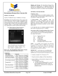
Download Assay Kit (CPRG)
Transcript
Assay Kit (CPRG)
Cat. No.
A10100K
Content
5X Lysis Buffer
Standard Dilution Buffer
Substrate Buffer
Stop Buffer
10X CPRG Substrate Stock
(Chlorophenol red-β-D-galactopyranoside)
β-gal enzyme standard, 40 units
Shipping Condition
A Division of Gene Therapy Systems, Inc
Qty
55 ml
55 ml
55 ml
55 ml
5 x 1 ml
Storage
4°C
4°C
4°C
4°C
-20°C
100 µl
-20°C
RELATED PRODUCTS
GenePORTER™ Transfection Reagent, 75 reactions
GenePORTER™ Transfection Reagent, 150 reactions
GenePORTER™ Transfection Reagent, 750 reactions
GenePORTER™ 2 Transfection Reagent, 75 reactions
GenePORTER™ 2 Transfection Reagent, 150 reactions
GenePORTER™ 2 Transfection Reagent, 750 reactions
gWiz™ β-galactosidase Expression Vector, 25 µg
β-galactosidase Assay Kit (ONPG)
X-Gal Staining Kit
Blue ice.
Catalog #
T201007
T201015
T201075
T202007
T202015
T202075
P010200
A10200K
A10300K
INTRODUCTION
LacZ is a commonly used reporter gene in transfection experiments because the gene product, β-galactosidase, is
very stable, resistant to proteolytic degradation, and easy to assay. Levels of active β-galactosidase expression can be quickly measured by
its catalytic hydrolysis of Chlorophenol red-β-D-galactopyranoside (CPRG) substrate to a dark red product. The assay kit provides all the
required reagents, and offers a rapid, simple and sensitive method to quantify the enzyme expression in transfected cells (e.g., transfected
with Genlantis’ gWiz β-gal vector). The high sensitivity improves the measurement of β-gal activity when the reporter gene expression is low.
USAGE
• Transfect cells with a plasmid expressing LacZ gene.
• Lyse cells using lysis buffer.
• Transfer the lysate to a fresh tube or a microtiter plate. Dilute the lysate if needed.
• Prepare a β-galactosidase standard curve with standard dilution buffer.
• Add the substrate and monitor the color development at 570-595 nm.
• Calculate the expression levels based on a standard curve.
EXAMPLE PROTOCOLS
NOTE: A quick freeze/thaw cycle (freeze 1-2 hours at
–20°C or –70°C then thaw at room temperature) of the dish
can be performed to improve lysis. Keep dish frozen at
–70oC until ready to proceed to the colorimetric assay.
OPTIONAL: Before proceeding to the colorimetric assay,
the plate or dish can be centrifuged for 2-3 minutes to pellet
the insoluble material. Use the supernatant for the assay.
NOTE: Dilute 5X Lysis buffer to 1X with distilled deionized water
before use. Unused 1X Lysis Buffer may be stored at 4°C for
future use.
A. Protocol For Adherent Cells
1. Aspirate the growth medium from the culture dish 24-72
hours after transfection (use non-transfected cells for
control). Optionally, wash cells 1 time with 1X PBS.
2. Add 1X Lysis Buffer to the culture dish. Use the following
recommended volumes depending on your culture dish:
Type of Dish
96-well plate
24-well plate
12-well plate
6-well plate
60 mm dish
100 mm dish
3.
4.
B.
1.
1X Lysis Buffer Volume(µl/well or dish)
50
250
500
1000
2500
5000
2.
3.
Incubate the dish 10-15 minutes at room temperature by
swirling it slowly several times to ensure complete lysis.
The culture dishes can be observed under a microscope to
confirm that the cells are lysed completely.
Proceed to Section C. Below.
VKM021607
10190 Telesis Court. San Diego, CA 92121
4.
Protocol For Suspension Cells
Aspirate the supernatant 24-72 hours post-transfection after
centrifuging cells at 250 x g for 5 minutes. Optionally, wash
cells 1 time with 1X PBS.
Resuspend the cell pellet in 1X Lysis Buffer. The amount of
Lysis Buffer depends on the size of the culture dishes used
for transfection (i.e., cell pellet size); we recommend using
between 50 to 2000 µl.
Incubate the cell lysate 10-15 minutes at room temperature
by gently swirling the dishes several times to ensure
complete lysis. Proceed to the colorimetric assay or freeze
the plate at –70°C until ready.
NOTE: See NOTE above.
OPTIONAL: See OPTIONAL above.
Proceed to Section C. Below.
Genlantis
Page 1 of 2
Toll Free: (888) 428-0558 • (858) 457-1919 • Web: http://www.genlantis.com
Enhanced β-Galactosidase Assay Kit (CPRG) – Cat#A10100K
C. Colorimetric CPRG Assay
NOTE: Dilute 10X CPRG stock to 1X with Substrate Buffer
just before performing the colorimetric assay. Unused 1X
CPRG may be stored at –20°C for future use. We
recommend using 1X CPRG solution only 2 times after a
freeze/thaw cycle.
CAUTION: Wear Gloves for manipulating the CPRG since it
will stain exposed skin.
6.
7.
96-well Microtiter Plate Assay*
1.
2.
3.
Thaw the dish, tube or plate of lysed cells at room
temperature. If the transfection is performed with a 96well plate, perform the assay directly on the plate (flat
bottom only).
Add 50 µl of Standard Dilution Buffer to the wells of a 96well plate except control wells, which are set aside for a
standard curve (see below).
Prepare a serial dilution of β-galactosidase (E. coli)
standards using the Standard Dilution Buffer separately.
A 50 µl aliquot of each point on the standard curve is
transferred to the control wells of the plate - the highest
recommended amount of β-galactosidase is 100
milliunits (100,000~200,000 pg). 2X serial dilution of
standard curve consisting of 8 points is recommended. A
dilution protocol example is shown in the following table:
β-gal
Standard
(miliunits)
Standard
Dilution Buffer
Volume
β-gal Standard Volume
100
50
25
12.5
6.25
3.125
1.562
0.78
995 µl
200 µl
200 µl
200 µl
200 µl
200 µl
200 µl
200 µl
5 µl of β-gal standard stock
200µl 100 mu β-gal standard
200 µl of 50 mu β-gal standard
200 µl of 25 mu β-gal standard
200 µl of 12.5 mu β-gal standard
200 µl of 6.25 mu β-gal standard
200 µl of 3.125 mu β-gal standard
200 µl of 1.562 mu β-gal standard
NOTES: Adjust the standard curve to suit the specific
experimental conditions, such as cell type, transfection
reagent, or plasmid vector. The dilutions for the standard
curve must be prepared fresh each the assay.
4.
5.
Add 50 µl of each sample/well.
NOTE: It may be necessary to dilute the cell lysate in 1X
Lysis Buffer when transfection efficiency is very high. In
contrast, when transfection efficiency is low, reduce the
volume of lysis buffer used to harvest the cells (see
description above) or use up to 150 µl of cell extract for
the colorimetric assay. If the transfection is performed in
a 96-well plate, perform the assay directly on the plate.
Prepare a blank by adding 50 µl of lysis buffer to a well.
Add also 50 µl of cell lysate from non-transfected cells
(negative control) to a well to control endogenous βgalactosidase activity.
VKM021607
10190 Telesis Court. San Diego, CA 92121
8.
Add 100 µl of 1X CPRG Substrate Solution to each well.
Incubate plate at room temp until dark red color develops
(~10 minutes to 4 hours depending on cell type).
Read the absorbance at 570-595 nm with a microtiter
spectrophotometer. Stop solution is not required for this
format since all wells are read simultaneously without a
time gap. Be sure that there are no bubbles present in
the wells while reading because they interfere with the
absorbance reading (remove bubbles with a fine gauge
needle, tips, or very weak gas flow).
Quantify β-galactosidase expression based on a linear
standard curve.
Macro assay
1.
2.
3.
4.
5.
5.
6.
Thaw the cell lysate and transfer 100 µl to a fresh tube,
or 50 µl to a 96-well plate. If a 96-well plate is used,
follow the protocol described above.
NOTE: It may be necessary to dilute the cell lysate in 1X
Lysis Buffer when transfection efficiency is very high. In
contrast, when transfection efficiency is low, reduce the
volume of lysis buffer used to harvest the cells (see
description above) or use up to 150 µl of cell extract for
the colorimetric assay.
Prepare a blank by adding 100 µl of lysis buffer to a tube.
Add also 100 µl of cell lysate from non-transfected cells
(negative control) to a tube to control endogenous βgalactosidase activity.
Add 50 µl of Standard Dilution Buffer to each tube.
Prepare a serial dilution of β-galactosidase (E. coli)
standards with Standard Dilution Buffer separately.
Transfer 50 µl of each standard to a fresh tube
containing 100 µl cell lysate from a mock transfection.
The highest recommended amount of beta-galactosidase
is 200,000 pg. (100 milliunits). Adjust the standard curve
to suit the specific experimental conditions, such as cell
type, transfection reagent, or plasmid vector. 2X serial
dilution of standard curve consisting of 8 points is
recommended. A dilution protocol example is shown in
the section of 96-well plate assay.
Add 300 µl of 1X CPRG Substrate Solution to each tube.
Incubate the tubes at room temperature till the red color
develops (from approximately 10 minutes to 4 hours
depending on the cell type). Add 500 µl of Stop Solution
to stop the reaction. Final volume is 950 µl.
Read the absorbance at 570-595 nm with a
spectrophotometer.
Quantify β-galactosidase expression based on a linear
standard curve.
*Felgner, J.H. et al. Enhanced gene delivery and mechanism studies
with a novel series of cationic lipid formulations. J. Biol. Chem. 269,
2550-2561 (1994).
Genlantis
Page 2 of 2
Toll Free: (888) 428-0558 • (858) 457-1919 • Web: http://www.genlantis.com


