1
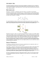
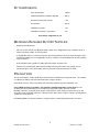
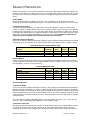
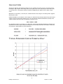
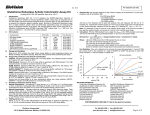
B-Bridge International, Inc.
Glutathione
Colorimetric Detection Kit
User Manual
Catalog # K3006-C
1
B-Bridge International, Inc.
Glutathione 110628
TABLE OF CONTENTS
Intended Use
3
Background
3
Assay Principle
3
Kit Components
4
Materials Required
4
Precautions
4
Reagent Preparation
5
Sample Preparation
6
Assay Protocol- End Point
7
Assay Protocol- Kinetic
7
Calculations
8
Typical Standard Curve: Example
8
Notice to Purchaser
This product is to be used for Research Purposes Only. It is not to be used for Drug or
Diagnostic Purposes, nor is it intended for Human Use. B-Bridge products may not be
resold, modified for resale, or used to manufacture commercial products without the
express written consent of B-Bridge International, Inc.
EXCEPT AS OTHERWISE EXPRESSLY SET FORTH IN THIS USER MANUAL, B-BRIDGE
DOES NOT MAKE ANY REPRESENTATION OR WARRANTIES OR CONDITIONS OF ANY
KIND, EITHER EXPRESSED OR IMPLIED, WITH RESPECT TO THE PRODUCTS, OR
INFORMATION DISCLOSED HEREUNDER, INCLUDING, BUT NOT LIMITED TO, THE IMPLIED
WARRANTIES OF MERCHANTABILITY, FIT FOR A PARTICULAR PURPOSE, OR
NONINFRINGEMENT OF THE INTELLECTUAL PROPERTY RIGHTS OF THIRD PARTIES.
B-Bridge International, Inc. All Rights Reserved.
2
B-Bridge International, Inc.
Glutathione 110628
INTENDED USE
The B-Bridge Glutathione Colorimetric Detection Kit (cat.# K3006-C) quantitatively measures Glutathione
(GSH) and oxidized glutathione (GSSG) in Human Whole Blood, Serum, Plasma, Erythrocytes, Urine, Cell
Lysates and Tissue Samples. GSH is identical across species and we expect this kit may measure GSH from
sources other than human.
BACKGROUND
Glutathione (L-γ-glutamyl-L-cysteinylglycine; GSH) is the highest concentration non-protein thiol in
mammalian cells and is present in concentrations of 0.5 – 10 mM. GSH plays a key role in many biological
processes, including the synthesis of proteins and DNA, the transport of amino acids, and the protection of
cells against oxidation. Harmful hydrogen peroxide cellular levels are minimized by the enzyme glutathione
peroxidase (GP) using GSH as a reductant.
The oxidized GSH dimer, GSSG, is formed from GSH and peroxide by the GP reaction (see below). An
important role of GSSG in the NFκß activating signal cascade is suggested by the facts that the potent NFΚß
inducer, tetradecanoyl phorbol acetate, increases intracellular GSSG levels and GSSG/GSH ratios.
Glutathione S-transferases (GST) are an important group of enzymes that catalyze the nucleophilic addition
of GSH to electrophiles. They are encoded by 5 gene families; 4 encode cytosolic GST and one encodes the
microsomal form of GST. They have been implicated in a number of diseases. In asthma arachidonic acid is
converted to unstable leukotriene A4 (LTA4). LTA4 is either hydrated to form LTB4 or it is conjugated to GSH
by a GST, leukotriene C4 synthase, to form leukotriene C4. LTC4 and its derivative LTD4 are important
molecules in bronchial asthma. Leukotriene C4 synthase is therefore an important therapeutic target. It has
also been shown that increased expression of GSTs can lead to drug resistance. Three glutathione adducts
of the drug melphalan, used to treat ovarian cancer and multiple myeloma, have been isolated from reactions
involving human microsomal GSTs.
ASSAY PRINCIPLE
The Glutathione Colorimetric Detection Kit is designed to quantitatively measure glutathione (GSH), and
oxidized glutathione (GSSG) present in a variety of samples.
The kit utilizes a colorimetric substrate that reacts with the free thiol group on GSH to yield a highly colored
product. Supplied reagents are in solution and require simple dilution for use in the assay. By using
2-Vinylpyridine (not supplied) to block any free GSH in the sample, Oxidized Glutathione (GSSG) can be
determined. Any samples that have not been treated with 2-Vinylpyridine will yield Total GSH levels. The
Free GSH concentration in the sample is calculated from the difference between the Total GSH determined
and the GSH generated from Oxidized Glutathione for the 2-Vinylpyridine treated samples.
Our Fluorescent Glutathione Detection kit (Catalog Number K3006-1) allows for the measurement of both
Free and Oxidized Glutathione with higher sensitivity in the same sample in the same well without using
2-Vinylpyridine.
3
B-Bridge International, Inc.
Glutathione 110628
KIT COMPONENTS
Clear 96-well plate
4 plates
Oxidized Glutathione Standard (250 µM)
200 µL
Detection Reagent Concentrate
1 mL
Assay Buffer
225 mL
NADPH Concentrate
1 mL
Glutathione Reductase Concentrate
1mL
Store above components at 4°C
MATERIALS REQUIRED BUT NOT SUPPLIED
-
Deionized or distilled water
-
Aqueous 5-sulfo-salicylic acid dihydrate (SSA) solution at 5% weight/volume (1g of SSA per 20 mL of
water) for treating samples to remove protein.
-
2-Vinylpyridine (2VP) is used to block any free GSH or other thiols present in the treated samples. 2VP is
prepared by adding 27 µL of 2-vinylpyridine to 98 µL of ethanol. Use immediately and discard remaining
unused solutions.
-
A 96 well plate reader capable of reading optical absorption at 405-412 nm.
-
Software for converting raw optical density readings from the plate reader and carrying out four
parameter logistic curve (4PLC) fitting. Contact your plate reader manufacturer for details
PRECAUTIONS
This kit should only be used by qualified personnel who have had laboratory safety instruction. The complete
User Manual should be read and understood before using this product.
Sulfosalicylic acid is a strong acid solution and should be treated like any other laboratory acid.
2VP is TOXIC and may cause burns. 2VP solutions should be prepared in a fume hood. Use immediately and discard remaining unused solutions by mixing with copious amounts of water.
Dimethyl sulfoxide is a powerful aprotic organic solvent that has been shown to enhance the rate of skin
absorption of skin-permeable substances. Wear protective gloves when using the solvent especially when it
contains dissolved chemicals.
In all cases, please consult your institution’s safety procedures for working with hazardous chemicals.
4
B-Bridge International, Inc.
Glutathione 110628
REAGENT PREPARATION
Allow the kit reagents to come to room temperature for 30 minutes. We recommend that all standards and
samples be run in duplicate to allow the end user to accurately determine GSH concentrations. Ensure that
all samples have reached room temperature and have been diluted as appropriate prior to running them in
the kit.
Sample Diluent
Prepare the Sample Diluent by diluting one part 5% SSA 1:5 with four parts Assay Buffer and vortex
thoroughly. The pH of the Sample Diluent must be > 6. Sample Diluent can be stored at 4°C for one month.
2-Vinylpyridine Treatment
To measure Oxidized Glutathione, free GSH must be blocked by alkylation. To 250 µL of SSA treated
samples, standards or Sample Diluent add 5 µL of the ethanolic solution of 2VP (see page 4) and allowed to
incubate at room temperature for 1 hour. The 2VP treated samples and standards should then be diluted in
Assay Buffer and Sample Diluent according to the dilutions recommended for each sample type prior to using
in the assay. The 2VP treated Sample Diluent is used for the zero standard. Samples treated with 2VP
should be read off a standard curve generated with 2VP treated standards.
Colorimetric Detection Reagent
Prepare the Colorimetric Detection Reagent by diluting one part Colorimetric Detection Reagent Concentrate
1:10 with nine parts Assay Buffer. See Colorimetric Detection Reagent Dilution Table for suitable volumes.
Colorimetric Detection Reagent Dilution Table
Whole
Reagent
Half plate
plate
Colorimetric Detection Concentrate
140 µl
260 µl
Assay Buffer
1.26 mL
2.34 mL
Total Colorimetric Reagent Volume
1.4 mL
1.6 mL
Two
Plates
500 µl
4.5 mL
5 mL
Four
Plates
1 mL
9 mL
10 mL
Reaction Mixture
Prepare the Reaction Mixture by diluting one part each NADPH and Glutathione Reductase Concentrates
1:10 into eight parts Assay Buffer. See Reaction Mix Dilution Table for suitable volumes. Store any unused
Reaction Mixture at 4°C for no more than 2 days.
Reaction Mix Dilution Table
Reagent
NADPH Concentrate
Glutathione Reductase Concentrate
Assay Buffer
Total Reaction Mix Volume
Half plate
140 µl
140 µl
1.12 mL
1.4 mL
Whole
plate
260 µl
260 µl
2.08 mL
1.6 mL
Two
Plates
500 µl
500 µl
4 mL
5 mL
Four
Plates
1 mL
1 mL
8 mL
10 mL
Standard Preparation
To Determine GSSG
For the measurement of Oxidized Glutathione (GSSG), a 50 µL aliquot of the 250 µM Oxidized Glutathione
Standard should be treated with 1 µL of2VP as outlined on page 9. 2VP-treated Standards are prepared by
labeling six test tubes as #1 through #6. Pipet 475 µL of Sample Diluent into tube #1 and 250 µL into tubes #2
to #6. Carefully add 25 µL of the 2VP-treated Standard to tube #1 and vortex completely. Take 250 µL of the
solution in tube #1 and add it to tube #2 and vortex completely. Repeat for tubes #3 through #6 as indicated
in the table below.
The concentration of Oxidized Glutathione in tubes 1 through 6 will be 12.5, 6.25, 3.125, 1.56, 0.781 and
0.391 µM. 2VP treated Sample Diluent must be used as a 0 µM standard.
To Determine Total GSH
Standards are prepared by labeling six test tubes as #1 through #6. Pipet 475 µL of Sample Diluent into tube
#1 and 250 µL into tubes #2 to #6. Carefully add 25 µL of the supplied Standard to tube #1 and vortex
completely. Take 250 µL of the solution in tube #1 and add it to tube #2 and vortex completely. Repeat for
5
B-Bridge International, Inc.
Glutathione 110628
tubes #3 through #6 according to the table below.
The concentration of Total GSH in tubes 1 through 6 will be 25, 12.5, 6.25, 3.125, 1.56, and 0.781 µM after
addition of the Reaction Mixture. Sample Diluent must be used as a 0 µM standard.
Reagent
Sample Diluent
Glutathione Standard
Standard 1
Standard 2
Standard 3
Standard 4
Standard 5
GSSG Concentration
Total GSH Concentration
Standard
1
475 µl
25 µl
Standard
2
250 µl
Standard
3
250 µl
Standard
4
250 µl
Standard
5
250 µl
Standard
6
250 µl
250 µl
250 µl
250 µl
250 µl
12.5 µM
25 µM
6.25 µM
12.5 µM
3.125 µM
6.25 µM
1.56 µM
3.125 µM
0.781 µM
1.56 µM
250 µl
0.391 µM
0.781 µM
Use all Standards within 2 hours of preparation.
SAMPLE PREPARATION
Sample Diluent
Prepare the Sample Diluent by diluting one part 5% SSA 1:5 with four parts Assay Buffer and vortex
thoroughly. The pH of the Sample Diluent must be > 6. Sample Diluent can be stored at 4°C for one month
All samples and standards must be in Sample Diluent before starting the assay
To measure Oxidized Glutathione in samples, reduced Glutathione (GSH) in the sample must be blocked by
treatment with 2-vinylpyridine, 2VP (see page 6 for preparation). SSA treated samples should be treated with
2VP by addition of 5 µL of 2VP solution for every 250 µL of sample (see page 9). 2VP treated samples must
be read off a standard curve made with 2VP-treated standards. Use all samples within 2 hours of dilution.
Whole Blood, EDTA or Heparin Plasma, or Urine
Thoroughly mix sample with an equal volume of cold 5% SSA. Incubate for 10 minutes at 4°C. Centrifuge at
14,000 rpm for 10 minutes at 4°C. Collect the supernatant. If the supernatent contains particulates,
re-centrifuge the supernatant for 15 minutes and collect the clarified second supernatant. Samples can be
stored in aliquots at ≥ -70°C or analyzed immediately. At this point the SSA concentration will be 2.5%.
The supernatant must be diluted 1:2.5 with Assay Buffer by mixing one part with 1.5 parts of Assay Buffer to
bring the SSA concentration to 1%. The sample will have been diluted 1:5 at this point.
All final dilutions are made in Sample Diluent. Treated Whole Blood must be further diluted at least 1:20 for a
recommended final dilution of ≥ 1:100. For Treated Plasma and Treated Urine a final dilution of ≥ 1:5 is
recommended, but further dilutions in Sample Diluent may be necessary.
Tissue Samples
Fresh tissue is washed with ice cold PBS to remove blood then blotted on filter paper before recording wet
weight. NOTE: Samples that have been frozen will contain lysed cells. The PBS wash may contain
substantial amounts of GSH and/or GSSG.
• For Samples Where a Protein Determination is to be Obtained: Homogenize at 10 mg/250 µL in ice cold
100mM phosphate buffer, pH 7. Centrifuge at 14,000 rpm for 10 minutes at 4°C and remove an aliquot of the
supernatant for protein determination. Thoroughly mix a second aliquot of the supernatant with an equal
volume of cold 5% SSA. Incubate for 10 minutes at 4°C. Centrifuge at 14,000 rpm for 10 minutes at 4°C to
remove precipitated protein. Collect the supernatant. The supernatant must be diluted 1:2.5 with Assay
Buffer by mixing one part with 1.5 parts of Assay Buffer. The SSA concentration will be 1%.
• For Samples Not Requiring a Protein Determination: Homogenize at 10 mg/250 µL in ice cold 5% SSA,
incubate at 10 minutes at 4°C, then centrifuge at 14,000 rpm for 10 minutes at 4°C to remove precipitated
6
B-Bridge International, Inc.
Glutathione 110628
protein. Collect the supernatant. The supernatant must be diluted 1:5 with Assay Buffer by mixing one part
with 4 parts of Assay Buffer. The SSA concentration will be 1%.
Further sample dilutions must be determined by the end-user since it will be dependent upon the tissue type
and the amount of tissue used. These dilutions must be made in the prepared Sample Diluent.
Erythrocytes, Red Blood Cells (RBC’s)
Collect blood with heparin or EDTA. Centrifuge the sample, remove and discard the plasma and white cell
layer. Wash the RBC’s 2 times by suspending in 3 volumes of isotonic saline (0.9%), centrifuging at 600 x g
for 10 minutes and discarding the saline wash.
After the 2 washes, mix 250µL RBC’s with 1mL of cold 5% SSA. Incubate for 10 minutes at 4°C and
centrifuge at 14,000 rpm for 10 minutes at 4°C. Collect the supernatant. At this point the SSA concentration
will be 4%. The supernatant must be diluted 1:4 with Assay Buffer by mixing one part with 3 parts of Assay
Buffer. The SSA concentration will now be 1% and the sample will have been diluted 1:20 at this point.
Further dilutions are made in Sample Diluent for a recommended final dilution of ≥ 1:40.
Cell Lysates
Washed cell pellets are resuspended at 1-10x106 cells/mL in cold 5% SSA (we used Jurkats at 5x10⁶
cells/mL) and are lysed and deproteinized by vigorous vortexing, freeze/thaw cycling or other suitable
disruption method. Incubate cells at 4°C for 10 minutes followed by centrifugation for 10 minutes at 14,000
rpm and 4°C. NOTE: Samples that have been frozen will contain lysed cells. The PBS wash may
contain substantial amounts of GSH and/or GSSG.
The deproteinized supernatants must be diluted 1:5 with Assay Buffer by mixing one part with 4 parts of
Assay Buffer. The SSA concentration will be 1%. The sample will have been diluted 1:5 at this point. Further
sample dilutions must be done in Sample Diluent and need to be determined by the end-user since it will be
dependent upon the cell type and number of cells used. The recommended final dilution is ≥ 1:20.
Use all samples within 2 hours of dilution.
ASSAY PROTOCOL- END POINT
For Oxidized Glutathione (GSSG) use the 2VP treated standards, 2VP treated Sample Diluent and 2VP
treated samples diluted with Sample Diluent as described previously.
For Total Glutathione use the standards and samples diluted with Sample Diluent as described previously.
1. Pipet 50 µL of either 2VP treated or untreated samples or standards into duplicate wells in the plate.
2. Pipet 50 µL of either 2VP treated or untreated Sample Diluent into duplicate wells as the Zero Standard.
3. Add 25 µL of the Colorimetric Detection Reagent to each well using a repeater or multichannel pipet.
4. Add 25 µL of the Reaction Mixture to each of the wells using a repeater or multichannel pipet.
5. Gently tap the sides of the plate to ensure adequate mixing of the reagents.
6. Incubate at room temperature for 20 minutes.
7. Read the optical density at 405 nm. These data will be used to determine either
Oxidized Glutathione or Total Glutathione concentration.
ASSAY PROTOCOL- KINETIC
1. Carry out steps 1-3 above.
2. Gently tap the sides of the plate to ensure adequate mixing of the reagents.
3. Add 25 µL of the Reaction Mixture to each of the wells using a repeater and immediately place plate in
reader and read optical density at 405 nm every minute for at least
10 minutes. These data will be used to determine Total or Oxidized Glutathione
concentration kinetically.
7
B-Bridge International, Inc.
Glutathione 110628
CALCULATIONS
Average the duplicate optical density readings for each standard and sample. Create a standard curve by
reducing the data using the 4PLC fitting routine on the plate reader, after subtracting the mean ODs for the
zero standard. The concentrations obtained should be multiplied by the dilution factor to obtain sample
values.
Glutathione concentrations (see below) are calculated from the data using the curve fitting routine supplied
with the plate reader.
Oxidized Glutathione concentrations of the samples are determined from the data obtained from 2VP-treated
samples read off a 2VP-treated standard curve. The concentration of Oxidized Glutathione (GSSG) in the
samples would be half of the GSH concentration read off the curve.
Note: 1 GSSG = 2 GSH
Free glutathione (GSH) concentrations are obtained by subtracting the Oxidized Glutathione (GSSG) levels
obtained from the 2VP treated standard and samples from non-treated standards and samples (Total GSH).
Concentrations obtained will be in µM of Glutathione.
TYPICAL STANDARD CURVE: EXAMPLE ONLY
8
B-Bridge International, Inc.
Glutathione 110628