1
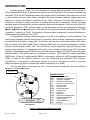
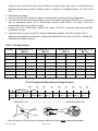
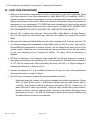
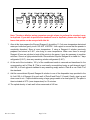
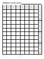
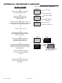
Adipocyte Lipolysis Assay Kit for 3T3-L1 Cells: Glycerol Detection Cat# LIP-1-L1; LIP-1-NCL1, LIP-1-L1-F, LIP-1-NCL1DIF INSTRUCTION MANUAL ZBM0008.05 STORAGE CONDITIONS 96-well plate cultured 3T3-L1 preadipocytes (LIP-1-L1 only): 37°C incubator Cryopreserved 3T3-L1 preadipocytes ( LIP-1-L1-F only): liquid nitrogen o This product is shipped on dry ice. Remove and store in liquid nitrogen storage immediately upon arrival. Glycerol Reagent A & Buffers: 4°C Use reconstituted Glycerol Reagent A within 7 days! Glycerol Standard & Controls: -20°C Media: 4°C or -20°C [see label for details] All Zen-Bio Inc products are for research use only. Not approved for human or veterinary use or for use in diagnostic or clinical procedures. LIMITED PRODUCT WARRANTY This warranty limits our liability to replacement of this product. No other warranties of any kind, expressed or implied, including without limitation, implied warranties of merchantability or fitness for a particular purpose, are provided by Zen-Bio, Inc. Zen-Bio, Inc. shall have no liability for any direct, indirect, consequential, or incidental damages arising out of the use, the results of use, or the inability to use this product. ORDERING INFORMATION AND TECHNICAL SERVICES Zen-Bio, Inc. 3200 East North Carolina Highway 54 Suite 100 PO Box 13888 Research Triangle Park, NC 27709 Telephone (919) 547-0692 Facsimile (FAX) (919) 547-0693 Toll Free 1-866-ADIPOSE Electronic mail (e-mail) World Wide Web Rev. 01.11.2012 (866)-234-7673 [email protected] http://www.zen-bio.com Page 1 of 12 INTRODUCTION Lipolysis plays a central role in the regulation of energy balance. Lipolysis is the process in which triglycerides (TG) are hydrolyzed into glycerol and free fatty acids. This process releases free fatty acids (FFA) into the bloodstream where they may be either re-esterified by the adipocyte or travel to other tissues and exert other effects throughout the body. Elevated adipocyte lipolysis has been observed in obese and diabetic individuals (Arner 1996). Excessive free fatty acid production is believed to contribute to insulin resistance in skeletal muscle that is observed in obesity. Hormone sensitive lipase is the rate-limiting enzyme catalyzing triglyceride breakdown. Perilipins, one of the PAT (perilipins, adipophilin, TIP47 proteins) family of lipid-associated proteins, are implicated in adipocyte lipolysis by mediating the interaction of HSL with the triacylglycerol molecule (Brasaemle et al. 2004; reviewed in, Tansey et al. 2004.) The presence of these proteins corresponds to lipolytic stimulation in cultured adipocytes (Braemle et al. 2004). The sympathetic nervous system also plays a key role in the regulation of lipid mobilization. The main lipolytic pathway involves beta-agonists (-agonists), which activate -adrenergic receptors via the intracellular Gs proteins in adipocytes. This leads to the activation of adenylate cyclase (AC), which then increases cyclic AMP (cAMP) levels. Elevated cAMP acts as a second messenger to activate hormone sensitive lipase (HSL). HSL, the rate-limiting enzyme regulating adipocyte lipolysis, then catalyzes the hydrolysis of triglycerides and results in the release of glycerol and FFA (increased lipolysis). Phosphodiesterases (PDE) are enzymes that hydrolyze cAMP to 5’-AMP (5 prime adenosine monophosphate). This action results in a decrease in lipolysis. PDE inhibitors increase intracellular cAMP levels. 3-isobutyl-1-methylxanthine (IBMX), a non-specific inhibitor of cAMP phosphodiesterases (PDE), is used as the positive control if your test compounds are suspected PDE inhibitors. Isoproterenol, a non-specific -adrenergic agonist is used as the positive control if your test compounds affect lipolysis via -adrenergic receptors (Robidoux et al. 2004). This lipolysis assay kit provides the tool to study chemical compounds that may influence lipolysis in cultured adipocytes. EPINEPHRINE 1, 2, 3 AR NOREPINEPHRINE ABBREVIATIONS: AC adenylate cyclase AMP adenosine monophosphate AR adrenergic receptors ATP adenosine triphosphate Gs G protein coupled receptor IR insulin receptor FFA free fatty acids PDE phosphodiesterase PKA protein kinase Per perilipins TG triglyceride AMP adenosine monophosphate ATP adenosine triphosphate IR insulin receptor PDE phosphodiesterase Per perilipins AC Gs IR PDE ATP P cAMP 5’-AMP PKA TG Per HSL FFA + glycerol FFA + glycerol bloodstream Figure 1. Overview of adipocyte lipolysis Rev. 01.11.2012 Page 2 of 12 PRINCIPLE OF THE ASSAY Lipolytic activity is assessed by the measurement of glycerol released into the medium from triglyceride breakdown. Glycerol released to the medium is phosphorylated by adenosine triphosphate (ATP) forming glycerol-1-phosphate (G-1-P) and adenosine-5’-diphosphate (ADP) in the reaction catalyzed by glycerol kinase. G-1-P is then oxidized by glycerol phosphate oxidase to dihydroxyacetone phosphate (DAP) and hydrogen peroxide (H2O2). A quinoneimine dye is produced by the peroxidase catalyzed coupling of 4-aminoantipyrine (4AAP) and sodium N-ethytl-N-(3-sulfopropyl)m-anisidine (ESPA) with H2O2, which shows an absorbance maximum at 540nm. The increase in absorbance at 540nm is directly proportional to glycerol concentration of the sample. GLYCEROL + ATP G-1-P + O2 G-1-P + ADP DAP + H2O2 H2O2 +4-AAP + ESPA Quinoneimine dye + H2O ITEMS INCLUDED IN THE KIT ITEM PLATE QTY 1 STORAGE 37°C VIAL 1 Liquid N2 PLATE 2 ----- 3T3-L1 Preadipocyte Medium 50ml (LIP-1-L1, LIP-1-L1-F, LIP-1-NCL1DIF ONLY) BOTTLE 1 4°C 3T3-L1 Adipocyte Differentiation); 15ml (LIP-1-L1, LIP-1-L1-F, LIP-1-NCL1DIF ONLY BOTTLE 1 4°C 3T3-L1 Adipocyte Maintenance; 100ml (LIP-1-L1, LIP-1-L1-F, LIP-1-NCL1DIF ONLY BOTTLE 1 4°C (cat# AM-1-L1) Assay Buffer 100 ml BOTTLE 1 4°C Wash Buffer 50 ml BOTTLE 1 4°C Vehicle (green cap) Positive Control (blue cap) 0.1% DMSO in Assay Buffer READY TO USE. 1 ml / VIAL 10 l / 1 -20°C 1 -20°C Alternate Positive Control (red cap) 3-Isobutyl-1-methylxanthine (IBMX), 100 mM in DMSO Dilute to 100 M in Assay Buffer before use! (i.e. 1 l in 1 ml Assay Buffer) Reconstitute with 11.0 ml deionized water prior to use. 1 -20°C BOTTLE 1 4°C EACH 2 ----- 100 l / 1 -20°C Plate A Assay Plates, blank Preadipocyte Medium (cat# PM-1-L1) Differentiation Medium (cat# DM-2-L1 Adipocyte Medium Glycerol Reagent A (cat# RGTA-10) Tray Glycerol standard (cat# LIP-GLYSTAN) Rev. 01.11.2012 DESCRIPTION UNIT 96 well plate 3T3-L1 preadipocytes, subconfluent (LIP-1-L1 ONLY) Cryopreserved 3T3-L1 preadipocytes, 500,000 cells, Pass 8 96-well assay plate, blank (for samples + standards) Isoproterenol, 10 mM in DMSO. Dilute to 1 M in Assay Buffer before use! (i.e.1 l in 10 ml Assay Buffer) VIAL 10 l / VIAL Use within 7 days! For multi-channel pipetters, clear polyvinyl Glycerol @ 1mM [Reconstitute with 400 l Wash Buffer to make the 200 M glycerol standard; see page 6 for recommended dilution scheme] Page 3 of 12 VIAL Other equipment/reagents required but not provided with the kit: Multi-channel pipet , single channel pipet and pipet tips Sterile trays for multi-channel pipetters during differentiation of cells Plate reader with a filter of 540 nm Incubator at 37oC Large gauge needle Option – Step 5 of Assay Procedure: 96 well plate, blank Tubes for diluting glycerol standards NOTE: THIS KIT IS DESIGNED FOR THE ASSAY OF A 96 WELL PLATE (100 ASSAY POINTS). IF YOU WISH TO TEST ANOTHER PLATE FORMAT, PLEASE CONTACT ZEN-BIO TO PURCHASE ADDITIONAL REAGENTS TO COMPLETE YOUR STUDY. ASSAY PROCEDURE A. DIFFERENTIATION PROCEDURE 1. Preparing 3T3-L1 preadipocytes: 1.1. Cat# LIP-1-L1: 1.1.1. Preadipocytes are plated sub-confluent in 3T3-L1 Preadipocyte Medium (cat# PM-1-L1) and shipped the next day via overnight delivery. Check the seal for each plate. Call Zen-Bio if there is any problem with the shipment. Place the package into a sterile environment. THIS IS VERY IMPORTANT SINCE BREAKING THE VACUUM SEAL MAY POTENTIALLY INTRODUCE CONTAMINATION INTO THE PLATE. Use scissors to snip open the bag at any end to release the vacuum seal. 1.1.2. In a sterile environment, remove the plate from the bag, taking care to not disturb the cover top from the plate. Open the lid and remove the white liner using sterile forceps or a hemostat and discard. Carefully remove the clear adhesive seal by grabbing the edge with sterile forceps or hemostat and lifting the film slowly towards the other end. Discard adhesive film in appropriate biohazard waste container. 1.1.3. Remove 100-150µl per well of the excess medium added to each well for shipping should prior to incubation in a CO2 incubator. Replace lid on plate. 1.1.4. Incubate cells until they are 100% confluent (in about 4-5 days). Cells will need to be fed every other day with PM-1-L1 during this time. See Table 1 for feeding volumes. See Step 2 for further differentiation instructions. 1.2. Cat# LIP-1-L1-F: 1.2.1. Store cells in liquid nitrogen until ready for use. Remove cells from liquid nitrogen and place immediately into a 37C water bath with agitation. Be careful not to submerge the cap of the vial into water. Do not leave the vials in water bath after most of the content has thawed. Rinse the vials with 70% ethanol before taking them to the culture hood. 1.2.2. Upon thawing, add the cells to a sterile conical bottom centrifuge tube, containing 10 ml of 3T3-L1 Preadipocyte Medium (PM-1-L1). Centrifuge at 280 x g, 20C, 5 minutes. 1.2.3. Aspirate the medium and resuspend cells in a volume of PM-1-L1 appropriate for counting the cells. Count using a hemacytometer. Place approximately 5,000-10,000 cells/cm2 in 150µ/ per well volume in a 96-well tissue culture treated cultureware using 3T3-L1 Preadipocyte Medium (PM1-L1). Rev. 01.11.2012 Page 4 of 12 Cells will need to be fed every other day with PM-1-L1 during this time. See Table 1 for feeding volumes. Maintain cells until they are 100% confluent (in about 4-5 days) in a humidified incubator, 37C, with 5-10% CO2. 2. Differentiation procedure: 3. Once the cells are 100% confluent, incubate an additional 48 hours before initiating differentiation. 4. Two days after the cells have been confluent, remove the Preadipocyte Medium (cat# PM-1-L1) and replace with an appropriate volume 3T3-L1 Differentiation Medium (cat# DM-2-L1; see table 1 below for recommended volumes). Incubate for 3 days. 5. Remove the 3T3-L1 Differentiation Medium and replace with 3T3-L1 Adipocyte Maintenance Medium. Incubate for 2-3 days. 6. Feed cells every 2-3 days using 3T3-L1 Adipocyte Maintenance Medium until ready for assay. 3T3-L1 adipocytes are suitable for most assays 7-14 days post differentiation (see Table 1 and 3T3-L1 Growth and Differentiation Feeding Schedule) Table 1. Feeding Volumes Format Change PM-1-L1 to PM-1-L1 OUT IN Change PM-1-L1 to DM-2-L1 OUT IN Change DM-2-L1 to AM-1-L1 OUT IN Change AM-1-L1 to AM-1-L1 OUT IN 96 well plate 90l/well 90l/well 150l/well 150 l / well 90 l /well 120l /well 90 l /well 120l /well 48 well plate 300 l /well 300 l /well 500l /well 500 l /well 300 l /well 400 l /well 300 l /well 400 l /well 24 well plate 0.6 ml/well 0.6 ml/well 1.0 ml/well 1.0 ml/well 0.6 ml/well 0.8 ml/well 0.6 ml/well 0.8 ml/well 12 well plate 1.2 ml/well 1.2 ml/well 2.0 ml/well 2.0 ml/well 1.2 ml/well 1.6 ml/well 1.2 ml/well 1.6 ml/well 6 well plate 1.8 ml/well 1.8 ml/well 3.0 ml/well 3.0 ml/well 1.8 ml/well 2.4 ml/well 1.8 ml/well 2.4 ml/well T-75 flask 12 ml/flask 12 ml/flask 20 ml/flask 20 ml/flask 12 ml/flask 16 ml/flask 12 ml/flask 16 ml/flask T-25 flask 4.2 ml/flask 4.2 ml/flask 7 ml/flask 7 ml/flask 4.2 ml/flask 5.6 ml/flask 4.2 ml/flask 5.6 ml/flask 3T3-L1 Growth and Differentiation Feeding Schedule proliferation Feed PM-1-L1 Feed PM-1-L1 DAY DAY DAY DAY -2 0 3 5 48 hrs Feed 100% PM-1-L1 confluent * Feed DM-2-L1 Feed AM-1-L1 DAY DAY 7** Feed AM-1-L1 Feed AM-1-L1 DAY DAY DAY 11 13 15 9 Feed AM-1-L1 Feed AM-1-L1 Feed AM-1-L1 PREADIPOCYTE MATURE ADIPOCYTE nucleus Lipid droplets nucleus * Once the cells are 100% confluent, incubate an additional 48 hours before initiating differentiation. Rev. 01.11.2012 Page 5 of 12 ** 3T3-L1 adipocytes are suitable for most assays 7-14 days post differentiation B. LIPOLYSIS PROCEDURE 1. Make your stock solution using whatever vehicle is appropriate for your test compounds. Dilute your stock solutions to their final concentration in Assay Buffer (100 ml is available). NOTE: if desired, maintain a constant concentration of solvent by preparing all compound dilutions in the highest concentration of that solvent. Dilute your controls in assay buffer. Prepare all vehicles as appropriate for your compounds, 0.1% DMSO has been included as the vehicle for the positive controls. Include the Assay Buffer alone as a vehicle control. PLEASE NOTE: ZEN-BIO DOES NOT RECOMMEND THE USE OF SOLVENTS AT CONCENTRATIONS ABOVE 1%. 2. Remove 120 l medium from each well. Gently add 200 l Wash Buffer to all wells. Remove 200 l of the media and Wash Buffer from each well and replace with another 200 l Wash Buffer. 3. Remove all the media and Wash Buffer from the cells in triplicate wells. Treat the cells with 150 l of the test compounds resuspended in Assay Buffer three (3) wells at a time. Treat with the diluted IBMX and Isoproterenol as positive controls. Use the Assay Buffer alone as one of the vehicle controls. Please be sure to include both the vehicle provided in the kit and your vehicle (if your test compounds are not dissolved in DMSO). The assay should be performed in triplicate. 4. OPTION: to determine if the compound alone reacts with the Glycerol Reagent A, prepare a fresh plate (not included in kit) containing 100 l of the compound. This plate can be incubated at 37oC with the treated cells. When performing the assay, add 100 l of Glycerol Reagent A following the instructions in Steps 9 and 10. ° 5. Incubate the plates at 37 C in a humidified incubator for 3 hours (for time course experiments the longest time point is usually 24 hours). 6. One hour prior to the assay, prepare the glycerol standards as follows: Briefly spin down the contents of the glycerol standard tube before reconstitution. Pipette 400 l of Wash Buffer into the 1 mM glycerol standard tube provided and mix well by vortexing. This produces a diluted stock glycerol standard of 200 M. Pipette 250 l of wash buffer into 6 tubes (not provided). Using the newly diluted stock glycerol solution, prepare a dilution series as depicted below. Mix each new dilution thoroughly before proceeding to the next. The 200 M stock dilution serves as the highest standard, and the wash buffer serves as the zero standard. Rev. 01.11.2012 Page 6 of 12 400 l Wash Buffer 250 l 250 l 250 l 250 l 250 l 250 l Std 200 M 100 M 50 M 25 M 12.5 M 6.25 M 3.125 M Note: The above dilution series generates enough volume to perform the standard curve in duplicate. If you wish to perform the standard curve in duplicate, please note that eight fewer data points can be assayed with this kit. 7. Also at this time prepare the Glycerol Reagent A by adding 11.0 ml room temperature deionized water per bottle and gently invert. DO NOT VORTEX! Use a pipet to ensure that the powder is completely dissolved. Store at room temperature. If using a Reagent A solution previously prepared and stored at 2-8C, also bring to room temperature. Make sure there is enough Reagent A from one solution to treat all the points in the assay. It may be necessary to combine solutions. Store in a light protected bottle. Reconstituted Glycerol Reagent A is stable for 7 days refrigerated (2-8C); store any remaining solution refrigerated (2-8C). 8. At the end of the incubation, 100 l of the conditioned media is removed and transferred to the corresponding well of Plate B. [This is most easily accomplished using a multi-channel pipet.] Add 100 l of each glycerol standard to any remaining empty wells in Plate B or use Plate C for the standards. 9. Add the reconstituted Glycerol Reagent A solution to one of the disposable trays provided in the kit. Add 100 l of Reagent A to each well of Plate B and Plate C (if used). Gently, pipet up and down once to mix. Pop the bubbles using a large gauge needle or a clean pipet tip. The plate is then incubated at 25oC (room temperature) for 15 minutes. 10. The optical density of each well is then measured at 540 nm. Rev. 01.11.2012 Page 7 of 12 GLYCEROL STANDARD CURVE Generate standard curve: see example below [DO NOT use this standard curve to generate your data. This is an example.] Subtract the OD value of the 0M standard from all OD values including the standard curve. uM glycerol 0 3.125 6.25 12.5 25 50 100 200 OD 0.044 0.054 0.062 0.083 0.126 0.205 0.372 0.698 OD 0.041 0.053 0.063 0.084 0.125 0.208 0.374 0.697 ODblank ODblank 0.012 0.020 0.041 0.084 0.163 0.330 0.656 0.011 0.021 0.042 0.083 0.166 0.332 0.655 Avg ODblank 0.043 0.011 0.020 0.041 0.083 0.164 0.331 0.655 Slope 0.003 Intercept 0.001 R2 1.000 y = observed O.D. minus the blank x = concentration of glycerol in M To calculate x for each y, (i.e. to change the observed O.D. into glycerol concentration) use the following equation: y=(slope) times (x) plus intercept y=mx+b so x=(y-b)/m x=(y – 0.001)/0.003 where 0.003= slope of the line and 0.001= y intercept. Be careful to enter the proper sign for the y intercept value as it may be a negative number. Any OD values greater than the highest standard (200 M) should be suspect. The compound should be re-assayed using a lower dose of the compound at treatment OR a dilute solution of the conditioned medium at the time of the assay. The R2 value should be equal or greater then 0.98 for the standard curve to be valid. Any R2 values below 0.98, must have the standard curve run again. Data are expressed as M glycerol released. OPTION: express data as Fold induction over appropriate vehicle Fold induction = M glycerol SAMPLE M glycerol VEHICLE Rev. 01.11.2012 Page 8 of 12 TROUBLESHOOTING Problem High background or the glycerol reagent A turns purple before the assay begins. Suggestions Use clean tray and tips Change pipet tips frequently Use Glycerol Reagent A before the expiration date No response to either positive control Visually observe adequate differentiation of the cultured adipocytes prior to assay. Edge effects Ensure a saturated humidity in the incubator to prevent evaporation from the outside wells Inconsistent OD reading The Assay Buffer contains bovine serum albumin (BSA). Be careful when pipetting to avoid bubbles. If bubbles persist, burst the bubbles using a large gauge needle and read the plate again. FREQUENTLY ASKED QUESTIONS 1. I want to perform a lipolysis time course experiment. How many time points can I complete? We do not recommend performing more than 2 time points per assay. For time course experiments, add 250 l assay medium with treatments per well. Remove 100 l for each time point. 2. I have more samples plus standards to run than can fit on 1 96 well plate. Can I compare data obtained from multiple plates? The lipolysis kit is designed for the assay of a single plate. You may purchase 2 or more kits of the same lot number. You may then use one plate that includes the blank, vehicle(s), and positive and negative controls. Additional plates may then be used for the assay of the remainder of your samples. In order to obtain comparable data, all plates must be assayed on the same day using kits from the same lot number. Plate C is provided for the assay of glycerol standards. 3. I do not have time to perform the assay. Can I freeze the conditioned media in PLATE B? How long can I store the samples before I complete the assay? Yes. The conditioned media in PLATE B can be immediately stored at -80C for a maximum of 7 days. Bring the conditioned media in PLATE B to room temperature BEFORE adding the Glycerol Reagent A and completing the assay. 4. Can I buy the reagents separately? The Glycerol Standard, cat# LIP-GLYSTAN and Glycerol Reagent A, cat# RGTA-10 are sold separately. Assay Buffer is not sold separately. 5. I need to know the concentration of the BSA in the Assay Buffer? ZenBio, Inc does not provide the concentrations of the components of our media and buffers. If knowledge of the BSA concentration is critical to your experiment, you may order Assay Buffer WITHOUT BSA for no additional charge. Please note it on your order. Rev. 01.11.2012 Page 9 of 12 APPENDIX A: PLATE LAYOUT A B C D E F G H 1 2 3 4 5 6 7 8 9 10 11 12 Rev. 01.11.2012 Page 10 of 12 APPENDIX B: PROCEDURE FLOWCHART Make test compound dilutions in ONallDAY OF ASSAY Assay Buffer. Plate A = plate of mature 3T3-L1 adipocytes Plate A Remove 120 l media from all wells. Add 200 l Wash Buffer to all wells. 120 l media OOOOOOOOOOOO OOOOOOOOOOOO OOOOOOOOOOOO OOOOOOOOOOOO OOOOOOOOOOOO 200 l Wash Buffer Remove 200 l media & Wash Buffer. Add another 200 l Wash Buffer to all wells. Plate A 200 l Wash Buffer OOOOOOOOOOOO OOOOOOOOOOOO OOOOOOOOOOOO OOOOOOOOOOOO OOOOOOOOOOOO Add another 200 l Wash Buffer Plate A Remove all media & Wash Buffer. Add 150 l treatments/controls to 3 wells at a time. OPTION: Add compounds to a fresh plate without cells. OOOOOOOOOOOO OOOOOOOOOOOO OOOOOOOOOOOO OOOOOOOOOOOO OOOOOOOOOOOO Remove 3 wells at a time Add treatments 3 wells at a time o Incubate 5-24 hours at 37 C. One hour prior to assay, reconstitute Glycerol Reagent A and prepare standards. Keep all at room temp. Plate A Remove 100 l/well conditioned media from Plate A to Plate B. Add 100 l glycerol standards to empty wells OOOOOOOOOOOO OOOOOOOOOOOO OOOOOOOOOOOO OOOOOOOOOOOO OOOOOOOOOOOO Plate B 100 l OOOOOOOOOOOO OOOOOOOOOOOO OOOOOOOOOOOO OOOOOOOOOOOO OOOOOOOOOOOO Blank assay plate Add 100 l/well reconstituted Glycerol Reagent A to Plate B (including the glycerol standards at 100l/well in the blank assay plate(s) and optional plate without cells. Blank assay plate GLYCEROL REAGENT A OOO OOO OOO OOO OOO Plate C may be necessary for the assay of glycerol standards if al 96 wells of Plate A are used. o Incubate at 25 C (room temperature) for 15 minutes. Pop the bubbles in each well. Measure the optical density of each well at 540 nm using a spectrophotometer plate reader. Rev. 01.11.2012 OOOOOOOOOOOO OOOOOOOOOOOO OOOOOOOOOOOO OOOOOOOOOOOO OOOOOOOOOOOO Page 11 of 12 REFERENCES 1. Arner P (1996) Diabetes Rev 4(4):450-463. 2. Botion LM & Green A. Diabetes (1999) 48:1691-1697 3. Brasaemle DL, Dolios G, Shapiro L, Wang R. (2004) J Biol Chem 279(45): 46835-42. 4. Cooper DMF, Schlegel W, Lin MC, Rodbell M. (1979) J Biol Chem 254(18):8927-8931. 5. Dyck DJ Can J Appl Physiol (2000) 25(6):495-523. 6. Kordik CP & Reitz AB. J Medicinal Chem (1999) 42(2):181-201. 7. Rieusset J, Chambrier C, Bouzakri K, Dussere E, Auwerx J, Riou J-P, Laville M, Vidal H. Diabetologia (2001) 44:544-554. 8. Robidoux J, Martin TL, Collins S. (2004) Ann Rev Chem 253: 7570-7578. 9. Scriba D, Aprath-Husmann I, Blum WF, Hauner H. Eur J Endocrinol (2000) 143:439-445 10. Snyder PB Emerging Therapeutic Targets (1999) 3(4): 587-599. 10. Tansey JT, Sztalryd C, Hlavin EM, Kimmel AR, Londos C. (2004) IUBMB Life 56(7): 379-85. Rev. 01.11.2012 Page 12 of 12