1
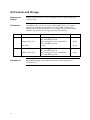
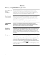
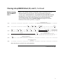
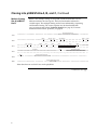
pUB6/V5-His A, B, and C Catalog no. V250-01 and V250-20 Rev. date: 30 November 2009 Manual part no. 25-0251 MAN0000089 Corporate Headquarters Invitrogen Corporation 1600 Faraday Avenue Carlsbad, CA 92008 T: 1 760 603 7200 F: 1 760 602 6500 E: [email protected] For country-specific contact information visit our web site at www.invitrogen.com User Manual ii Table of Contents Kit Contents and Storage........................................................................................................................... iv Introduction .............................................................................................................. 1 Product Overview ........................................................................................................................................1 Methods .................................................................................................................... 2 Cloning into pUB6/V5-His A, B, and C ....................................................................................................2 Transfection and Analysis...........................................................................................................................6 Creating Stable Cell Lines ...........................................................................................................................8 Appendix ................................................................................................................ 11 Human UbC Promoter...............................................................................................................................11 pUB6/V5-His Vector..................................................................................................................................12 pUB6/V5-His/lacZ.....................................................................................................................................14 Blasticidin ....................................................................................................................................................15 Recipe...........................................................................................................................................................16 Accessory Products ....................................................................................................................................17 Technical Support.......................................................................................................................................18 Purchaser Notification ...............................................................................................................................19 References....................................................................................................................................................20 iii Kit Contents and Storage Shipping and Storage pUB6/V5-His A, B, and C vectors are shipped on wet ice. Upon receipt, store vectors at –20°C. Kit Contents The pUB6/V5-His A, B, and C vectors and the pUB6/V5-His /lacZ control plasmid are supplied with each product (Cat. no. V250-01, V250-20). The pUB6/V5-His with Blasticidin Kit (Cat. no. V250-01) also includes blasticidin antibiotic. The amount of each reagent provided is listed below. Cat. no. Vector V250-01 pUB6/V5-His A, B, and C 40 μL of 0.5 μg/μL vector in 10 mM TrisHCl, 1 mM EDTA, pH 8.0 20 μg pUB6/V5-His /lacZ 40 μL of 0.5 μg/μL vector in 10 mM TrisHCl, 1 mM EDTA, pH 8.0 20 μg Blasticidin powder 50 mg pUB6/V5-His A, B, and C 40 μL of 0.5 μg/μL vector in 10 mM TrisHCl, 1 mM EDTA, pH 8.0 20 μg pUB6/V5-His /lacZ 40 μL of 0.5 μg/μL vector in 10 mM TrisHCl, 1 mM EDTA, pH 8.0 20 μg V250-20 Intended Use iv Composition Amount For research use only. Not intended for human or animal diagnostic or therapeutic uses. Introduction Product Overview Description of the System pUB6/V5-His A, B, and C are 5.5 kb vectors designed for overproduction of recombinant proteins in mammalian cell lines. Features of the vectors allow purification and detection of expressed proteins (see pages 12–13 for more information). High-level stable and transient expression can be carried out in most mammalian cells. The vectors contain the following elements: Human ubiquitin C promoter (hUbC) for high-level expression across a broad range of species and cell types (Schorpp et al., 1996; Wulff et al., 1990) (see page 11 for more information). Three reading frames to facilitate in-frame cloning with a C-terminal peptide encoding the V5 epitope and a polyhistidine (6×His) metal-binding tag. Blasticidin resistance gene (bsd) for selection of stable cell lines (see page 15 for more information). Episomal replication in cell lines that are latently infected with SV40 or that express the SV40 large T antigen (e.g. COS-1, COS-7). The control plasmid, pUB6/V5-His/lacZ, is included for use as a positive control for transfection, expression, and detection in the cell line of choice. Experimental Outline Use the following outline to clone and express your gene of interest in pUB6/V5His. 1. Consult the multiple cloning sites described on pages 3–5 to determine which vector (A, B, or C) should be used to clone your gene in frame with the C-terminal V5 epitope and polyhistidine tag. 2. Ligate your insert into the appropriate vector and transform into E. coli. Select transformants on 50 to 100 μg/mL ampicillin or 50 μg/mL blasticidin. 3. Analyze your transformants for the presence of insert by restriction digestion. 4. Select a transformant with the correct restriction pattern and use sequencing to confirm that your gene is cloned in frame with the C-terminal peptide. 5. Transfect your construct into the cell line of choice. Generate a stable cell line, if desired. 6. Test for expression of your recombinant gene by western blot analysis or other functional assay. For antibodies to the V5 epitope or the polyhistidine, C-terminal tag, see the page 17. 7. To purify your recombinant protein, you may use metal-chelating resin such as ProBond™. ProBond™ resin is available separately (see page 17 for ordering information). 1 Methods Cloning into pUB6/V5-His A, B, and C General Molecular Biology Techniques For help with DNA ligations, E. coli transformations, restriction enzyme analysis, purification of single-stranded DNA, DNA sequencing, and DNA biochemistry, refer to Molecular Cloning: A Laboratory Manual (Sambrook et al., 1989) or Current Protocols in Molecular Biology (Ausubel et al., 1994). E. coli Strain for Transformation Many E. coli strains are suitable for the propagation of this vector, including TOP10F´, JM109, and INVF´. We recommend that you propagate vectors containing inserts in E. coli strains that are recombination deficient (recA) and endonuclease A deficient (endA). For your convenience, TOP10F´ is available as chemically competent or electrocompetent cells from Invitrogen (see page 17). Transformation Method You may use any method of your choice for transformation. Chemical transformation is the most convenient for most researchers. Electroporation is the most efficient and the method of choice for large plasmids. Maintaining pUB6/V5-His To propagate and maintain the pUB6/V5-His vectors, use a small amount of the supplied 0.5 μg/μL stock solution in TE, pH 8.0 to transform a recA, endA E. coli strain like TOP10F´, DH5, JM109, or equivalent. Select transformants on LB plates containing 50–100 μg/mL ampicillin or 50 g/mL blasticidin. Be sure to prepare a glycerol stock of plasmid-containing E. coli strain for long-term storage (see page 5). Cloning Considerations Your insert should contain a Kozak consensus sequence with an ATG initiation codon for proper initiation of translation (Kozak, 1987; Kozak 1990). An example of a Kozak consensus sequence is provided below. Other sequences are possible, but the G or A at position –3 and the G at position +4 (shown in bold) illustrates the most commonly occurring sequence with strong consensus. Replacing one of the two bases at these positions provides moderate consensus, while having neither results in weak consensus. The ATG initiation codon is shown underlined. (G/A)NNATGG If you wish to express your protein WITHOUT the C-terminal peptide, be sure to include a stop codon. Continued on next page 2 Cloning into pUB6/V5-His A, B, and C, Continued Multiple Cloning Site of pUB6/V5His A Below is the multiple cloning site for pUB6/V5-His A. Restriction sites are labeled to indicate the cleavage site. The boxed nucleotides indicate the variable region. Note that there is a stop codon between the BamH I site and the BstX I site. The multiple cloning site has been confirmed by sequencing and functional testing. The vector sequence may be downloaded from www.invitrogen.com or from Technical Support (see page 18). For more information on the hUbC promoter, see page 11. UB forward priming site 1121 TTTAGGCACC TTTTGAAATG TAATCATTTG GGTCAATATG TAATTTTCAG TGTTAGACTA GTAAATTGTC CGCTAAATTC 1201 TGGCCGTTTT TGGCTTTTTT GTTAGAC GAA GCT TGG TAC CGA GCT CGG ATC CAC TAG TCC AGT GTG GTG Glu Ala Trp Tyr Arg Ala Arg Ile His *** Ser Ser Val Val Hind III EcoR I Pst I EcoR V BstX I* Asp718 I Not I Kpn I Sac I BamH I Xba I Xho I BstX I* Apa I BstB I 1270 GAA TTC TGC AGA TAT CCA GCA CAG TGG CGG CCG CTC GAG TCT AGA GGG CCC TTC GAA GGT AAG CCT Glu Phe Cys Arg Tyr Pro Ala Gln Trp Arg Pro Leu Glu Ser Arg Gly Pro Phe Glu Gly Lys Pro 1336 ATC CCT AAC CCT CTC CTC GGT CTC GAT TCT ACG CGT ACC GGT CAT CAT CAC CAT CAC CAT TGA GT Ile Pro Asn Pro Leu Leu Gly Leu Asp Ser Thr Arg Thr Gly His His His His His His *** 1401 TTAAACCCGC TGATCAGCCT CGACTGTGCC TTCTAGTTGC CAGCCATCTG TTGTTTGCCC CTCCCCCGTG CCTTCCTTGA 1481 CCCTGGAAGG TGCCACTCCC ACTGTCCTTT CCTAATAAAA TGAGGAAATT GCATCGCATT GTCTGAGTAG GTGTCATTCT Age I V5 epitope Pme I Polyhistidine region BGH reverse priming site BGH polyadenylation signal * Note that there are two BstX I sites in the polylinker. Continued on next page 3 Cloning into pUB6/V5-His A, B, and C, Continued Multiple Cloning Site of pUB6/V5His B Below is the multiple cloning site for pUB6/V5-His B. Restriction sites are labeled to indicate the cleavage site. The boxed nucleotides indicate the variable region. The multiple cloning site has been confirmed by sequencing and functional testing. The vector sequence may be downloaded from www.invitrogen.com or from Technical Support (see page 18). For more information on the hUbC promoter, see page 11. UB forward priming site 1121 TTTAGGCACC TTTTGAAATG TAATCATTTG GGTCAATATG TAATTTTCAG TGTTAGACTA GTAAATTGTC CGCTAAATTC 1201 TGGCCGTTTT TGGCTTTTTT GTTAGACG AAG CTT GGT ACC GAG CTC GGA TCC ACT AGT CCA GTG TGG TGG Lys Leu Gly Thr Glu Leu Gly Ser Thr Ser Pro Val Trp Trp 1271 AAT TCT GCA GAT ATC CAG CAC AGT GGC GGC CGC TCG AGT CTA GAG GGC CCG CGG TTC GAA GGT AAG Asn Ser Ala Asp Ile Gln His Ser Gly Gly Arg Ser Ser Leu Glu Gly Pro Arg Phe Glu Gly Lys 1337 CCT ATC CCT AAC CCT CTC CTC GGT CTC GAT TCT ACG CGT ACC GGT CAT CAT CAC CAT CAC CAT TGA Pro Ile Pro Asn Pro Leu Leu Gly Leu Asp Ser Thr Arg Thr Gly His His His His His His *** 1403 GTTTAAAC CCGCTGATCA GCCTCGACTG TGCCTTCTAG TTGCCAGCCA TCTGTTGTTT GCCCCTCCCC CGTGCCTTCC 1481 TTGACCCTGG AAGGTGCCAC TCCCACTGTC CTTTCCTAAT AAAATGAGGA AATTGCATCG CATTGTCTGA GTAGGTGTCA Asp718 I Kpn I Hind III Pst I EcoR V BstX I* Not I Xho I Xba I Age I V5 epitope Pme I Sac I BamH I BstX I* EcoR I Apa I BstB I Polyhistidine region BGH reverse priming site BGH polyadenylation signal * Note that there are two BstX I sites in the polylinker. Continued on next page 4 Cloning into pUB6/V5-His A, B, and C, Continued Multiple Cloning Site of pUB6/V5His C Below is the multiple cloning site for pUB6/V5-His C. Restriction sites are labeled to indicate the cleavage site. The boxed nucleotides indicate the variable region. The multiple cloning site has been confirmed by sequencing and functional testing. The vector sequence may be downloaded from www.invitrogen.com or from Technical Support (see page 18). UB forward priming site 1121 TTTAGGCACC TTTTGAAATG TAATCATTTG GGTCAATATG TAATTTTCAG TGTTAGACTA GTAAATTGTC CGCTAAATTC 1201 TGGCCGTTTT TGGCTTTTTT GTTAGACGA AGC TTG GTA CCG AGC TCG GAT CCA CTA GTC CAG TGT GGT GGA Ser Leu Val Pro Ser Ser Asp Pro Leu Val Gln Cys Gly Gly 1272 ATT CTG CAG ATA TCC AGC ACA GTG GCG GCC GCT CGA GGT CAC CCA TTC GAA GGT AAG CCT ATC CCT Ile Leu Gln Ile Ser Ser Thr Val Ala Ala Ala Arg Gly His Pro Phe Glu Gly Lys Pro Ile Pro 1338 AAC CCT CTC CTC GGT CTC GAT TCT ACG CGT ACC GGT CAT CAT CAC CAT CAC CAT TGA GTTTAA Asn Pro Leu Leu Gly Leu Asp Ser Thr Arg Thr Gly His His His His His His *** 1401 ACCCGCTGAT CAGCCTCGAC TGTGCCTTCT AGTTGCCAGC CATCTGTTGT TTGCCCCTCC CCCGTGCCTT CCTTGACCCT 1481 GGAAGGTGCC ACTCCCACTG TCCTTTCCTA ATAAAATGAG GAAATTGCAT CGCATTGTCT GAGTAGGTGT CATTCTATTC Hind III Pst I EcoR V BstX I* Not I Asp718 I Kpn I Xho I Age I Sac I BamH I BstE II BstX I* EcoR I BstB I V5 epitope Polyhistidine region Pme I BGH Reverse priming site BGH polyadenylation signal * Note that there are two BstX I sites in the polylinker. MEND ION AT RECOM Transforming Ligation Mixtures Preparing a Glycerol Stock Transform your ligation mixtures into a competent recA, endA E. coli strain (e.g., TOP10F´, DH5) and select on LB plates containing 50–100 μg/mL ampicillin or 50 μg/mL blasticidin. Select 10–20 clones and analyze for the presence and orientation of your insert. We recommend that you sequence your construct with the UB Forward and BGH Reverse primer sequences to confirm that your gene is fused in frame with the V5 epitope and the C-terminal polyhistidine tag. Refer to the diagram on pages 3–5 for the sequence and location of the primer binding sites. Once you have identified the correct clone, purify the colony and make a glycerol stock for long-term storage. It is also a good idea to keep a DNA stock of your plasmid at –20°C. 1. Streak the original colony out for single colonies on an LB plate containing 50 μg/mL ampicillin (or 50 g/mL blasticidin). Incubate the plate at 37°C overnight. 2. Isolate a single colony and inoculate into 1–2 mL of LB containing 50 μg/mL ampicillin (or 50 g/mL blasticidin). 3. Grow the culture to mid-log phase (OD600 = 0.5–0.7). 4. Mix 0.85 mL of culture with 0.15 mL of sterile glycerol and transfer to a cryovial. 5. Store at –80°C. 5 Transfection and Analysis Introduction Once you have confirmed that your construct is in the correct orientation and fused to the C-terminal peptide (if desired), then you are ready to transfect your cell line of choice. We recommend that you include the positive control vector and a mock transfection to evaluate your results. Plasmid Preparation Plasmid DNA for transfection into eukaryotic cells must be very clean and free from phenol and sodium chloride. Contaminants will kill the cells and salt will interfere with lipid complexing, decreasing transfection efficiency. We recommend isolating plasmid DNA using the PureLink™ HiPure Miniprep Kit or the PureLink™ HiPure Midiprep Kit (see page 17 for ordering information). Methods of Transfection For established cell lines (e.g., HeLa), consult original references or the supplier of your cell line for the optimal method of transfection. It is recommended that you follow exactly the protocol for your cell line. Pay particular attention to medium requirements, when to pass the cells, and at what dilution to split the cells. Further information is provided in Current Protocols in Molecular Biology. Methods for transfection include calcium phosphate (Chen and Okayama, 1987; Wigler et al., 1977), lipid-mediated (Felgner et al., 1987; Felgner et al., 1989), and electroporation (Chu et al., 1987; Shigekawa and Dower, 1988). Invitrogen offers the Lipofectamine™ 2000 Reagent for mammalian transfection (see page 17 for ordering). Positive Control pUB6/V5-His/lacZ is provided as a positive control vector for mammalian transfection and expression (see page 14). It may be used to optimize transfection conditions for your cell line. The gene encoding -galactosidase is expressed in mammalian cells under the control of the human ubiquitin C (hUbC) promoter. A successful transfection will result in -galactosidase expression that can be easily assayed (see below). Assay for -galactosidase Activity You may assay for -galactosidase expression by activity assay using cell-free lysates (Miller, 1972) or by staining the cells for activity. Invitrogen offers the -Gal Assay Kit and the -Gal Staining Kit for fast and easy detection of -galactosidase expression (see page 17 for ordering information). Continued on next page 6 Transfection and Analysis, Continued Detecting Fusion Proteins Several antibodies are available from Invitrogen that can be used to detect expression of your fusion protein from pUB6/V5-His (see page 17 for ordering information). To detect the fusion protein by western blot, you will need to prepare a cell lysate from transfected cells. We recommend that you perform a time course to optimize expression of the fusion protein (e.g. 24, 48, 72 hours, etc. after transfection). To lyse cells: 1. Wash cell monolayers (~106 cells) once with phosphate-buffered saline (PBS). 2. Scrape cells into 1 mL PBS and pellet the cells at 1,500 × g for 5 minutes. 3. Resuspend in 50 μL Cell Lysis Buffer (see recipe below). Other lysis buffers are suitable. 4. Incubate cell suspension at 37°C for 10 minutes to lyse the cells. 5. Vortex the cell lysate and centrifuge at 10,000 × g for 10 minutes to pellet nuclei. Transfer the supernatant to a fresh tube. Assay the supernatant for protein concentration. Note: Do not use protein assays utilizing Coomassie® Blue or other dyes. NP-40 interferes with the binding of the dye with the protein. 6. Add SDS-PAGE sample buffer to a final concentration of 1X and boil the sample for 5 minutes. 7. Load 20 μg of lysate onto an SDS-PAGE gel and electrophorese. Use the appropriate percentage of acrylamide to resolve your fusion protein. 7 Creating Stable Cell Lines Introduction The pUB6/V5-His vectors contain the blasticidin resistance gene (bsd) to allow for selection of stable cell lines using blasticidin (Kimura et al., 1994). We recommend that you test the sensitivity of your mammalian host cell to blasticidin as natural resistance varies among cell lines. General information and guidelines are provided below for your convenience. Possible Sites for Linearization To obtain stable transfectants, you may choose to linearize your vector before transfection. While linearizing your vector may not improve the efficiency of transfection, it increases the chances that the vector does not integrate in a way that disrupts the gene of interest. The table below lists some unique sites that may be used to linearize your construct prior to transformation. Other restriction sites are possible. Note that for the enzymes listed below, the cleavage site is indicated for versions A, B, and C of pUB6/V5-His. Be sure that your insert does not contain the restriction enzyme site you wish to use to linearize your vector. Enzyme Location Supplier Bgl II Upstream of hUbC promoter Many Bst1107 I End of SV40 polyA AGS*, Fermentas, Takara Sap I Backbone New England Biolabs BspLU 11I Backbone Boehringer-Mannheim AlwN I pMB1 origin Amersham, New England Biolabs, Life Technologies Eam1105 I Ampicillin gene AGS*, Fermentas, Takara Bgl I Ampicillin gene Many Fsp I Ampicillin gene Many Sca I Ampicillin gene Many Ssp I Backbone Many Continued on next page 8 Creating Stable Cell Lines, Continued Selection in Mammalian Cell Lines Selecting Stable Integrants To generate a stable cell line expressing your protein, you need to determine the minimum concentration of blasticidin required to kill your untransfected host cell line. Typically, concentrations between 2 and 10 μg/mL blasticidin are sufficient to kill the untransfected host cell line. Test a range of concentrations (see below) to ensure that you determine the minimum concentration necessary for your cell line. See page 15 for details on handling and preparing Blasticidin solution. 1. Seed cells at 20–25% confluency for each time point (~6 time points) and allow the cells to adhere overnight. 2. The next day, substitute culture medium with medium containing varying concentrations of blasticidin (e.g., 0, 1, 3, 5, 7.5, and 10 μg/mL blasticidin). 3. Replenish the selective medium every 3–4 days. Cells sensitive to blasticidin will round up and detach from the plate. Dead cells will accumulate in the medium. 4. Count the number of viable cells at regular intervals to determine the appropriate concentration of blasticidin that prevents growth. Once the appropriate concentration of blasticidin is determined, you can generate a stable cell line with your construct. 1. Transfect your cells using the optimal protocol for your cell line. Include a sample of untransfected cells as a negative control. 2. 48 hours after transfection, split the cells into fresh medium containing blasticidin at the appropriate concentration for your cell line. Split the cells such that they are no more than 25% confluent. 3. Replenish selective medium every 3–4 days until blasticidin-resistant colonies are detected. 4. Pick and expand colonies. Continued on next page 9 Creating Stable Cell Lines, Continued Preparing Cells for Lysis Lysis of Cells Use the procedure below to prepare cells for lysis prior to purification of your protein on ProBond™. You will need 5 × 10⁶ to 1 × 10⁷ cells for purification of your protein on a 2 mL ProBond™ column (see ProBond™ Protein Purification manual). 1. Seed cells (from a stable cell line) in five T-75 flasks or 2 to 3 T-175 flasks. 2. Grow the cells in selective medium until they are 80–90% confluent. 3. Harvest the cells by treating with trypsin-EDTA for 2 to 5 minutes or by scraping the cells in PBS. 4. Inactivate the trypsin by diluting with fresh medium (if necessary) and transfer the cells to a sterile microcentrifuge tube. 5. Centrifuge the cells at 1,500 × g for 5 minutes. Resuspend the cell pellet in PBS. 6. Centrifuge the cells at 1,500 × g for 5 minutes. You may lyse the cells immediately or freeze in liquid nitrogen and store at –80°C until needed. If you are using ProBond™ resin, refer to the ProBond™ Protein Purification manual for details about sample preparation for chromatography. If you are using other metal-chelating resin, refer to the manufacturer's instruction for recommendations on sample preparation. 10 Appendix Human UbC Promoter Description The human UbC promoter allows high-level expression of recombinant protein in most mammalian cell lines (Wulff et al., 1990) and in virtually all tissues tested in transgenic mice (Schorpp et al., 1996). The diagram below shows the features of the UbC promoter used in pUB6/V5-His (Nenoi et al., 1996). Features are marked as per Nenoi, et al., 1996. 5´ end of UbC promoter 11 GAGATCTGGC CTCCGCGCCG GGTTTTGGCG CCTCCCGCGG GCGCCCCCCT CCTCACGGCG AGCGCTGCCA CGTCAGACGA 91 AGGGCGCAGG AGCGTCCTGA TCCTTCCGCC CGGACGCTCA GGACAGCGGC CCGCTGCTCA TAAGACTCGG CCTTAGAACC 171 CCAGTATCAG CAGAAGGACA TTTTAGGACG GGACTTGGGT GACTCTAGGG CACTGGTTTT CTTTCCAGAG AGCGGAACAG 251 GCGAGGAAAA GTAGTCCCTT CTCGGCGATT CTGCGGAGGG ATCTCCGTGG GGCGGTGAAC GCCGATGATT ATATAAGGAC 331 GCGCCGGGTG TGGCACAGCT AGTTCCGTCG CAGCCGGGAT TTGGGTCGCG GTTCTTGTTT GTGGATCGCT GTGATCGTCA Sp 1 Sp 1 TATA box Start of Transcription Exon 1 5´ end of Intron 1 411 CTTGGTGAGT AGCGGGCTGC TGGGCTGGCC GGGGCTTTCG TGGCCGCCGG GCCGCTCGGT GGGACGGAAG CGTGTGGAGA 491 GACCGCCAAG GGCTGTAGTC TGGGTCCGCG AGCAAGGTTG CCCTGAACTG GGGGTTGGGG GGAGCGCAGC AAAATGGCGG 571 CTGTTCCCGA GTCTTGAATG GAAGACGCTT GTGAGGCGGG CTGTGAGGTC GTTGAAACAA GGTGGGGGGC ATGGTGGGCG 651 GCAAGAACCC AAGGTCTTGA GGCCTTCGCT AATGCGGGAA AGCTCTTATT CGGGTGAGAT GGGCTGGGGC ACCATCTGGG 731 GACCCTGACG TGAAGTTTGT CACTGACTGG AGAACTCGGT TTGTCGTCTG TTGCGGGGGC GGCAGTTATG CGGTGCCGTT 811 GGGCAGTGCA CCCGTACCTT TGGGAGCGCG CGCCCTCGTC GTGTCGTGAC GTCACCCGTT CTGTTGGCTT ATAATGCAGG 891 GTGGGGCCAC CTGCCGGTAG GTGTGCGGTA GGCTTTTCTC CGTCGCAGGA CGCAGGGTTC GGGCCTAGGG TAGGCTCTCC 971 TGAATCGACA GGCGCCGGAC CTCTGGTGAG GGGAGGGATA AGTGAGGCGT CAGTTTCTTT GGTCGGTTTT ATGTACCTAT 1051 CTTCTTAAGT AGCTGAAGCT CCGGTTTTGA ACTATGCGCT CGGGGTTGGC GAGTGTGTTT TGTGAAGTTT TTTAGGCACC 1131 TTTTGAAATG TAATCATTTG GGTCAATATG TAATTTTCAG TGTTAGACTA GTAAATTGTC CGCTAAATTC TGGCCGTTTT 1211 TGGCTTTTTT GTTAGACGAA GCTTGG.... UB Forward priming site 3´ end of Intron 1 5´ end of Exon 2 11 pUB6/V5-His Vector The figure below summarizes the features of the pUB6/V5-His vectors. The sequences for pUB6/V5-His A, B, and C are available for downloading from www.invitrogen.com or from Technical Support (see page 18). Age I Map of pUB6/ V5-His BGH pA bC PU f1 6xHis Term Pme I Hind III Asp718 I Kpn I Sac I BamH I BstX I EcoR I Pst I EcoR V BstX I Not I Xho I BstE II* Xba I* Apa I* BstB I V5 epitope or i 0 ori SV4 Comments for pUB6/V5-His A: 5463 nucleotides pU C o ri UbC promoter: bases 18-1227 UB forward priming site: bases 1167-1188 Multiple cloning site: bases 1229-1326 V5 epitope: bases 1327-1368 Polyhistidine tag: bases 1378-1395 BGH reverse priming site: bases 1418-1435 BGH polyadenylation signal: bases 1421-1648 f1 origin: bases 1694-2122 SV40 promoter and origin: bases 2149-2458 EM-7 promoter: bases 2506-2561 Blasticidin resistance gene: bases 2580-2978 SV40 polyadenylation signal: bases 3136-3266 pUC origin: bases 3649-4322 Ampicillin resistance gene: bases 4467-5327 S V4 0 p A bs d n 5.5 kb EM-7 Ampicilli pUB6/V5-His A, B, C * Frame-dependent variations. BstE II is only found in version C. In addition, there are no Xba I or Apa I sites in version C. Continued on next page 12 pUB6/V5-His Vector, Continued Features of pUB6/V5-His pUB6/V5-His A (5463 bp), pUB6/V5-His B (5467 bp), and pUB6/V5-His C (5459 bp) contain the following elements. All features have been functionally tested. Feature Benefit Human ubiquitin C (hUbC) promoter Allows overexpression of your recombinant protein in a broad range of mammalian cell types (Hershko and Ciechanover, 1982; Wulff et al., 1990; Schorpp et al., 1996). Multiple cloning site in three reading frames Allows insertion of your gene and facilitates cloning in frame with the V5 epitope and the C-terminal polyhistidine tag. V5 epitope (Gly-Lys-Pro-Ile-Pro-Asn-ProLeu-Leu-Gly-Leu-Asp-Ser-Thr) Allows detection of your recombinant protein with the Anti-V5 Antibodies (Southern et al., 1991). C-terminal polyhistidine tag Allows purification of your recombinant protein on metal-chelating resin such as ProBond™. In addition, the C-terminal polyhistidine tag is the epitope for the Anti-His(C-term) Antibodies (Lindner et al., 1997). Bovine growth hormone (BGH) polyadenylation signal Efficient transcription termination and polyadenylation of mRNA (Goodwin and Rottman, 1992). f1 origin Allows rescue of single-stranded DNA. SV40 early promoter and origin Allows efficient, high-level expression of the blasticidin resistance gene and episomal replication in cells expressing the SV40 large T antigen. EM-7 promoter For expression of the blasticidin resistance gene in E. coli. Blasticidin resistance gene (bsd) Selection of stable transfectants in mammalian cells (Kimura et al., 1994). SV40 polyadenylation signal Efficient transcription termination and polyadenylation of mRNA. pUC origin High-copy number replication and growth in E. coli. Ampicillin resistance gene (β-lactamase) Selection of vector in E. coli. 13 pUB6/V5-His/lacZ pUB6/V5-His/lacZ is a 8510 bp control vector containing the gene for -galactosidase. This vector was constructed by ligating a 3190 bp Hind III-Age I fragment containing the lacZ gene and the V5 epitope to a 5320 bp Hind III-Age I fragment containing the hUbC promoter, polyhistidine tag and blasticidin resistance gene from pUB6/V5-His A. Map of Control Vector The figure below summarizes the features of the pUB6/V5-His/lacZ vector. The nucleotide sequence for pUB6/V5-His/lacZ is available for downloading from www.invitrogen.com or by contacting Technical Support. (see page 18). bC PU BGH pA f1 Pme I lacZ 6xHis Not I Xho I BstE II BstB I Hind III BamH I Pst I V5 epitope Age I Description or i 0 ori SV4 lacZ Comments for pUB6/V5-His/lacZ: 8510 nucleotides UbC promoter: bases 18-1227 UB forward priming site: bases 1167-1188 LacZ ORF: bases 1290-4346 V5 epitope: bases 4374-4415 Polyhistidine tag: bases 4425-4442 BGH reverse priming site: bases 4465-4482 BGH polyadenylation signal: bases 4468-4695 f1 origin: bases 4741-5169 SV40 promoter and origin: bases 5196-5505 EM-7 promoter: bases 5553-5608 Blasticidin resistance gene: bases 5627-6025 SV40 polyadenylation signal: bases 6183-6313 pUC origin: bases 6696-7369 Ampicillin resistance gene: bases 7514-8374 14 pU C o ri S V4 0 p A bs d n 8.5 kb EM - 7 A m p i c il li pUB6/V5-His/ Term Blasticidin Description Blasticidin S HCl is a nucleoside antibiotic isolated from Streptomyces griseochromogenes which inhibits protein synthesis in both prokaryotic and eukaryotic cells (Takeuchi et al., 1958; Yamaguchi et al., 1965). Resistance is conferred by expression of either one of two blasticidin S deaminase genes: BSD from Aspergillus terreus (Kimura et al., 1994) or bsr from Bacillus cereus (Izumi et al., 1991). These deaminases convert blasticidin S to a non-toxic deaminohydroxy derivative (Izumi et al., 1991). Handling Blasticidin Always wear gloves, mask, goggles, and protective clothing (e.g., a laboratory coat) when handling blasticidin. Weigh out blasticidin and prepare solutions in a hood. Preparing and Storing Stock Solutions Blasticidin is soluble in water. Water is generally used to prepare stock solutions of 5 to 10 mg/mL. Molecular Weight, Formula, and Structure Dissolve blasticidin in sterile water and filter-sterilize the solution. Aliquot in small volumes suitable for one time use (see last point below) and freeze at –20°C for long-term storage or store at 4°C for short term storage. Aqueous stock solutions are stable for 1–2 weeks at 4°C and 6–8 weeks at -20°C. pH of the aqueous solution should not exceed 7 to prevent inactivation of blasticidin. Do not subject stock solutions to freeze/thaw cycles (do not store in a frostfree freezer). Upon thawing, use what you need and discard the unused portion. The formula for blasticidin is C17H26N8O5-HCl, and the molecular weight is 458.9. The diagram below shows the structure of blasticidin. NH2 N N HOOC NH N NH O -HCl CH3 H2N O NH2 O 15 Recipe Cell Lysis Buffer 50 mM Tris 150 mM NaCl 1% Nonidet P-40 pH 7.8 1. This solution can be prepared from the following common stock solutions. For 100 mL, combine: 1 M Tris base 5 M NaCl 5 mL 3 mL Nonidet P-40 1 mL 2. Bring the volume to 90 mL with deionized water and adjust the pH to 7.8 with HCl. 3. Bring the volume up to 100 mL. Store at room temperature. Note: Protease inhibitors may be added at the following concentrations: 1 mM PMSF 1 μg/mL Pepstatin 1 μg/mL Leupeptin 16 Accessory Products Introduction The following products may be used with the pUB6/V5-His vectors. For details, visit www.invitrogen.com or contact Technical Support (page 18). Amount Catalog no. 6 × 2 mL precharged, prepacked ProBond™ resin columns and buffers for native and denaturing purification K850-01 50 mL R801-01 150 mL R801-15 PureLink™ HiPure Plasmid Midiprep Kit 25 preps K2100-04 Lipofectamine™ 2000 Reagent 0.75 mL 11668-027 5 × 80 μL C665-55 20×50 μL C3030-03 80 mL K1455-01 1 kit K1465-01 Item ProBond™ Purification System ProBond™ Resin ™ Electrocomp TOP10F´ ® One Shot TOP10F´ Chemically Competent E. coli -Gal Assay Kit -Gal Staining Kit Antibodies If you do not have an antibody specific to your protein, Invitrogen offers the Anti-V5, or Anti-His(C-term) antibodies to detect your recombinant fusion protein. Horseradish peroxidase (HRP)- and alkaline phosphatase (AP)conjugated antibodies are available for convenient one-step detection. Antibody Anti-V5 Anti-V5-HRP Anti-V5-AP Anti-His(C-term) Anti-His(C-term)-HRP Anti-His(C-term)-AP Primers Epitope Catalog no. Detects a 14 amino acid epitope derived from the P and V proteins of the paramyxovirus, SV5 (Southern et al., 1991): GKPIPNPLLGLDST R960-25 Detects the C-terminal polyhistidine tag (requires the free carboxyl group for detection) (Lindner et al., 1997): HHHHHH-COOH R930-25 R961-25 R962-25 R931-25 R932-25 For your convenience, Invitrogen offers a custom primer synthesis service. Visit www.invitrogen.com for more details. 17 Technical Support Web Resources Contact Us Visit the Invitrogen website at www.invitrogen.com for: Technical resources, including manuals, vector maps and sequences, application notes, SDSs, FAQs, formulations, citations, handbooks, etc. Complete technical support contact information Access to the Invitrogen Online Catalog Additional product information and special offers For more information or technical assistance, call, write, fax, or email. Additional international offices are listed on our website (www.invitrogen.com). Corporate Headquarters: 5791 Van Allen Way Carlsbad, CA 92008 USA Tel: 1 760 603 7200 Tel (Toll Free): 1 800 955 6288 Fax: 1 760 602 6500 E-mail: [email protected] Japanese Headquarters: LOOP-X Bldg. 6F 3-9-15, Kaigan Minato-ku, Tokyo 108-0022 Tel: 81 3 5730 6509 Fax: 81 3 5730 6519 E-mail: [email protected] European Headquarters: Inchinnan Business Park 3 Fountain Drive Paisley PA4 9RF, UK Tel: +44 (0) 141 814 6100 Tech Fax: +44 (0) 141 814 6117 E-mail: [email protected] SDS Safety Data Sheets (MSDSs) are available on our website at www.invitrogen.com/sds. Certificate of Analysis The Certificate of Analysis provides detailed quality control and product qualification information for each product. Certificates of Analysis are available on our website. Go to www.invitrogen.com/support and search for the Certificate of Analysis by product lot number, which is printed on the box. Limited Warranty Invitrogen (a part of Life Technologies Corporation) is committed to providing our customers with high-quality goods and services. Our goal is to ensure that every customer is 100% satisfied with our products and our service. If you should have any questions or concerns about an Invitrogen product or service, contact our Technical Support Representatives. All Invitrogen products are warranted to perform according to specifications stated on the certificate of analysis. The Company will replace, free of charge, any product that does not meet those specifications. This warranty limits the Company’s liability to only the price of the product. No warranty is granted for products beyond their listed expiration date. No warranty is applicable unless all product components are stored in accordance with instructions. The Company reserves the right to select the method(s) used to analyze a product unless the Company agrees to a specified method in writing prior to acceptance of the order. Invitrogen makes every effort to ensure the accuracy of its publications, but realizes that the occasional typographical or other error is inevitable. Therefore the Company makes no warranty of any kind regarding the contents of any publications or documentation. If you discover an error in any of our publications, please report it to our Technical Support Representatives. Life Technologies Corporation shall have no responsibility or liability for any special, incidental, indirect or consequential loss or damage whatsoever. The above limited warranty is sole and exclusive. No other warranty is made, whether expressed or implied, including any warranty of merchantability or fitness for a particular purpose. 18 Purchaser Notification Limited Use Label License No. 22: Vectors and Clones Encoding Histidine Hexamer This product is licensed under U.S. Patent Nos. 5,284,933 and 5,310,663 and foreign equivalents from Hoffmann-LaRoche, Inc., Nutley, NJ and/or Hoffmann-LaRoche Ltd., Basel, Switzerland and is provided only for use in research. Information about licenses for commercial use is available from QIAGEN GmbH, Max-Volmer-Str. 4, D-40724 Hilden, Germany. Limited Use Label License No. 51: Blasticidin and the Blasticidin Selection Marker Blasticidin and the blasticidin resistance gene (bsd) are the subject of U.S. Patent No.5,527,701 sold under patent license for research purposes only. For information on purchasing a license to this product for purposes other than research, contact Licensing Department, Life Technologies Corporation, 5791 Van Allen Way, Carlsbad, California 92008. Phone (760) 603-7200. Fax (760) 602-6500. email: [email protected] 19 References Ausubel, F. M., Brent, R., Kingston, R. E., Moore, D. D., Seidman, J. G., Smith, J. A., and Struhl, K. (1994). Current Protocols in Molecular Biology (New York: Greene Publishing Associates and WileyInterscience). Chen, C., and Okayama, H. (1987). High-Efficiency Transformation of Mammalian Cells by Plasmid DNA. Mol. Cell. Biol. 7, 2745-2752. Chu, G., Hayakawa, H., and Berg, P. (1987). Electroporation for the Efficient Transfection of Mammalian Cells with DNA. Nuc. Acids Res. 15, 1311-1326. Felgner, P. L., Gadek, T. R., Holm, M., Roman, R., Chan, H. W., Wenz, M., Northrop, J. P., Ringold, G. M., and Danielsen, M. (1987). Lipofectin: A Highly Efficient, Lipid-mediated DNA-transfection Procedure. Proc. Natl. Acad. Sci. USA 84, 7413-7417. Felgner, P. L., Holm, M., and Chan, H. (1989). Cationic Liposome Mediated Transfection. Proc. West. Pharmacol. Soc. 32, 115-121. Goodwin, E. C., and Rottman, F. M. (1992). The 3´-Flanking Sequence of the Bovine Growth Hormone Gene Contains Novel Elements Required for Efficient and Accurate Polyadenylation. J. Biol. Chem. 267, 16330-16334. Hershko, A., and Ciechanover, A. (1982). Mechanisms of Intracellular Protein Breakdown. Ann. Rev. Biochem. 51, 335-364. Izumi, M., Miyazawa, H., Kamakura, T., Yamaguchi, I., Endo, T., and Hanaoka, F. (1991). Blasticidin SResistance Gene (bsr): A Novel Selectable Marker for Mammalian Cells. Exp. Cell Res. 197, 229-233. Kimura, M., Takatsuki, A., and Yamaguchi, I. (1994). Blasticidin S Deaminase Gene from Aspergillus terreus (BSD): A New Drug Resistance Gene for Transfection of Mammalian Cells. Biochim. Biophys. Acta 1219, 653-659. Kozak, M. (1987). An Analysis of 5´-Noncoding Sequences from 699 Vertebrate Messenger RNAs. Nuc. Acids Res. 15, 8125-8148. Kozak, M. (1991). An Analysis of Vertebrate mRNA Sequences: Intimations of Translational Control. J. Cell Biol. 115, 887-903. Kozak, M. (1990). Downstream Secondary Structure Facilitates Recognition of Initiator Codons by Eukaryotic Ribosomes. Proc. Natl. Acad. Sci. USA 87, 8301-8305. Lindner, P., Bauer, K., Krebber, A., Nieba, L., Kremmer, E., Krebber, C., Honegger, A., Klinger, B., Mocikat, R., and Pluckthun, A. (1997). Specific Detection of His-tagged Proteins With Recombinant Anti-His Tag scFv-Phosphatase or scFv-Phage Fusions. BioTechniques 22, 140-149. Miller, J. H. (1972). Experiments in Molecular Genetics (Cold Spring Harbor, New York: Cold Spring Harbor Laboratory). Nenoi, M., Mita, K., Ichimura, S., Cartwright, I. L., Takahashi, E., Yamaguchi, M., and Tsuji, H. (1996). Heterogeneous Structure of the Polyubiquitin Gene UbC of HeLa S3 Cells. Gene 175, 179-185. Sambrook, J., Fritsch, E. F., and Maniatis, T. (1989). Molecular Cloning: A Laboratory Manual, Second Edition (Plainview, New York: Cold Spring Harbor Laboratory Press). Schorpp, M., Jäger, R., Schellander, K., Schenkel, J., Wagner, E. F., Weiher, H., and Angel, P. (1996). The Human Ubiquitin C Promoter Directs High Ubiquitous Expression of Transgenes in Mice. Nuc. Acids Res. 24, 1787-1788. Continued on next page 20 References, Continued Shigekawa, K., and Dower, W. J. (1988). Electroporation of Eukaryotes and Prokaryotes: A General Approach to the Introduction of Macromolecules into Cells. BioTechniques 6, 742-751. Southern, J. A., Young, D. F., Heaney, F., Baumgartner, W., and Randall, R. E. (1991). Identification of an Epitope on the P and V Proteins of Simian Virus 5 That Distinguishes Between Two Isolates with Different Biological Characteristics. J. Gen. Virol. 72, 1551-1557. Takeuchi, S., Hirayama, K., Ueda, K., Sakai, H., and Yonehara, H. (1958). Blasticidin S, A New Antibiotic. The Journal of Antibiotics, Series A 11, 1-5. Wigler, M., Silverstein, S., Lee, L.-S., Pellicer, A., Cheng, Y.-C., and Axel, R. (1977). Transfer of Purified Herpes Virus Thymidine Kinase Gene to Cultured Mouse Cells. Cell 11, 223-232. Wulff, B. S., O'Hare, M. M., Boel, E., Theill, L. E., and Schwartz, T. W. (1990). Partial Processing of the Neuropeptide Y Precursor in Transfected CHO Cells. FEBS Lett. 261, 101-105. Yamaguchi, H., Yamamoto, C., and Tanaka, N. (1965). Inhibition of Protein Synthesis by Blasticidin S. I. Studies with Cell-free Systems from Bacterial and Mammalian Cells. J. Biochem. (Tokyo) 57, 667-677. ©2009 Life Technologies Corporation. All rights reserved. 21 Notes 22 Corporate Headquarters 5791 Van Allen Way Carlsbad, CA 92008 T: 1 760 603 7200 F: 1 760 602 6500 E: [email protected] For country-specific contact information, visit our web site at www.invitrogen.com User Manual