1
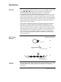
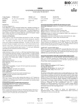
Instruction Manual
LUX™ Fluorogenic Primers
For real-time PCR and RT-PCR
Version F
13 September 2004
25-0546
ii
Table of Contents
Introduction ......................................................................................................................1
Designing and Ordering Custom LUX™ Primers ..............................................................3
Storing and Reconstituting Primers..................................................................................5
Certified LUX™ Primer Sets for Housekeeping Genes .....................................................6
Real-Time qPCR..............................................................................................................7
Multiplex Real-Time qPCR.............................................................................................11
Two-Step Real-Time qRT-PCR......................................................................................12
One-Step Real-Time qRT-PCR .....................................................................................16
Troubleshooting .............................................................................................................21
Accessory Products .......................................................................................................23
Purchaser Notification....................................................................................................24
Technical Service...........................................................................................................25
References ....................................................................................................................27
iii
iv
Introduction
Overview
LUX™ (Light Upon eXtension) Primers are an easy to use, highly sensitive, and
efficient method for performing real-time quantitative PCR (qPCR) and
RT-PCR (qRT-PCR). Each primer pair in the LUX™ system includes a
fluorogenic primer with a fluorophore attached to its 3′ end and a
corresponding unlabeled primer. The fluorogenic primer has a short sequence
tail of 4–6 nucleotides on the 5′ end that is complementary to the 3′ end of the
primer. The resulting hairpin secondary structure provides optimal quenching
of the fluorophore (see the figure below). When the primer is incorporated into
double-stranded DNA during PCR, the fluorophore is dequenched and the
signal increases by up to 10-fold.
LUX™ Primers combine high specificity with simple design and streamlined
protocols. LUX™ Primers require no special probes or quenchers and are
compatible with melting curve analysis of real-time qPCR products, which
allows for the differentiation of amplicons and primer dimer artifacts by their
melting temperatures. LUX™ Primers are available with two different reporter
dyes, which provides for multiplexing capability. You can custom-design
LUX™ Primers™ from a target DNA sequence using Web- or desktop-based
software, or order predesigned and validated Certified LUX™ Primer Sets for
Housekeeping Genes.
LUX™ Primer
Reaction
Relative fluorescence:
0.1
Hairpin primer
0.4
Single-stranded primer
1.0
Extended primer
(double-stranded DNA)
Labeling
Each fluorogenic LUX™ primer is labeled with one of two reporter dyes—FAM
(6-carboxy-fluorescein) or JOE (6-carboxy-4', 5'-dichloro-2', 7'-dimethoxyfluorescein).
Continued on next page
Introduction, Continued
Applications
LUX™ Primers can be used in real-time quantitative PCR and RT-PCR to
quantify 100 or fewer copies of a target gene in as little as 1 pg of template
DNA or RNA. They have a broad dynamic range of 7–8 orders. See the
guidelines and sample protocols for qPCR on pages 7–10 and guidelines and
sample protocols for qRT-PCR on pages 12–20.
Multiplex applications use separate FAM and JOE-labeled primer sets to
detect two different genes in the same sample. Typically, a custom-designed
FAM-labeled primer set would be used to detect the gene of interest, and a
JOE-labeled Certified LUX™ Primer Set would be used to detect a
housekeeping gene as an internal control. See the optimization guidelines for
multiplex qPCR on page 11.
Instrument
Compatibility
LUX™ Primers are compatible with a wide variety of real-time qPCR
instruments, including but not limited to the ABI PRISM® 7700, 7000, and 7900
and GeneAmp® 5700; the Bio-Rad iCycler™; the Stratagene Mx4000™ and
Mx3000™; the Cepheid Smart Cycler®; the Corbett Research Rotor-Gene; and
the Roche LightCycler®.
At a minimum, the real-time qPCR instrument should:
•
Detect fluorescence at each PCR cycle
•
Excite and detect FAM-labeled LUX™ Primers near their
excitation/emission wavelengths of 490/520 nm, and/or
•
Excite and detect JOE-labeled LUX™ Primers near their
excitation/emission wavelengths of 520/550 nm
Refer to the specific instrument’s user manual for operating instructions.
ABI PRISM is a registered trademark of Applera Corporation. GeneAmp is a registered trademark of
Roche Molecular Systems, Inc. LightCycler is a registered trademark of Idaho Technologies, Inc.
iCycler, Mx4000, Mx3000, Rotor-Gene, and Smart Cycler are trademarks of their respective
companies.
Designing and Ordering Custom LUX™ Primers
LUX™ Designer
Primer Design
Software
To design and order custom LUX™ Primers for your genes of interest, visit the
Invitrogen LUX™ Web site at www.invitrogen.com/LUX and follow the link to
the LUX™ Designer software. The software is available as either a Web-based
application or a Microsoft® Windows®-compatible download. Follow the stepby-step instructions in the software to submit your target sequence and
generate primer designs.
LUX™ Designer will automatically generate one or more primer designs based
on each sequence you submit and the selected design parameters. The design
software includes algorithms to minimize primer self-complementarity and
interactions between primers. It also assigns rankings to the generated
designs—based on primer melting temperature, hairpin structure, selfannealing properties, etc.—to aid in selection.
When the designs have been generated, you can review them, select a design,
select the fluorophore labels, and place your order.
Guidelines for
Submitting a
Target Sequence
When you submit a target sequence containing your gene of interest, keep in
mind the following design criteria:
•
The optimal amplicon length for real-time qPCR ranges from 80 to 200
bases. You can specify a minimum, optimal, and maximum amplicon
length when you submit the sequence.
•
The target sequence should be at least 10 bases longer than the minimum
amplicon size you select. The longer the sequence, the more likely that
an optimal primer design can be developed.
•
The sequence must contain only standard IUPAC (International Union of
Pure and Applied Chemistry) letter abbreviations.
•
When you select the design parameters, the default melting temperature
range is 60–68oC. Do not change this default unless the design engine finds
no primers in this range. For primers in this range, PCR annealing
temperatures from 55o to 64oC are appropriate.
When you first submit a sequence, the Disable Score-Based Rejection checkbox
should not be checked; the resulting scores provide an important measure of
primer suitability. Scores in the range of 0.0–4.0 are acceptable. If no primers
with a score of 4.0 or lower can be generated from a sequence, you can disable
score-based rejection and redesign the primers. See the LUX™ Designer Help
for additional guidance.
Selecting a Primer
Design
After you submit your sequence, LUX™ Designer will first generate one or
more designs for the labeled primer. The labeled primer can be either the
forward or the reverse primer. After you select a design for the labeled primer,
you will be prompted to select a design for the corresponding unlabeled
primer.
Continued on next page
3
Designing and Ordering Custom LUX™ Primers, Continued
Selecting Labels
After you have selected a primer set (labeled and unlabeled) for a particular
sequence, you can specify the particular label and synthesis scale. Custom
LUX™ Primers are provided in 50 nM or 200 nM synthesis scale.
When selecting labels in a multiplex reaction, we recommend using the FAM
label for your gene of interest and the JOE label for the housekeeping gene that
you will use as the internal control. Certified LUX™ Primer Sets for
Housekeeping Genes are recommended for the JOE-labeled control gene.
Placing the Order
After you have selected the label and synthesis scale, you can submit your
order to Invitrogen using the Web site or by e-mail or fax. Each primer order
will be shipped directly from Invitrogen’s Custom Primer Facilities. Labeled
primers are supplied in an amber tube; unlabeled primers are supplied in a
clear tube.
Each primer ordered from Invitrogen’s Custom Primer Facilities comes with a
Certificate of Analysis (COA) verifying the amount and sequence.
Product
Qualification
Custom LUX™ Primers are tested post-synthesis by optical density (OD) ratio
measurements and mass spectroscopy to ensure efficient dye labeling and
correct molecular weight and composition.
See the Certificate of Analysis shipped with each primer for more information.
Storing and Reconstituting Primers
Primer Storage and
Stability
Store primers at –20oC in the dark. LUX™ Primers are stable for:
•
>12 months when stored at –20oC in lyophilized form.
•
>6 months when stored at –20oC in solution.
Stability can be extended by storing at –70oC.
Important
Be careful to minimize the exposure of labeled LUX™ Primers to direct light, as
this can reduce their fluorescent intensity.
Reconstituting
Primers
Custom LUX™ Primers are provided lyophilized in 50-nmole or 200-nmole
synthesis scale. To reconstitute primers, centrifuge the tube for a few seconds
to collect the oligonucleotide in the bottom of the tube. Carefully open, add an
appropriate volume of TE buffer or ultrapure water, close the tube, rehydrate
for 5 minutes, and vortex for 15 seconds.
We recommend that you rehydrate primers at concentrations greater than
10 µM. To prepare a 100 µM primer stock solution, multiply the primer
amount in nmoles by ten to determine the volume of diluent in µl.
After reconstitution, store the primer stock at –20oC in the dark, where it will
be stable for 6 months or more.
5
Certified LUX™ Primer Sets for Housekeeping Genes
Certified LUX™
Primer Sets for
Housekeeping
Genes
Certified LUX™ Primer Sets for Housekeeping Genes are predesigned primer
sets for genes that are commonly used as internal controls for normalizing
real-time qRT-PCR experiments. These primer sets have been optimized and
functionally validated to provide accurate, reproducible results using standard
LUX™ protocols. They are supplied ready to use in TE buffer.
Each Certified LUX™ Primer Set includes a FAM- or JOE-labeled LUX™ primer
and a corresponding unlabeled primer. Each primer (labeled and unlabeled is
supplied at 100 µl and a concentration of 10 µM. Available sets are listed
below. For additional information, visit www.invitrogen.com/LUX.
Product
GenBank
Accession no.
Forward/
Reverse Label
Cat. no.
FAM label
Cat. no.
JOE label
Relative
expression
CDS
Location
PCR Product
Size Range
Human genes
18S rRNA
X03205
Forward
115HM-01
115HM-02
hβ-ACTIN
NM_001101
Forward
101H-01
101H-02
+++
++++
n/a
101-150 bp
Exons 2/3
101-150 bp
hATPSase
NM_001686
Forward
108H-01
108H-02
hB2M
NM_004048
Forward
113H-01
113H-02
+++
n/a
101-150 bp
+++
Exons 1/2
101-150 bp
hGAPDH
NM_002046
Forward
100H-01
100H-02
+++
Exons 4/5
151–200 bp
hPGK1
NM_000291
Forward
109H-01
109H-02
+++
n/a
50-100 bp
hPPIA
NM_021130
Forward
106H-01
106H-02
+++
Exons 2/3
50-100 bp
hRPL4
NM_000968
Reverse
103H-01
103H-02
+++
Exons 8/9
101-150 bp
hEEF1G
NM_001404
Forward
107H-01
107H-02
++
n/a
50-100 bp
hHPRT1
NM_000194
Reverse
105H-01
105H-02
++
Exons 5/6
50-100 bp
hSDHA
NM_004168
Forward
102H-01
102H-02
++
Exons 12/13
50-100 bp
hTFRC
NM_003234
Forward
111H-01
111H-02
++
Exons 10/11
101-150 bp
hGUS
NM_000181
Forward
112H-01
112H-02
+
Exons 7/8
101-150 bp
hHMBS
NM_000190
Forward
110H-01
110H-02
+
Exons 2/3
50-100 bp
hTBP
NM_003194
Forward
104H-01
104H-02
+
Exons 3/4
101-150 bp
hUBC
NM_021009
Forward
114H-01
114H-02
+
n/a
50-100 bp
X03205
Forward
115HM-01
115HM-02
++++
n/a
101-150 bp
mβ-ACTIN
NM_007393
Forward
101M-01
101M-02
+++
Exons 2/3
101-150 bp
mB2M
X01838
Forward
113M-01
113M-02
+++
n/a
50-100 bp
mEEF1G
AF321126
Forward
107M-01
107M-02
+++
n/a
101-150 bp
mGAPDH
NM_008084
Forward
100M-01
100M-02
+++
Exons 4/5
151–200 bp
mPGK1
NM_008828
Forward
109M-01
109M-02
+++
Exons 1/2
101-150 bp
mPPIA
NM_008907
Reverse
106M-01
106M-02
+++
Exons 1/2
50-100 bp
mRPL4
NM_022510
Forward
103M-01
103M-02
+++
Exons 2/3
151-200 bp
Mouse/rat genes
18S rRNA
mHPRT1
NM_013556
Forward
105M-01
105M-02
++
Exons 6/7
50-100 bp
mSDHA
AF095938
Forward
102M-01
102M-02
++
Exons 6/7
50-100 bp
mATPSase
NM_016774
Forward
108M-01
108M-02
+
n/a
50-100 bp
mGUS
NM_010368
Forward
112M-01
112M-02
+
Exons 7/8
50-100 bp
mHMBS
XM_129404
Reverse
110M-01
110M-02
+
Exons 4/5
50-100 bp
mTBP
NM_013684
Forward
104M-01
104M-02
+
Exons 3/4
101-150 bp
mTFRC
NM_011638
Forward
111M-01
111M-02
+
Exons 2/3
101-150 bp
n/a
101-150 bp
Exons 2/3
50–100 bp
Drosophila genes
d18S rRNA
AY037174
Reverse
115D-01
115D-02
++++
dActin
NM_079486
Forward
101D-01
101D-02
+++
Real-Time qPCR
Introduction
Real-time qPCR uses genomic DNA, cDNA, or plasmid DNA as starting
material. This section provides guidelines and example protocols for
performing real-time qPCR using LUX™ Primers.
Template
Specifications
The target template for real-time qPCR is linear single-stranded or doublestranded DNA, cDNA, or circular DNA (such as plasmids) that has been
linearized. The amount of DNA typically ranges from 102 to 107 copies or 1 pg
to 10 µg of template.
See page 12 for instructions on generating cDNA using reverse transcription as
part of two-step real-time qRT-PCR.
Primer
Concentration
For optimal PCR conditions, primer titrations of 50–500 nM per primer are
recommended. The sample reactions on pages 9–10 use 200 nM of each primer,
equivalent to 1 µl of a 10-µM primer solution in a 50-µl reaction.
Magnesium
Concentration
The optimal Mg++ concentration for a given target/primer/polymerase
combination can vary between 1 mM and 10 mM, but is usually in the range of
3 mM. See the sample reactions on pages 9–10.
dNTP
Concentration
The optimal concentration of dATP, dCTP, dGTP, and dTTP is 200 µM each. If
dUTP is used in place of dTTP, its optimal concentration is 400 µM.
DNA Polymerase
We recommend using a “hot-start” DNA polymerase, preferably one that has
been optimized for real-time qPCR. Platinum® Quantitative PCR SuperMix
UDG (Catalog no. 11730-017) is a 2X-concentrated, ready-to-use mixture
containing all components except primers and template. It uses Platinum® Taq
DNA polymerase and has been specifically formulated to provide optimal
performance in real-time qPCR systems.
ROX Reference
Dye
We recommend using ROX Reference Dye (Cat. no. 12223-023) to normalize
the fluorescent reporter signal in real-time qPCR for instruments that are
compatible with this option. ROX Reference Dye can be used to adjust for nonPCR-related fluctuations in fluorescence between reactions, and provides a
stable baseline in multiplex reactions. It is composed of a glycine conjugate of
5-carboxy-X-rhodamine, succinimidyl ester (25 µM) in 20 mM Tris-HCl (pH
8.4), 0.1 mM EDTA, and 0.01% Tween® 20.
ROX is supplied at 50X concentration. Add 1 µl of ROX for every 50 µl of
reaction volume. For convenience and to reduce pipetting errors, you can
premix a solution of ROX and Platinum® Quantitative PCR SuperMix-UDG.
Add 1 µl of ROX for every 25 µl of SuperMix-UDG. Store mixture at either
-20°C or 4°C in the dark.
Continued on next page
7
Real-Time qPCR, Continued
Bovine Serum
Albumin (BSA)
Bovine serum albumin (BSA) is required in qPCR reactions on the Roche
LightCycler® because the glass capillaries in the LightCycler® have a high
surface-to-volume ratio and the glass surface binds molecules like Taq DNA
polymerase, effectively removing them from the reaction. The addition of BSA
blocks this surface binding.
Nonacetylated BSA is strongly recommended because acetylated BSA will
inhibit PCR at the concentrations required in LightCycler® reactions. This
inhibition is most likely due to the transfer of acetyl groups to essential
components of the PCR, like the Taq DNA polymerase.
To ensure that the BSA does not contain RNA or DNA, we recommend using
ultrapure, molecular biology-grade nonacetylated BSA from
Panvera/Invitrogen (Cat. nos. P2489 and P2046).
Melting Curve
Analysis
Melting curve analysis is strongly recommended during qPCR to identify the
presence of primer dimers and analyze the specificity of the reaction. Program
your real-time instrument to perform this analysis after thermocycling.
Melting curve analysis identifies the change in fluorescent signal that occurs as
double-stranded DNA (dsDNA) dissociates or “melts” into single-stranded
DNA. By identifying the temperature at which the dsDNA dissociates, you can
distinguish smaller artifacts like primer dimers with a lower annealing
temperature from larger amplicons with a higher annealing temperature.
The presence of primer dimers in samples containing template decreases PCR
efficiency and obscures analysis and determination of cycle thresholds. The
formation of primer dimers most often occurs in no-template controls, where
the polymerase enzyme is essentially idle, and in this case the quantitative
analysis of the template samples is not affected. Melting curve analysis of notemplate controls can discriminate between primer dimers and spurious
amplification due to contaminating nucleic acids in reagent components.
Instrument
Settings
Follow the manufacturer’s instructions for configuring your real-time qPCR
instrument for use with LUX™ Primers. Note the following general settings:
•
Program your instrument to perform melting curve analysis at the end of
thermocycling, if this option is available.
•
The quencher setting on the instrument should reflect the fact that LUX™
Primers do not contain a quencher.
•
We recommend using ROX Reference Dye, if your instrument is
compatible with this option. Adjust the instrument settings accordingly.
Additional guidelines and settings for specific instruments are available at
www.invitrogen.com/lux; click on Instrument Protocols.
Continued on next page
Real-Time qPCR, Continued
Protocol for
Instruments Using
PCR Tubes or
Plates
The following protocol uses Platinum® Quantitative PCR SuperMix-UDG with
ROX reference reagent. It has been optimized for use with real-time qPCR
instruments that use PCR tubes or plates. A protocol for the Roche
LightCycler® is provided on the following page.
Note: The following protocol uses a 50-µl reaction volume; smaller volumes
may be used, depending on the requirements of your instrument. Before
proceeding, see the real-time qPCR guidelines on the previous pages. For
multiplex reactions, see the guidelines on page 11.
1.
To reduce well-to-well variation, prepare a Master Mix of all the reaction
ingredients except template. The following table provides Master Mix
volumes for one reaction and 50 reactions (scale up or down as needed):
Component
Platinum® Quantitative PCR SuperMix-UDG1
ROX Reference Dye (optional)
Labeled LUX™ Primer (10 µM)
Unlabeled primer (10 µM)
Sterile distilled water2
Vol/1 rxn
25 µl
1 µl
1 µl
1 µl
to 40 µl
Vol/50 rxns
1250 µl
50 µl
50 µl
50 µl
to 2000 µl
1
Final concentration: 0.03 U/µl Platinum® Taq DNA polymerase, 20 mM Tris-HCl
(pH 8.4), 50 mM KCl, 3 mM MgCl2, 200 µM dGTP, 200 µM dATP, 200 µM dCTP,
400 µM dUTP, 1 U UDG
2
2.
or use DNase/RNase Free Distilled Water (Cat. No. 10977-015).
Program the real-time qPCR instrument as follows:
3-Step Cycling (recommended)
50oC, 2 min hold (UDG treatment)
95oC, 2 min hold
45 cycles of:
95oC, 15 s
55oC, 30 s
72oC, 30 s
2-Step Cycling (optional)
50oC, 2 min hold (UDG treatment)
95oC, 2 min hold
45 cycles of:
95oC, 15 s
60-65oC, 30-45 s
Melting Curve Analysis (recommended)
Refer to instrument documentation
3.
Add 40 µl of the Master Mix to an optical PCR tube or each well of a
96-well PCR plate.
4.
Add 10 µl of template in TE or sterile dH2O to each reaction vessel. Cap or
seal the tube/plate.
5.
Gently mix and make sure that all components are at the bottom of the
tube/plate wells. Centrifuge briefly if needed.
6.
Place reaction in the real-time qPCR instrument and run the program.
Collect and analyze results.
Continued on next page
9
Real-Time qPCR, Continued
Protocol for the
Roche LightCycler®
The following protocol uses Platinum® Quantitative PCR SuperMix-UDG and
has been optimized for the Roche LightCycler®. Consult the LightCycler®
documentation for detailed instructions on preparing the capillary tubes and
operating the instrument. FAM-labeled LUX™ Primers are also compatible
with Roche enzyme mixes.
Note: JOE-labeled LUX™ Primers are not compatible with the current version
of the LightCycler®; use FAM-labeled primers only. The following protocol
uses a 20-µl reaction volume. Before proceeding, see the real-time qPCR
guidelines on the previous pages.
1.
To reduce well-to-well variation, prepare a Master Mix of all the reaction
ingredients except template. The following table provides volumes for one
reaction and 34 reactions (scale as needed):
Component
Platinum® Quantitative PCR SuperMix-UDG1
FAM-labeled LUX™ Primer (10 µM)
Unlabeled primer (10 µM)
Bovine serum albumin (5 mg/ml)2
Platinum® Taq DNA Polymerase3
Sterile distilled water4
Vol/1 rxn
10 µl
1 µl
1 µl
1 µl
0.12 µl
to 18 µl
Vol/34 rxns
340 µl
34 µl
34 µl
34 µl
4 µl
to 612 µl
1
Final concentration: 0.03 U/µl Platinum® Taq DNA polymerase, 20 mM Tris-HCl
(pH 8.4), 50 mM KCl, 3 mM MgCl2, 200 µM dGTP, 200 µM dATP, 200 µM dCTP,
400 µM dUTP, 1 U UDG
2
Validated with non-acetylated Ultrapure BSA (10% solution) from Panvera (Cat.
nos. P2489 and P2046).
3
Total units of Platinum® Taq DNA Polymerase in the reaction is 1.2 (including 0.6 U
from Platinum® Quantitative PCR SuperMix-UDG)
4
or use DNase/RNase Free Distilled Water (Cat. No. 10977-015).
2.
Set the fluorescence on the Roche LightCycler® to the F1 channel.
3.
Program the instrument as follows:
Thermal Cycling
Melting Curve Analysis (optional)
Program choice: Amplification
Program choice: Melting curve
Analysis mode: Quantification
Analysis mode: Melting curves
Cycling:
Cycling:
95oC, 0 s
50oC, 2 min hold (UDG treatment)
55oC, 15 sec
95oC, 2 min hold
45 cycles of:
95oC, 0 (increase 0.1oC/s with
o
continuous acquisition)
94 C, 5 s
40oC, 0 s
55oC, 10 s (single acquire)
72oC, 10 s
4. Add 18 µl of Master Mix to each capillary tube of the LightCycler®.
5.
Add 2 µl of template to each tube, and cap the tube.
6.
Centrifuge the tubes at 700 × g for 5 seconds.
7.
Place the reaction tubes in the rotor of the LightCycler® and run the
program. Collect and analyze results.
Multiplex Real-Time qPCR
Multiplex
Real-Time qPCR
In multiplex real-time qPCR, different sets of primers with different
fluorogenic labels are used to amplify separate genes in the template DNA.
Multiplexing with LUX™ Primers offers simplified PCR kinetics and increased
reaction efficiency when compared with probe-based technologies, because
only two oligos are used per target.
LUX™ Primers have been tested in multiplex reactions using a FAM-labeled
primer set for the gene of interest and a JOE-labeled set for a housekeeping
gene used as an internal control to normalize between different reactions. We
recommend using Certified LUX™ Primer Sets for Housekeeping Genes for the
internal control (see page 6).
Note: We recommend selecting a housekeeping gene that matches the relative
expression level of your gene of interest. The relative expression levels of
predesigned, certified LUX™ Primer Sets are shown on page 6.
In a standard multiplex reaction, you can include the additional primers at the
same volumes and concentration as the primers in a singleplex reaction, as
shown in the example mixture below:
Component
Platinum® Quantitative PCR SuperMix-UDG (2X)
ROX Reference Dye (50X)
Template
Forward primer 1 (FAM label) (10 µM)
Reverse primer 1 (10 µM)
Forward primer 2 (JOE label) (10 µM)
Reverse primer 2 (10 µM)
Sterile distilled water
Volume
25 µl
1 µl
10 µl
1 µl
1 µl
1 µl
1 µl
to 50 µl
All other reaction volumes remain the same.
Follow the thermal cycling guidelines provided in Protocol for Instruments
Using PCR Tubes or Plates on page 9. If you have difficulty performing the
multiplex reaction using these guidelines, see the optimization hints below.
Optimizing
Multiplex
Conditions
If you notice a decline in PCR efficiency in your multiplex real-time qPCR, you
can optimize the reaction by performing the steps listed below.
Note: We recommend that you perform one optimization step and then repeat
the reaction to test for efficiency before moving on to the next step:
1.
Reduce the primer concentration of the gene with the highest expression
levels (typically the housekeeping gene) to 1/2 the primer concentration of
the other gene. For example, in a standard 50-µl reaction, you would add
the primers for the less abundant gene at 1 µl each, and add the primers
for the more abundant gene at 0.5 µl each.
2.
Increase the MgCl2 in the reaction from 3 mM to 6 mM.
3.
Double the amount of polymerase enzyme (to 0.06 U/µl of reaction
volume). If you are using Platinum® Quantitative PCR SuperMix-UDG,
add Platinum® Taq DNA polymerase stand-alone enzyme (Catalog no.
10966-018) to double the amount of enzyme.
4.
Increase the dNTP concentrations in the reaction to 400 µM each.
11
Two-Step Real-Time qRT-PCR
Introduction
Real-time qRT-PCR uses RNA as starting material in a reverse transcription
reaction to generate first-strand cDNA. The cDNA is then quantified in a
separate real-time qPCR reaction.
In two-step qRT-PCR, first-strand synthesis is performed, and then the
reaction is transferred to a separate tube for the qPCR reaction. This section
provides guidelines for two-step qRT-PCR and an optimized protocol using
the SuperScript™ III Platinum® Two-Step qRT-PCR Kit.
For the real-time qPCR portion of the two-step protocol, see additional
guidelines on primers, magnesium, dNTPs, ROX Reference Dye, BSA, melting
curve analysis, and instrument settings on pages 7–8.
Template
Specifications
The target template for two-step qRT-PCR is total RNA or mRNA. Highquality, intact RNA is essential for full-length, high-quality cDNA synthesis
and accurate quantification. Starting material can range from 10 pg to 1 µg
total RNA. Then use 1 pg to 10 µg of the cDNA from the first-strand reaction
in the qPCR step.
The purity and integrity of the starting RNA have a direct impact on results.
RNase and genomic DNA contamination are the most common problems, and
purification methods should include RNase inhibitors and DNase digestion to
minimize these.
We recommend using the Micro-to-Midi Total RNA Purification System
(Catalog no. 12183-018) or TRIzol® reagent (Catalog no. 15596-026) to isolate
total RNA. High-quality total RNA can be purified from as little as 100 cells up
to 107 cells or 200 mg of tissue.
Isolation of mRNA is typically not required, but can be performed using the
FastTrack® 2.0 mRNA Isolation Kit (Catalog no. K1593-02).
Enzyme
Specifications
For two-step qRT-PCR, we recommend using a high-specificity, high-yield
reverse transcriptase such as SuperScript™ III Reverse Transcriptase and a
“hot-start” DNA polymerase such as Platinum® Taq DNA Polymerase. The
SuperScript™ III Platinum® Two-Step qRT-PCR Kit (Catalog nos. 11734-050 and
11734-068) includes SuperScript™ III RT, Platinum® Taq DNA Polymerase, and
all the other necessary components for two-step qRT-PCR except the RNA. See
the example protocol on page 13.
For first-strand cDNA synthesis alone, we recommend the SuperScript™ III
First-Strand Synthesis System for RT-PCR (Catalog no. 18080-051).
Continued on next page
Two-Step Real-Time qRT-PCR, Continued
Removing
Genomic DNA from
RNA Samples
We strongly recommend that you decrease the genomic DNA content in the
RNA sample by performing a digest with DNase I, Amplification Grade
(Catalog no. 18068-015), as described below. The DNase I digest is designed
for up to 1 µg of RNA; for larger amounts of RNA, increase volumes
accordingly.
Combine the following in a tube on ice:
Component
RNA template
DNase reaction buffer
DNase I, Amplification Grade
DEPC-treated ddH20
Reverse
Transcription
Protocol
Conc.
—
10X
1 U/µl
Volume
x µl
1 µl
1 µl
to 10 µl
1.
Incubate at room temperature for 15 min.
2.
Add 1 µl of 25-mM EDTA solution to the reaction mixture and incubate at
65oC for 10 min to inactivate the DNase I.
The following protocol for generating first-strand cDNA uses components
from the SuperScript™ III Platinum® Two-Step qRT-PCR Kit (Catalog nos.
11734-050 and 11734-068). The RT Enzyme Mix contains SuperScript™ III RT and
RNaseOUT™. The 2X RT Reaction Mix contains oligo(dT)20 (2.5 µM), random
hexamers (2.5 ng/µl), 10 mM MgCl2, and dNTPs.
Note: The E. coli RNase H digestion step is included to remove the RNA
template from the cDNA:RNA hybrid molecule after first-strand synthesis.
This has been shown to increase PCR sensitivity.
1.
Combine the following kit components in a tube on ice. For multiple
reactions, a master mix without RNA may be prepared:
2X RT Reaction Mix
RT Enzyme Mix
RNA (10 pg to 1 µg)
DEPC-treated water
10 µl
2 µl
x µl
to 20 µl
2.
Gently mix tube contents and incubate at 25oC for 10 min.
3.
Incubate tube at 42oC for 50 min.
4.
Terminate the reaction at 85oC at 5 min, and then chill on ice.
5.
Add 1 µl (2 U) of E. coli RNase H and incubate at 37oC for 20 min.
Store the reaction at –20oC until use. Proceed to the real-time qPCR protocol on
the following page.
Continued on next page
13
Two-Step Real-Time qRT-PCR, Continued
Real-Time qPCR
Protocol for
Instruments Using
PCR Tubes or
Plates
The following real-time qPCR protocol uses components from the
SuperScript™ III Platinum® Two-Step qRT-PCR Kit (Catalog nos. 11734-050 and
11734-068). It has been optimized for use with real-time qPCR instruments that
use tubes or plates. See the guidelines on pages 7–8. A protocol for the Roche
LightCycler® is provided on the following page.
1.
To reduce well-to-well variation, prepare a Master Mix of all the reaction
ingredients except template:
Component
Platinum® Quantitative PCR SuperMix-UDG1
ROX Reference Dye (optional)
Labeled LUX™ Primer (10 µM)
Unlabeled primer (10 µM)
Sterile distilled water
Vol/1 rxn
25 µl
1 µl
1 µl
1 µl
to 45 µl
Vol/50 rxns
1250 µl
50 µl
50 µl
50 µl
to 2250 µl
1
Final concentration: 0.06 U/µl Platinum® Taq DNA polymerase, 20 mM Tris-HCl
(pH 8.4), 50 mM KCl, 3 mM MgCl2, 200 µM dGTP, 200 µM dATP, 200 µM dCTP,
400 µM dUTP, 0.04 U/µl UDG
2.
Program the real-time qPCR instrument as follows:
Thermal Cycling
50oC, 2 min hold (UDG treatment)
95oC, 2 min hold
45 cycles of:
95oC, 15 s
55oC, 30 s
72oC, 30 s
Melting Curve Analysis
Refer to instrument documentation
3.
Add 45 µl of the Master Mix to an optical PCR tube or each well of a
96-well PCR plate.
4.
Add ∼5 µl (102 to 107 copies or 1 pg to 10 µg) of the cDNA from the firststrand synthesis reaction (step 5, page 13) to each reaction vessel. Cap or
seal the tube/plate.
5.
Gently mix and make sure that all components are at the bottom of the
tube/plate wells. Centrifuge briefly if needed.
6.
Place reaction in the real-time qPCR instrument and run the program.
Collect and analyze results.
Continued on next page
Two-Step Real-Time qRT-PCR, Continued
Protocol for the
Roche LightCycler®
The following protocol uses components from the SuperScript™ III Platinum®
Two-Step qRT-PCR Kit (Catalog nos. 11734-050 and 11734-068) and has been
optimized for the Roche LightCycler®. Consult the LightCycler®
documentation for detailed instructions on preparing the capillary tubes and
operating the instrument. FAM-labeled LUX™ Primers are also compatible
with Roche enzyme mixes.
Note: JOE-labeled LUX™ Primers are not compatible with the current version
of the LightCycler®; use FAM-labeled primers only. The following protocol
uses a 20-µl reaction volume. Before proceeding, see the real-time qPCR
guidelines on pages 7–8.
1.
To reduce well-to-well variation, prepare a Master Mix of all the reaction
ingredients except template. The following table provides volumes for one
reaction and 34 reactions (scale as needed):
Component
Platinum® Quantitative PCR SuperMix-UDG1
FAM-labeled LUX™ Primer (10 µM)
Unlabeled primer (10 µM)
BSA, UltraPure (5 mg/ml)
Platinum® Taq DNA Polymerase2
Sterile distilled water
Vol/1 rxn
10 µl
1 µl
1 µl
1 µl
0.12 µl
to 18 µl
Vol/34 rxns
340 µl
34 µl
34 µl
34 µl
4 µl
to 612 µl
1
Final concentration: 0.06 U/µl Platinum® Taq DNA polymerase, 20 mM Tris-HCl
(pH 8.4), 50 mM KCl, 3 mM MgCl2, 200 µM dGTP, 200 µM dATP, 200 µM dCTP,
400 µM dUTP, 0.04 U/µl UDG
2
Total units of Platinum® Taq DNA Polymerase in the reaction is 1.2 (including 0.6 U
from Platinum® Quantitative PCR SuperMix-UDG).
2.
Set the fluorescence on the Roche LightCycler® to the F1 channel.
3.
Program the instrument as follows:
Thermal Cycling
Melting Curve Analysis (optional)
Program choice: Amplification
Program choice: Melting curve
Analysis mode: Quantification
Analysis mode: Melting curves
Cycling:
Cycling:
95oC, 0 s
50oC, 2 min hold (UDG treatment)
55oC, 15 sec
95oC, 2 min hold
45 cycles of:
95oC, 0 (increase 0.1oC/s with
continuous acquisition)
94oC, 5 s
o
o
40 C, 0 s
55 C, 10 s (single acquire)
72oC, 10 s
4. Add 18 µl of Master Mix to each capillary tube of the LightCycler®.
5.
Add 2 µl (102 to 107 copies or 1 pg to 10 µg) of the cDNA from the firststrand synthesis reaction (step 5, page 13) to each tube, and cap the tube.
6.
Centrifuge the tubes at 700 × g for 5 seconds.
7.
Place the reaction tubes in the rotor of the LightCycler® and run the
program. Collect and analyze results.
15
One-Step Real-Time qRT-PCR
In one-step real-time qRT-PCR, cDNA synthesis and PCR are performed in a
single reaction tube using total RNA or mRNA as starting material. The
optimized enzyme mixture includes both a reverse transcriptase and a DNA
polymerase. This section provides guidelines and an example protocol for
performing one-step real-time qRT-PCR using LUX™ Primers. The example
protocol uses the SuperScript™ III Platinum® One-Step Quantitative RT-PCR
System for superior specificity and sensitivity with LUX™ Primers.
Primer
Concentration
For optimal one-step qRT-PCR, primer titrations of 50–500 nM per primer are
recommended. The 50-µl sample reaction on page 19 uses 200 nM of each
primer, equivalent to 1 µl of a 10 µM primer solution. Also see the note below.
MEND
ION
AT
RECOM
Introduction
Template
Specifications
In one-step qRT-PCR, the reverse primer drives the reverse transcription
reaction. We have found that doubling the concentration of the reverse primer
from 200 nM to 400 nM can in some cases decrease the cycle threshold for
detecting a given target concentration, and thus increase sensitivity. See
pages 3–4 for guidance on primer design.
The target template for one-step real-time qRT-PCR is RNA—usually total
cellular RNA or mRNA. The amount of template typically ranges from 1 pg to
100 ng per assay. The purity and integrity of the RNA have a direct impact on
results. RNase and genomic DNA contamination are the most common
problems, and purification methods should be designed to avoid these.
We recommend using the Micro-to-Midi Total RNA Purification System
(Catalog no 12183-018) or TRIzol® reagent (Catalog no. 15596-026) to isolate
total RNA. High-quality total RNA can be purified from as little as 100 cells up
to 107 cells or 200 mg of tissue.
To isolate mRNA, we recommend using the FastTrack® 2.0 mRNA Isolation
Kit (Catalog no. K1593-02).
Enzyme
Specifications
The one-step qRT-PCR enzyme mix should contain an optimized mixture of a
high-specificity, high-yield reverse transcriptase and a hot-start DNA
polymerase. We recommend using the SuperScript™ III Platinum® One-Step
Quantitative RT-PCR System (Catalog nos. 11732-020 and -088), which uses a
SuperScript™ III RT/Platinum® Taq enzyme mix. See the sample reactions on
pages 19–20.
Magnesium
Concentration
The optimal MgCl2 concentration for a given target/primer/polymerase
combination can vary between 1 mM and 10 mM, but is usually in the range of
3 mM (see the sample reaction on page 19).
dNTP
Concentration
The optimal concentration of dATP, dCTP, dGTP, and dTTP is 200 µM each. If
dUTP is used in place of dTTP, its optimal concentration is 400 µM.
Continued on next page
One-Step Real-Time qRT-PCR, Continued
ROX Reference
Dye
We recommend using ROX Reference Dye (Cat. no. 12223-023) to normalize
the fluorescent reporter signal for instruments that are compatible with this
option. ROX Reference Dye can be used to adjust for non-PCR-related
fluctuations in fluorescence between reactions, and provides a stable baseline
in multiplex reactions. It is composed of a glycine conjugate of 5-carboxy-Xrhodamine, succinimidyl ester (25 µM) in 20 mM Tris-HCl (pH 8.4), 0.1 mM
EDTA, and 0.01% Tween® 20.
ROX is supplied at 50X concentration. Add 1 µl of ROX for every 50 µl of
reaction volume.
Bovine Serum
Albumin (BSA)
Bovine serum albumin (BSA) is required in qPCR reactions on the Roche
LightCycler® because the glass capillaries in the LightCycler® have a high
surface-to-volume ratio and the glass surface binds molecules like Taq DNA
polymerase, effectively removing them from the reaction. The addition of BSA
blocks this surface binding.
Nonacetylated BSA is strongly recommended because acetylated BSA will
inhibit PCR at the concentrations required in LightCycler® reactions. This
inhibition is most likely due to the transfer of acetyl groups to essential
components of the PCR, like the Taq DNA polymerase.
To ensure that the BSA does not contain RNA or DNA, we recommend using
ultrapure, molecular biology-grade nonacetylated BSA from
Panvera/Invitrogen (Cat. nos. P2489 and P2046).
Melting Curve
Analysis
Melting curve analysis is strongly recommended during one-step qRT-PCR to
identify the presence of primer dimers and analyze the specificity of the
reaction. Program your real-time instrument to perform this analysis after
thermocycling.
Melting curve analysis identifies the change in fluorescent signal that occurs as
double-stranded DNA (dsDNA) dissociates or “melts” into single-stranded
DNA. By identifying the temperature at which the dsDNA dissociates, you can
distinguish smaller artifacts like primer dimers with a lower annealing
temperature from larger amplicons with a higher annealing temperature.
The presence of primer dimers in samples containing template decreases
reaction efficiency and obscures analysis and determination of cycle
thresholds. The formation of primer dimers most often occurs in no-template
controls, where the polymerase enzyme is essentially idle, and in this case the
quantitative analysis of the template samples is not affected. Melting curve
analysis of no-template controls can discriminate between primer dimers and
spurious amplification due to contaminating nucleic acids in reagent
components.
Continued on next page
17
One-Step Real-Time qRT-PCR, Continued
Instrument
Settings
Follow the manufacturer’s instructions for configuring your real-time qPCR
instrument for use with LUX™ Primers. Note the following general settings:
•
Program your instrument to perform melting curve analysis at the end of
thermocycling, if this option is available.
•
The quencher setting on the instrument should reflect the fact that LUX™
Primers do not contain a quencher.
•
We recommend using ROX Reference Dye, if your instrument is
compatible with this option. Adjust the instrument settings accordingly.
Additional guidelines and settings for specific instruments are available at
www.invitrogen.com/lux; click on Instrument Protocols.
Removing
Genomic DNA from
RNA Samples
We recommend that you decrease the genomic DNA content in the RNA
sample by performing a digest with DNase I, Amplification Grade (Catalog
no. 18068-015), as described below. The DNase I digest is designed for up to
1 µg of RNA; for larger amounts of RNA, increase volumes accordingly.
Combine the following in a tube on ice:
Component
RNA template
DNase reaction buffer
DNase I, Amplification Grade
DEPC-treated ddH20
Conc.
—
10X
1 U/µl
Volume
x µl
1 µl
1 µl
to 10 µl
1.
Incubate at room temperature for 15 min.
2.
Add 1 µl of 25-mM EDTA solution to the reaction mixture and incubate at
65oC for 10 min to inactivate the DNase I.
To verify the absence of genomic DNA in the RNA sample, prepare a control
reaction identical to the reactions on pages 19–20, using 2 U of Platinum® Taq
DNA polymerase (Catalog no. 10966-018) in place of the SuperScript™ III
RT/Platinum® Taq Mix.
Continued on next page
One-Step Real-Time qRT-PCR, Continued
Protocol for
Instruments Using
PCR Tubes or
Plates
The following protocol using the SuperScript™ III Platinum® One-Step
Quantitative RT-PCR System has been optimized for LUX™ Primers. Further
optimization may be required.
Note: Keep all components, reaction mixes and samples on ice. After
assembly, transfer the reaction to a thermal cycler preheated to the cDNA
synthesis temperature and immediately begin RT-PCR. We recommend
performing the cDNA synthesis reaction at 50°C, but higher temperatures (up
to 60°C) may be required for high GC content templates.
RNase inhibitor proteins, such as RNaseOUT™ (Catalog no. 10777-019), may be
added to the reaction to safeguard against degradation of RNA.
1.
The following table provides Master Mix volumes for a standard 50-µl
reaction size. Note that preparation of a master mix is crucial in
quantitative applications to reduce pipetting errors.
Component
SuperScript™ III RT/Platinum® Taq Mix
2X Reaction Mix1
ROX Reference Dye (optional)
Labeled LUX™ Primer (10 µM)
Unlabeled primer (10 µM)2
RNaseOUT™ (optional)
Sterile distilled water
2.
Vol/1 rxn Vol/100 rxns
1 µl
100 µl
25 µl
2500 µl
1 µl
100 µl
1 µl
100 µl
1 µl
100 µl
1 µl
100 µl
to 40 µl
to 4000 µl
1
Supplied at 2X concentration: includes 0.4 mM of each dNTP and 6 mM MgSO4
2
See the Important note on primer concentration on page 16.
Program the instrument with the following thermal cycling protocol (for
cDNA synthesis, use a 15-min incubation at 50°C as a starting point):
cDNA synthesis:
50oC for 15 min hold
PCR:
95oC for 2 min hold
40–50 cycles of:
95oC, 15 s
60oC, 30 s
Melting Curve Analysis (optional)
Program according to instrument instructions
3.
For each reaction, add 40 µl of the master mix to a 0.2-ml microcentrifuge
tube or each well of a 96-well PCR plate on ice.
4.
Add 10 µl of sample RNA (1 pg to 1 µg total RNA) to each tube/plate well,
and cap or seal.
5.
Gently mix and make sure that all components are at the bottom of the
tube/plate wells. Centrifuge briefly if needed.
6.
Place reactions in a preheated thermal cycler programmed as described
above. Collect data and analyze results.
Continued on next page
19
One-Step Real-Time qRT-PCR, Continued
Protocol for the
Roche LightCycler®
The following protocol using the SuperScript™ III Platinum® One-Step
Quantitative RT-PCR System has been optimized for LUX™ Primers and the
Roche LightCycler®. Further optimization may be required. FAM-labeled
LUX™ Primers are also compatible with Roche enzyme mixes.
Note: JOE-labeled primers are not compatible with the current version of the
LightCycler®; use FAM-labeled primers only.
After assembly, transfer the reaction to a thermal cycler preheated to the
cDNA synthesis temperature and immediately begin RT-PCR. We recommend
performing the cDNA synthesis reaction at 50°C, but higher temperatures (up
to 60oC) may be required for high GC content templates.
RNase inhibitor proteins, such as RNaseOUT™ (Catalog no. 10777-019), may be
added to the reaction to safeguard against degradation of RNA.
1.
The following table provides Master Mix volumes for a standard 20-µl
reaction size. Note that preparation of a master mix is crucial in
quantitative applications to reduce pipetting errors.
Component
SuperScript™ III RT/Platinum® Taq Mix
2X Reaction Mix1
FAM-labeled LUX™ Primer (10 µM)2
Unlabeled primer (10 µM)3
Bovine serum albumin (5 mg/ml)4
Sterile distilled water
Vol/1 rxn
0.8 µl
10 µl
1 µl
1 µl
1 µl
to 18 µl
Vol/34 rxns
27.2 µl
340 µl
34 µl
34 µl
34 µl
to 612 µl
1
Includes 0.4 mM of each dNTP and 6 mM MgSO4
In the LightCycler® reaction, the LUX™ Fluorogenic Primer must be FAM labeled.
3
See the Important note on primer concentration on page 16.
4
Validated with non-acetylated Ultrapure BSA (10% solution) from Panvera (Cat.
nos. P2489 and P2046)
2
2.
Set the fluorescence on the Roche LightCycler® to the F1 channel.
3.
Program the instrument as follows:
Thermal Cycling
Program choice: Amplification
Analysis mode: Quantification
Cycling:
45oC, 30 min hold (cDNA synthesis)
95oC, 2 min hold
50 cycles of:
95oC, 5 s
55oC, 10 s (single acquire)
72oC, 10 s
Melting Curve Analysis (optional)
Program choice: Melting curve
Analysis mode: Melting curves
Cycling:
95oC, 0 s
55oC, 15 sec
95oC, 0 (increase 0.1oC/s with
continuous acquisition)
40oC, 0 s
4.
Add 18 µl of Master Mix to each capillary tube of the LightCycler® on ice.
5.
Add 2 µl of sample RNA (1 pg to 1 µg total RNA) to each capillary tube
and cap the tube.
6.
Centrifuge the tubes at 700 × g for 5 seconds.
7.
Place the reaction tubes in the rotor of the LightCycler® and run the
program. Collect and analyze results.
Troubleshooting
Problem
Cause
Solution
Signals are present in notemplate controls, and/or
multiple peaks are present in
the melting curve graph
Template or reagents are
contaminated by nucleic acids
(DNA, cDNA)
Use melting curve analysis and/or run the PCR
products on a 4% agarose gel in an area separate
from the reaction assembly area to identify
contaminants.
To reduce the risk of contamination, take standard
precautions when preparing your PCR
reactions. Ideally, amplification reactions should
be assembled in a DNA-free environment. We
recommend using aerosol-resistant barrier tips.
Amplification of PCR
carryover products
Analyze the PCR product on a 4% agarose gel in
an area separate from the reaction assembly area
to identify contaminants.
We recommend using a UDG-based carryover
prevention system such as Platinum® Quantitative
PCR SuperMix-UDG or the SuperScript™ III
Platinum® Two-Step qRT-PCR Kit (protocols
provided on pages 9–10 and pages 12–15,
respectively). Since dUTP is substituted for dTTP
in the reaction cocktail, any amplified DNA will
contain uracil. UDG prevents reamplification of
PCR carryover products by removing uracil
residues from single or double stranded DNA.
dU-containing DNA that has been digested with
UDG is unable to serve as template in future
PCRs. UDG is inactivated at high temperature
during PCR thermal cycling, thereby allowing
amplification of genuine target sequence(s).
Primer dimers or other primer
artifacts are present
Use melting curve analysis of the PCR product to
identify primer dimers by their lower melting
point temperature. Confirm that your primer
designs have low scores (0.0-4.0) when generated
by the LUX™ Designer to minimize self-annealing.
Redesign primers if necessary.
If you are redesigning primers, you can first try
redesigning only the unlabeled primer to save the
cost of the LUX™ primer.
Primer contamination or truncated or degraded
primers can also lead to artifacts. Check the purity
of your primers by gel electrophoresis. If agarose
gels are used, we recommend cooling the gels
before visualization with intercalating dyes.
Continued on next page
21
Troubleshooting, Continued
Problem
Cause
Solution
No amplification curve
appears on the qPCR graph
There is no PCR product
Run the PCR product on a gel to determine
whether PCR worked. Then proceed to the
troubleshooting steps below.
No PCR product is evident,
either in the qPCR graph or
on a gel
The protocol was not followed
correctly
Verify that all steps have been followed and the
correct reagents, dilutions, volumes, and cycling
parameters have been used.
Template contains inhibitors,
nucleases, or proteases, or has
otherwise been degraded.
Purify or re-purify your template.
Primer designs are not optimal
Confirm that you are using the correct primers for
your sequence, the primer design scores are
within the 0.0-4.0 range in the LUX™ Designer,
and the optimal melting temperatures have been
specified. Redesign primers if necessary. When
redesigning primers, note that you can first try
redesigning only the unlabeled primer to save the
cost of the LUX™ primer.
qPCR instrument settings are
incorrect
Confirm that you are using the correct instrument
settings (dye selection, reference dye, filters,
acquisition points, etc.).
Problems with your specific
qPCR instrument
For instrument-specific tips and troubleshooting
using LUX™ Primers, see the instrument protocols
at www.invitrogen.com/lux.
PCR efficiency is above 110%
Template contains inhibitors,
nucleases, or proteases, or has
otherwise been degraded.
Purify or re-purify your template. Inhibitors in the
template may result in changes in PCR efficiency
between dilutions
PCR efficiency is below 90%
The PCR conditions are
suboptimal
Verify that the amount of primers you are using is
correct and that the labeled primer has not been
exposed to direct light.
PCR product is evident in the
gel, but not on the qPCR
graph
Verify that the reagents you are using have not
been freeze-thawed multiple times and have not
sat at room temperature for too long.
Reagent concentration in a
multiplex reaction may be
limiting the rate of the
reaction.
Perform a single-plex reaction using the same
primers and template to check efficiency. Then
determine which one of your primer sets should
be in limiting concentration. See the multiplex
guidelines on page 11.
Accessory Products
Products
The following products are available for use with LUX™ Primers in real-time
qPCR and qRT-PCR protocols:
Product
Amount
Catalog no.
100 rxns
11730-017
500 rxns
11730-025
100 PCRs
11734-050
500 PCRs
11734-068
SuperScript III Platinum One-Step Quantitative RTPCR System
100 rxns
11732-020
500 rxns
11732-088
Platinum® Taq DNA Polymerase
100 rxns
10966-018
250 rxns
10966-026
500 rxns
10966-034
®
Platinum Quantitative PCR SuperMix-UDG
™
®
SuperScript III Platinum Two-Step qRT-PCR Kit
™
®
Micro-to-Midi Total RNA Purification System
TRIzol® Reagent
5,000 rxns
10966-083
50 rxns
12183-018
100 ml
15596-026
200 ml
15596-018
Micro-FastTrack 2.0 mRNA Isolation Kit
20 rxns
K1520-02
ROX Reference Dye
500 µl
12223-023
DNase I, Amplification Grade (1 U/µl)
100 U
18068-015
RNaseOUT™ Recombinant Ribonuclease Inhibitor
(40 U/µl)
5,000 U
10777-019
10 mM dNTP Mix
100 µl
18427-013
DEPC-treated water
4 x 1.25 ml
10813-012
™
23
Purchaser Notification
Limited Use Label
License No. 114:
LUX™ Fluorogenic
Primer
The purchase of this product conveys to the buyer the non-transferable right to use the
purchased amount of the product and components of the product in research
conducted by the buyer (whether the buyer is an academic or for-profit entity). The
buyer cannot sell or otherwise transfer (a) this product (b) its components or (c)
materials made using this product or its components to a third party or otherwise use
this product or its components or materials made using this product or its components
for commercial purposes. The buyer may transfer information or materials made
through the use of this product to a scientific collaborator, provided that such transfer
is not for the commercial purposes of the buyer, and that such collaborator agrees in
writing (a) to not transfer such materials to any third party, and (b) to use such
transferred materials and/or information solely for research and not for commercial
purposes. Commercial purposes means any activity by a party for consideration and
may include, but is not limited to: (1) use of the product or its components in
manufacturing; (2) use of the product or its components to provide a service,
information, or data; (3) use of the product or its components for therapeutic,
diagnostic or prophylactic purposes; or (4) resale of the product or its components,
whether or not such product or its components are resold for use in research.
Invitrogen Corporation will not assert a claim against the buyer of infringement of
patents owned by Invitrogen based upon the manufacture, use or sale of a therapeutic,
clinical diagnostic, vaccine or prophylactic product developed in research by the buyer
in which this product or its components was employed, provided that neither this
product nor any of its components was used in the manufacture of such product. If the
purchaser is not willing to accept the limitations of this limited use statement,
Invitrogen is willing to accept return of the products with a full refund. For
information on purchasing a license to this product for purposes other than research,
contact Licensing Department, 1600 Faraday Avenue, Carlsbad, California 92008.
Phone (760) 603-7200. Fax (760) 602-6500.
Limited Use Label
License No. 4:
Products for PCR
which do not
include any rights
to perform PCR
This product is optimized for use in the Polymerase Chain Reaction (PCR) covered by
patents owned by Roche Molecular Systems, Inc. and F. Hoffmann-La Roche, Ltd.
(“Roche”). No license under these patents to use the PCR process is conveyed expressly
or by implication to the purchaser by the purchase of this product. A license to use the
PCR process for certain research and development activities accompanies the purchase
of certain reagents from licensed suppliers such as Invitrogen, when used in
conjunction with an Authorized Thermal Cycler, or is available from Applied
Biosystems. Further information on purchasing licenses to practice the PCR process
may be obtained by contacting the Director of Licensing at Applied Biosystems, 850
Lincoln Centre Drive, Foster City, California 94404 or at Roche Molecular Systems, Inc.,
1145 Atlantic Avenue, Alameda, California 94501.
Technical Service
World Wide Web
Visit the Invitrogen Web Resource using your World Wide Web browser. At the
site, you can:
•
Get the scoop on our hot new products and special product offers
•
View and download vector maps and sequences
•
Download manuals in Adobe® Acrobat® (PDF) format
•
Explore our catalog with full color graphics
•
Obtain citations for Invitrogen products
•
Request catalog and product literature
Once connected to the Internet, launch your Web browser (Internet Explorer 5.0
or newer or Netscape 4.0 or newer), then enter the following location (or URL):
http://www.invitrogen.com
...and the program will connect directly. Click on underlined text or outlined
graphics to explore. Don't forget to put a bookmark at our site for easy reference!
Contact Us
For more information or technical assistance, please call, write, fax, or email.
Additional international offices are listed on our Web page
(www.invitrogen.com).
Corporate Headquarters:
Invitrogen Corporation
1600 Faraday Avenue
Carlsbad, CA 92008
USA
Japanese Headquarters:
Invitrogen Japan K.K.
Nihonbashi Hama-Cho Park
Bldg. 4F
2-35-4, Hama-Cho, Nihonbashi
European Headquarters:
Invitrogen Ltd
3 Fountain Drive
Inchinnan Business Park
Paisley PA4 9RF, UK
Tel: 1 760 603 7200
Tel: 81 3 3663 7972
Tel: +44 (0) 141 814 6100
Tel (Toll Free): 1 800 955 6288
Fax: 81 3 3663 8242
Fax: 1 760 602 6500
E-mail: [email protected]
Tel (Toll Free in UK):
0800 5345 5345
E-mail:
[email protected]
MSDS Requests
Fax: +44 (0) 141 814 6287
E-mail: [email protected]
To request an MSDS, please visit our Web site (www.invitrogen.com) and follow
the instructions below.
1.
On the home page, go to the left-hand column under ‘Technical Resources’
and select ‘MSDS Requests’.
2.
Follow instructions on the page and fill out all the required fields.
3.
To request additional MSDSs, click the ‘Add Another’ button.
4.
All requests will be faxed unless another method is selected.
5.
When you are finished entering information, click the ‘Submit’ button. Your
MSDS will be sent within 24 hours.
Continued on next page
25
Technical Service, Continued
Limited Warranty
Invitrogen is committed to providing our customers with high-quality goods and
services. Our goal is to ensure that every customer is 100% satisfied with our
products and our service. If you should have any questions or concerns about an
Invitrogen product or service, please contact our Technical Service
Representatives.
Invitrogen warrants that all of its products will perform according to the
specifications stated on the certificate of analysis. The company will replace, free
of charge, any product that does not meet those specifications. This warranty
limits Invitrogen Corporation’s liability only to the cost of the product. No
warranty is granted for products beyond their listed expiration date. No
warranty is applicable unless all product components are stored in accordance
with instructions. Invitrogen reserves the right to select the method(s) used to
analyze a product unless Invitrogen agrees to a specified method in writing prior
to acceptance of the order.
Invitrogen makes every effort to ensure the accuracy of its publications, but
realizes that the occasional typographical or other error is inevitable. Therefore
Invitrogen makes no warranty of any kind regarding the contents of any
publications or documentation. If you discover an error in any of our
publications, please report it to our Technical Service Representatives.
Invitrogen assumes no responsibility or liability for any special, incidental,
indirect or consequential loss or damage whatsoever. The above limited
warranty is sole and exclusive. No other warranty is made, whether expressed
or implied, including any warranty of merchantability or fitness for a
particular purpose.
References
Ailenberg, M., and Silverman, M. (2000) Controlled hot start and improved specificity in carrying out
PCR utilizing touch-up and loop incorporated primers (TULIPS). BioTechniques 29, 1018–1024.
Bustin, S. A. (2000) Absolute quantification of mRNA using real-time reverse transcription polymerase
chain reaction assays. J. Mol. Endocrinol. 25, 169–193.
Cardullo, R. A., Agrawal, S., Flores, C., Zamecnik, P. C., and Wolf, D. E. (1988) Detection of nucleic acid
hybridization by nonradiative fluorescence resonance energy transfer. Proc. Natl. Acad. Sci. USA 85,
8790–8794.
Crockett, A.O., and Wittwer, C.T. (2001) Fluorescein-labeled oligonucleotides for real-time pcr: using
the inherent quenching of deoxyguanosine nucleotides. Anal. Biochem. 290, 89–97.
Higuchi, R., Fockler, C., Walsh, P.S., and Griffith, R. (1992) Simultaneous amplification and detection of
specific DNA sequences. Biotechnology 10, 413–417.
Higuchi, R., Fockler, C., Dollinger, G., and Watson, R. (1993) Kinetic PCR analysis: real-time monitoring
of DNA amplification reactions. Biotechnology 11, 1026–1030.
Holland et al. (1991) Detection of specific polymerase chain reaction product by utilizing the 5'-3'
exonuclease activity of Thermus aquaticus DNA polymerase. Proc. Natl. Acad. Sci. USA 88, 7276–
7280.
Kaboev, O. K., Luchkina, L. A., Tret'iakov, A. N., and Bahrmand, A.R. (2000) PCR hot start using
primers with the structure of molecular beacons (hairpin-like structure). Nucleic Acids Res. 28, e94.
Knemeyer, J.P., Marme, N., and Sauer, M. (2000) Probes for detection of specific DNA sequences at the
single-molecule level. Anal. Chem. 72, 3717–3724.
Murchie, A. I. H., Clegg, R. M., von Kitzing, E., Duckkett, D. R., Diekmann, S., and Lilley D. M. J. (1989)
Fluorescence energy transfer shows that the four-way DNA junction is a right-handed cross of
antiparallel molecules. Nature 341, 763–766.
Myakishev, M.V., Khripin, Y., Hu, S., and Hamer, D. H. (2001) High-throughput SNP genotyping by
allele-specific PCR with universal energy-transfer-labeled primers. Genome Res. 11, 163–169.
Nazarenko, I., Lowe, B., Darfler, M., Ikonomi, P., Schuster, D., and Rashtchian, A. (2002) Multiplex
quantitative PCR using self-quenched primers labeled with a single fluorophore. Nucl. Acids Res.
30, e37
Nazarenko, I., Pires, R., Lowe, B., Obaidy, M., and Rashtchian, A. (2002) Effect of primary and
secondary structure of oligodeoxyribonucleotides on the fluorescent properties of conjugated dyes.
Nucl. Acids Res. 30, 2089–2095
Nazarenko, I.A., Bhatnagar, S.K., and Hohman, R.J. (1997) A closed tube format for amplification and
detection of DNA based on energy transfer. Nucleic Acids Res. 25, 2516–2521.
Nuovo, G. J., Hohman, R. J., Nardone, G. A., and Nazarenko I. (1999) In situ amplification using
universal energy transfer-labeled primers. J. Histochem. Cytochem. 47, 273–279.
Todd, A. V., Fuery, C. J., Impey, H. L., Applegate, T. L. and Haughton, M.A. (2000) DzyNA-PCR: use of
DNAzymes to detect and quantify nucleic acid sequences in a real-time fluorescent format. Clin.
Chem. 46, 625–630.
Tyagi, S., and Kramer, F.R. (1996) Molecular beacons: probes that fluoresce upon hybridization. Nature
Biotechnol. 14, 303–308.
Wittwer, C.T., Herrmann, M.G., Moss, A.A., Rasmussen, R.P. (1997) Continuous fluorescence
monitoring of rapid cycle DNA amplification. BioTechniques 22, 130–138.
©2002–2004 Invitrogen Corporation. All rights reserved.
27
United States Headquarters:
Invitrogen Corporation
1600 Faraday Avenue
Carlsbad, California 92008
Tel: 1 760 603 7200
Tel (Toll Free): 1 800 955 6288
Fax: 1 760 603 7229
Email: [email protected]
European Headquarters:
Invitrogen Ltd
3 Fountain Drive
Inchinnan Business Park
Paisley PA4 9RF, UK
Tel (Free Phone Orders): 0800 269 210
Tel (General Enquiries): 0800 5345 5345
Fax: +44 (0) 141 814 6287
Email: [email protected]
International Offices:
Argentina 5411 4556 0844
Australia 1 800 331 627
Austria 0800 20 1087
Belgium 0800 14894
Brazil 0800 11 0575
Canada 800 263 6236
China 10 6849 2578
Denmark 80 30 17 40
France 0800 23 20 79
Germany 0800 083 0902
Hong Kong 2407 8450
India 11 577 3282
Italy 02 98 22 201
Japan 03 3663 7974
The Netherlands 0800 099 3310
New Zealand 0800 600 200
Norway 00800 5456 5456
Spain & Portugal 900 181 461
Sweden 020 26 34 52
Switzerland 0800 848 800
Taiwan 2 2651 6156
UK 0800 838 380
For other countries see our website
www.invitrogen.com