1
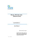
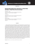
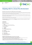
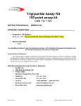
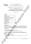
Signosis Innovative Plate Assay Solutions Human TGF-β1 ELISA Catalog Number EA-0208 (For Research Use Only) Introduction The transforming growth factor beta 1 (TGF-β1) gene codes a multifunctional cytokine that controls proliferation, differentiation, and other functions in many cell types, including cancer cells, the surrounding stromal cells, immune cells, endothelial and smooth-muscle cells. It causes immunosuppression and angiogenesis, which makes the cancer more invasive. TGF-β also converts effector T-cells, which normally attack cancer with an inflammatory (immune) reaction, into regulatory (suppressor) T-cells, which turn off the inflammatory reaction. TGF-β induces apoptosis in numerous cell types. TGF-β can act on adipocyte precursor cells (1). TGF- β1 has been shown to be a potent inhibitor of the differentiation of adipogenic cell lines (2). In addition, a differentiation-defective, insulin-independent cell linederived from the adipogenic cell line 1246 produces in its conditional medium a TGF- β1-like polypeptide which could modulate the cell ability to differentiate in an autocrine fashion. Increased TGF-b1 expression was associated with BMI and abdominal adipose tissue in morbid obesity (4). Diagram of ELISA Principle of the assay TGF-β1 ELISA is based on the principle of a solid phase enzyme-linked immunosorbent assay. The assay utilizes a mouse anti-human TGF-β1 antibody for immobilization on the microtiter wells and chicken anti-human TGF-β1 antibodies along with streptavidin conjugated to horseradish peroxidase (HRP) for detection. The test sample is allowed to react simultaneously with the two antibodies, resulting in the TGF-β1 molecules being sandwiched between the solid phase and enzyme-linked antibodies. After incubation, the wells are washed to remove unbound-labeled antibodies. A HRP substrate, TMB, is added to result in the development of a blue color. The color development is then stopped with the addition of Stop Solution changing the color to yellow. The concentration of TGF-β1 is directly proportional to the color intensity of the test sample. Absorbance is measured spectrophotometrically at 450 nm. Materials provided with the kit 96 well microplate coated with a mouse antihuman TGF-β1 antibody (4oC). Biotin labeled chicken anti-human TGF-β1 antibodies (-20oC). Streptavidin-HRP conjugate (4oC). Recombinant TGF-β1 standard (-20oC). 1X Diluent buffer (4oC). 5X Assay wash buffer (RT) Substrate (4oC). Stop Solution (4oC). Material required but not provided Microplate reader capable absorbance at 450 nm Deionized or distilled water. of measuring Signosis, Inc. • 1700 Wyatt Drive Suite 10-12 • Santa Clara, CA 95054 • Tel 408 747 0771 • Fax 408 864 2182 Reagent preparation before starting experiment Dilute the 5x Assay wash buffer to 1x buffer 40ml 5x Assay wash buffer 160ml ddH2O Dilute 50 times of human recombinant TGF-β1 (220ng/ml) with 1X Diluent buffer to 4400pg/ml and then 2-fold serial dilutions. Dilute 50 times by adding 4ul Human Recombinant TGF-β1 in 200ul 1X Diluent Buffer (See Step 2 in “Assay Procedure” for detailed procedure) Dilute 400 times of biotin labeled chicken antihuman TGF-β1 antibody with 1X Diluent buffer before use. Dilute 200 times of streptavidin-HRP with 1X Diluent buffer before use. Assay procedure 1. Cut the sealing film over the plate and remove it from the desired number of well strips. Make sure the rest of wells are well sealed. 2. See instruction and diagram below for standard preparation. a. b. c. d. Add 200ul 1X Diluent buffer to the 1st well. Add 100ul 1X Diluent Buffer to the rest wells of strip. Add appropriate amount of protein recombinant (follow instruction in “Reagent Preparation”) Mix dilutions in 1st well and transfer 100ul from the 1st well to the next dilution. (See picture) Incubate each well for 1 hr at room temperature with gentle shaking 7. Add 100 µl of diluted streptavidin-HRP conjugate to each well and incubate for 45 min at room temperature with gentle shaking. 8. Repeat the aspiration/wash as in step 4. 9. Add 100µl of substrate to each well and incubate for 10-30 minutes. 10. Add 50µl of Stop solution to each well. The color in the wells should change from blue to yellow. 11. Determine the optical density of each well with a microplate reader at 450 nm within 30 minutes. References (1) Petruschke T, Rohrig K, Hauner H. Transforming growth factor beta (TGFbeta) inhibits the differentiation of human adipocyte precursor cells in primary culture. Int J Obes Relat Metab Disord 1994;18:532–6. (2) lgnotz, R., and Massague, J. 1985. Type beta transforming growth factor controls the adipogenic differentiation of 3T3 fibroblasts. Proc. NatI. Acad. Sci. USA, 82: 8530-8540, 1985. (3) Yamada, Y., and Serrero, G. 1989 Characterization of transforming growth factors produced by the insulinindependent teratoma-derived cell line 1246-3A. J. Cell. Physiol., 140: 254-263. (4) Alessi MC, Bastelica D, Morange P, et al. Plasminogen activator inhibitor 1, transforming growth factor-beta 1, and BMI are closely associated in human adipose tissue during morbid obesity. Diabetes 2000;49:1374–80. Example of standard curve Use 100ul of Standard, 3. Add 100ul of sample per well and incubate for 1 hour control, or sample per well at room temperature with gentle shaking. and incubate for200µl 1 hr of 1X 4. Aspirate each well and wash by adding Assay wash buffer. Repeat the process three times for a total of three washes. Complete removal of liquid at each wash. After the last wash, remove any remaining liquid by inverting the plate against clean paper towels. 5. Add 100µl of diluted biotin-labeled anti-human TGFβ1 antibody to each well and incubate for 1 hour at room temperature with gentle shaking. 6. Repeat the aspiration/wash as in step 4. Signosis, Inc. • 1700 Wyatt Drive Suite 10-12 • Santa Clara, CA 95054 • Tel 408 747 0771 • Fax 408 864 2182