1
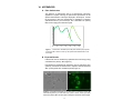
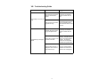
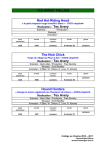
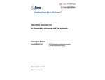
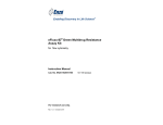
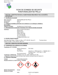
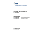
Enabling Discovery in Life Science® Cyto-ID™ Green Long-Term Cell Tracer Kit for flow cytometry and fluorescence microscopy Instruction Manual Cat. No. ENZ-51036-K025 For research use only. Rev. 1.0.1 June 2011 Notice to Purchaser The Cyto-ID™ Green Long-Term Cell Tracer Kit is a member of the CELLestial® product line, reagents and assay kits comprising fluorescent molecular probes that have been extensively benchmarked for live cell analysis applications. CELLestial® reagents and kits are optimal for use in demanding cell analysis applications involving confocal microscopy, flow cytometry, microplate readers and HCS/HTS, where consistency and reproducibility are required. This product is manufactured and sold by ENZO LIFE SCIENCES, INC. for research use only by the end-user in the research market and is not intended for diagnostic or therapeutic use. Purchase does not include any right or license to use, develop or otherwise exploit this product commercially. Any commercial use, development or exploitation of this product or development using this product without the express prior written authorization of ENZO LIFE SCIENCES, INC. is strictly prohibited. Limited Warranty These products are offered under a limited warranty. The products are guaranteed to meet appropriate specifications described in the package insert at the time of shipment. Enzo Life Sciences’ sole obligation is to replace the product to the extent of the purchase price. All claims must be made to Enzo Life Sciences, Inc. within five (5) days of receipt of order. Trademarks and Patents Enzo, CELLestial, and Cyto-ID are trademarks of Enzo Life Sciences, Inc. Several of Enzo’s products and product applications are covered by US and foreign patents and patents pending. Contents I. Introduction ............................................................... 1 II. Reagents Provided and Storage .............................. 1 III. Additional Materials That May Be Required ........... 2 IV. Safety Warnings and Precautions ........................... 2 V. Methods and Procedures ......................................... 3 A. REAGENT PREPARATION ................................................... 3 B. STAINING LIVE, SUSPENSION OR ADHERENT CELLS ............ 3 C. CELL ANALYSIS BY FLOURESCENCE/CONFOCAL MICROSCOPY ................................................................... 5 D. CELL ANALYSIS BY FLOW CYTOMETRY .............................. 5 VI. Appendices ............................................................... 6 A. FILTER SET SELECTION..................................................... 6 B. EXPECTED RESULTS ......................................................... 6 VII. References ................................................................ 7 VIII. Troubleshooting Guide ............................................ 8 I. Introduction The Cyto-ID™ Green Long-Term Cell Tracer Kit uses proprietary noncovalent cell labeling technology to stably incorporate a green fluorescent dye containing hydrophobic aliphatic chains into the cell membrane’s lipid bilayer. The dye may be loaded into cells by following the included protocol. The labeling buffer is isotonic for mammalian cells and contains no detergents or organic solvents. The appearance of labeled cells may vary depending upon the cell type from uniformly bright to punctuate. This difference is thought to relate to the extent of membrane internalization occurring after cell labeling. The Cyto-ID™ Green Tracer dye fluorescence is independent of pH within normally encountered physiologic ranges and fluorescence intensity per cell is typically unaffected by the ultimate pattern of dye distribution. The Cyto-ID™ Green Tracer dye is not toxic to cells, as determined using the benchmark MTT cell viability assay. The dye is well retained by cells for up to 96 hours after loading, and is passed to daughter cells upon mitosis. Since the dye does not covalently modify proteins within the cells, normal physiological responses are better preserved than with molecular probes based upon thiol-reactive chloromethyl-based or amine-reactive succinimidyl ester-based fluorescent dyes. Dual labeling is also possible using a variety of available CELLestial® dyes. Labeled cells can be visualized by epifluorescence or confocal fluorescence microscopy. Additionally, dye-labeled and unlabeled cell populations can be analyzed by flow cytometry. No transfer of fluorescence to adjacent cells was observed after a prolonged 96-hour incubation period. This is in stark contrast to Calcein AM and BCECF AM, which are only retained within viable cells for a few hours at physiological temperatures. The kit is suitable for a variety of applications including long term cell viability, cytotoxicity, cell adhesion, cell migration and cell-cell fusion studies. II. Reagents Provided and Storage All reagents are shipped on dry ice. Upon receipt, the kit should be stored upright at ≤-20°C, protected from light. When stored properly, these reagents are stable for one year upon receipt. Avoid repeated freezing and thawing. Reagents provided in the kit are sufficient for 25 reactions for flow cytometry or fluorescence microscopy. Reagent Quantity Cyto-ID™ Green Tracer Dye 4X Labeling Buffer 50 μl 12.5 mL 10X HBSS 25 mL 1 III. Additional Materials That May Be Required • Flow cytometer equipped with 488nm blue laser • Fluorescence microscope • 15 ml and 50 ml conical tubes • Adjustable speed centrifuge with a swinging bucket rotor • CO2 incubator (37°C), tissue culture plasticware, tissue culture reagents • 5 mL round bottom polystyrene tubes for holding cells • Calibrated, adjustable precision pipettors, preferably with disposable plastic tips • Glass microscope slides • Glass cover slips • Deionized water • Serum (e.g., Fetal Bovine Serum) • Growth medium (e.g., Dulbecco’s Modified Eagle Medium, D-MEM) IV. Safety Warnings and Precautions • This product is for research use only and is not intended for diagnotic purposes. • Some components of this kit may contain hazardous substances. Reagents can be harmful if ingested or absorbed through the skin and may cause irritation to the eyes. They should be treated as possible mutagens and should be handled with care and disposed of properly. • Observe good laboratory practices. Gloves, lab coat, and protective eyewear should always be worn. Never pipet by mouth. Do not eat, drink or smoke in the laboratory areas. All blood components and biological materials should be treated as potentially hazardous and handled as such. They should be disposed of in accordance with established safety procedures. • To avoid photobleaching, perform all manipulations in low light environments or protected from light by other means. • Methods are developed and optimized using kit components. Use only supplied buffers for optimal results. 2 V. Methods and Procedures NOTE: Allow all reagents to thaw at room temperature before starting with the procedures. Upon thawing, gently hand-mix or vortex the reagents prior to use to ensure a homogenous solution. Briefly centrifuge the vials at the time of first use, as well as for all subsequent uses, to gather the contents at the bottom of the tube. A. REAGENT PREPARATION 1. 2X Cyto-ID™ Green Tracer Dye Solution IMPORTANT: Prepare this reagent immediately before labeling cells. In an appropriate size container, mix, by vortexing, the following: 2 μL Cyto-ID™ Green Tracer Dye 1mL 1X Labeling Buffer (from step A2) 2. 1X Labeling Buffer Dilute each milliliter (mL) of 4X Labeling Buffer with 3 mL deionized water. 3. 1X HBSS Allow the 10X HBSS to warm to room temperature. Make sure that the reagent is free of any crystallization before dilution. For every 10 mL of 1X HBSS needed, dilute 1 mL of 10X HBSS with 9 mL deionized water. 4. Stop Buffer Prepare the Stop Buffer by adding 200 μL Fetal Bovine Serum (FBS) to 9.8 mL 1X HBSS (from step A3). B. STAINING LIVE, SUSPENSION OR ADHERENT CELLS NOTE: Cells are labeled by incorporating the dye into the cellular membrane. Best results are a factor of cell concentration and dye concentration. Loss of membrane integrity and poor cell recovery will result from over staining. Also, best results are obtained when adherent cells are dispersed into a cell suspension prior to staining. Perform all subsequent steps at ambient temperature (20-25°C). 1. Place a suspension containing 2 x 10⁷ cells in a 15 mL conical bottom polypropylene tube. Centrifuge cells at 400 x g and remove growth medium. Wash twice with 4 mL 1X HBSS. NOTE: Serum proteins and lipids also bind the dye, reducing the effective concentration available for cell labeling. 3 2. Centrifuge the cells at 400 x g for 5 minutes into a loose pellet. After centrifuging cells, carefully discard the supernatant, being careful not to remove any cells but leaving no more than 25 μL of supernatant. IMPORTANT: For reproducible results, it is important to minimize the amount of residual medium or buffer present when cells are re-suspended in the Labeling Buffer. 3. Prepare a 2X cell suspension by adding 1 mL of 1X Labeling Buffer to the cell pellet and suspending with gentle pipetting to insure complete dispersion. Do not vortex and do not let cells stand in Labeling Buffer for periods longer than 15 - 20 minutes. IMPORTANT: The presence of physiologic salts like Ca+2 and Mg+2 causes the dye to form micelles and substantially reduces staining efficiency. Therefore, it is important that the cells be suspended in 1X Labeling Buffer provided at the time the dye is added, not in medium or buffered salt solutions. 4. Immediately prior to staining, prepare a 2X Cyto-ID™ Green Tracer Dye Solution in Labeling Buffer as described in step A1 on page 3. 5. Rapidly add 1 mL of 2X cell suspension (from step B3, above) to 1 mL of freshly prepared 2X Cyto-ID™ Green Tracer Dye solution and immediately mix well with a pipetor or equivalent to disperse. IMPORTANT: Staining is nearly instantaneous. Brisk and consistent dispersion of the cells in the Cyto-ID™ Green Tracer Dye solution is vital for consistent labeling. 6. Incubate the cell-dye suspension from step B4 for 2–5 minutes with periodic mixing. Longer staining periods, not to exceed 7 minutes, will result in brighter cell staining. NOTE: Do not centrifuge the cells in Labeling Buffer before stopping the staining reaction. 7. Stop the staining by adding an equal volume (2 mL) of Stop Buffer and incubate for 1 minute to allow binding of excess dye. Do not dilute with 1X Labeling Buffer. 8. Centrifuge the cells at 400 x g for 5 minutes at 25°C and carefully remove the supernatant, being sure not to remove cells. To minimize carryover of residual dye bound to the tube walls, suspend cell pellet in 10 mL of complete (serum-containing) medium, transfer to a fresh sterile conical polypropylene tube and centrifuge at 400 X g for 5 minutes at 25°C, and wash the cell pellet 2 more times with 10 mL of complete medium each wash to ensure removal of unbound dye. NOTE: Staining efficiency is increased by transferring into a fresh tube at the first suspension step after staining. Do not use Labeling Buffer for washing steps. 4 9. After the final wash, suspend the cell pellet in 10 mL of complete medium and transfer to a T25 flask or slides and incubate at 37°C and 5% CO2 overnight for no less than 12 hours, to allow cells to recover. 10. Perform cell counting and viability assessment. After determining cellular recovery and viability, the following steps may be performed: a. Centrifuge the cell suspension and suspend to desired concentration of viable cells. b. Change the medium of the adherent cells. C. CELL ANALYSIS BY FLUORESCENCE/CONFOCAL MICROSCOPY SUSPENSION CELLS 1. Collect the stained cells by centrifugation for 5 minutes at room temperature at 400 x g and carefully discard the supernatant. Resuspend the cells with at least 20 μL of 1X HBSS. 2. Apply 20 µL of the cell suspension to a glass microscope slide and overlay with a coverslip. 3. Analyze the stained cells by wide-field fluorescence or confocal microscopy (60X magnification recommended). Use a standard FITC (Green) filter set for imaging the membrane signal. ADHERENT CELLS 1. Seed stained cells at your desired density (such as 1 x 10⁴ cells/ mL). Allow to adhere overnight. Remove growth medium and add 1X HBSS (step 3, page 3) as needed to maintain cell moisture and overlay with a coverslip. 2. Analyze the stained cells by wide-field fluorescence or confocal microscopy (60X magnification is recommended). Use a standard FITC (Green) filter set for imaging the membrane signal. D. CELL ANALYSIS BY FLOW CYTOMETRY The protocol described in this manual assumes that the user is familiar with the basic principles and practices of flow cytometry and is able to run samples according to the operator’s manual pertaining to the instrument being used. 1. Collect stained cells at a density not to exceed 1X 10⁶ cells/mL. NOTE: Adherent cells will need to be trypsinized prior to analysis on the flow cytometer. 2. Centrifuge at 400 x g for 5 minutes to pellet the cells. Carefully suspend the cells in 500 μL of 1X HBSS 3. Read on bench top flow cytometer by gating out cellular debris and using a 488 nm blue laser for excitation and FL1 for signal registration. 5 VI. APPENDICES A. Filter Set Selection The selection of optimal filter sets for a fluorescence microscopy applications requires matching the optical filter specifications to the spectral characteristics of the dyes employed in the analysis. Consult the microscope or filter set manufacturer for assistance in selecting optimal filter sets for your microscope. Use a standard FITC (Green) filter set for imaging the membrane signal. 300 350 400 450 500 550 600 Figure 1. Fluorescence excitation (359, 460) and emission (527) spectra for the Cyto-ID™ Green Tracer dye. All spectra were determined for cellbound dye. B. Expected Results Labeled cells can be visualized by epifluorescence microscopy using a standard FITC (Green) filter (Figure 2). Dye-labeled and unlabeled cell populations can be analyzed by flow cytometry. No transfer of fluorescence to adjacent cells was observed after a prolonged 96 hour incubation period (Figure 3). A B Figure 2: Composite bright-field (A) and fluorescence microscopy images (B) demonstrating staining of Jurkat cells with Cyto-ID™ Green Tracer dye. Stan- dard FITC (Green) filter set was used to image the membrane-bound signal. 6 Time Post-Mixing % of Stained Cells MFI Stained Cells % Unstained Cells MFI Unstained Cells 0 Hour 56.72 1466.11 40.47 2.97 24 Hours 52.92 451.65 44.39 4.08 48 Hours 50.63 101.84 46.59 3.91 120 Hours 51.59 12.3 46.23 3.24 Figure 3: Flow cytometry analysis of fluorescence of mixed population of Jurkat cells over time. Jurkat cells stained with Cyto-ID™ Green Tracer dye were mixed with an unstained population of Jurkat cells and incubated over a 120-hour period. VII. References 1. Horan, P.K., and Slezak, S.E., Stable Cell Membrane Labeling, Nature 340:167-168 (1989). 2. Horan, P.K. et al., Fluorescent Cell Labeling for in vivo and in vitro Cell Tracking Methods Cell Biol. 33:469-490(1990). 3. Poon, R.Y. et al., In Living Color: Flow Cytometry and Cell Sorting Protocols, Diamond, R. A., and DeMaggio, S., (eds.), SpringerVerlag, (New York, NY: 2000) pp.302-352. 4. Wallace, P.K., and Muirhead, K.A., Cell Tracking 2007: A proliferation of probes and applications, Immunol. Invest., 36:527-562 (2007). 7 VIII. Troubleshooting Guide Problem Potential Cause Suggestion Too much dye was incorporated into the cell membranes. Lower dye concentration and/or increase cell number. Cells remained in the labeling buffer too long. Decrease the time that cells are in the labeling buffer. The maximum time is 15-20 minutes. Labeled cells lysed, releasing dye to adjacent cells. Lower dye concentration and/or increase cell number. Excess dye in solution or on the walls of the tube. Wash cells 3-5 times after labeling. Transfer the samples to new tubes between washes. Be careful to remove as much liquid as possible during wash steps. Organic based solvents or detergents were used that may extract the dye. Use only aqueous-based fixatives and solvents and limit the use of detergents. Poor viability or recovery of cells. Cross-staining of cell populations 8 www.enzolifesciences.com Enabling Discovery in Life Science® NORTH/SOUTH AMERICA GERMANY UK & IRELAND ENZO LIFE SCIENCES INTERNATIONAL, INC. 5120 Butler Pike Plymouth Meeting, PA 19462-1202 USA T 1-800-942-0430/(610) 941-0430 F (610) 941-9252 E [email protected] www.enzolifesciences.com ENZO LIFE SCIENCES GMBH Marie-Curie-Strasse 8 DE-79539 Lörrach Germany T +49/0 7621 5500 526 Toll Free 0800 664 9518 F +49/0 7621 5500 527 E [email protected] www.enzolifesciences.com ENZO LIFE SCIENCES (UK) LTD. Palatine House Matford Court Exeter EX2 8NL UK T 0845 601 1488 (UK customers) T +44/0 1392 825900 (from overseas) F +44/0 1392 825910 E [email protected] www.enzolifesciences.com SWITZERLAND & REST OF EUROPE BENELUX FRANCE ENZO LIFE SCIENCES AG Industriestrasse 17, Postfach CH-4415 Lausen Switzerland T +41/0 61 926 89 89 F +41/0 61 926 89 79 E [email protected] www.enzolifesciences.com ENZO LIFE SCIENCES BVBA Frankrijklei 33 BE-2000 Antwerpen, Belgium T +32/0 3 466 04 20 F +33/0 437 484 239 E [email protected] www.enzolifesciences.com ENZO LIFE SCIENCES c/o Covalab s.a.s. 13, Avenue Albert Einstein FR -69100 Villeurbanne France T +33 472 440 655 F +33 437 484 239 E [email protected] www.enzolifesciences.com