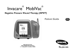
Download Wound Care Management Policy v3.Aug 15 (1)
Transcript
WOUND MANAGEMENT POLICY Version: 3 Ratified by: Senior Managers Operational Group Date ratified: August 2015 Title of originator/author: Tissue Viability Manager Title of responsible committee/group: Clinical Governance Group Date issued: August 2015 Review date: July 2018 Relevant Staff Group/s: All clinical staff involved in wound care This document is available in other formats, including easy read summary versions and other languages upon request. Should you require this please contact the Trust Equality and Diversity Lead on 01278 432000 Wound Management Policy V2.3 1 July 2015 DOCUMENT CONTROL Reference Version Status Author Aug15/WMP 3 Final Tissue Viability Manager Amended to reflect the acquisition of Somerset Community Health and changes to the Trusts governance structure and Amendments reporting arrangements. Further amended to reflect Epic 3 guidelines Document objectives: To guide staff in wound assessment and selection of dressings Intended recipients: All clinical staff involved in wound care (nursing, medical and allied health professionals) Committee/Group Consulted: Clinical Governance Group Monitoring arrangements and indicators: Clinical Governance Group Training/resource implications: 60 places are already offered per annum by Tissue Viability Service for wound management training Clinical Governance Approving body and date Date: August 2015 Group Formal Impact Assessment Impact Part 1 Date: August 2015 Clinical Audit Standards NO Date: N/A Ratification Body and date Senior Managers Operational Group Date: August 2015 Date of issue August 2015 Review date July 2018 Contact for review Tissue Viability Manager Lead Director Director of Nursing and Patient Safety CONTRIBUTION LIST Key individuals involved in developing the document Name Caroline Carrington, Charter House, Yeovil Sue Ramsden, Parkgate House, Yeovil Vanessa Redwood, Taunton & Somerset NHS Foundation Trust Members Members Designation or Group Tissue Viability Lead, East Somerset, Somerset Partnership NHS Foundation Trust Tissue Viability Lead, West Somerset, Somerset Partnership NHS Foundation Trust Tissue Viability Specialist Nurse, Taunton & Somerset NHS Foundation Trust Senior District Nurse Best Practice Group Members Community Hospital Best Practice Group Nurse Consultant, Minor Injuries Unit, Somerset Partnership NHS Foundation Trust Clinical Policy Review Group Infection Control team, Somerset Partnership NHS Foundation Trust ANNT task and finish Group Members Wound Formulary Group Michael Paynter, Bridgwater Hospital Members Infection Control Team Wound Management Policy V2.3 2 July 2015 CONTENTS Section Summary of Section Doc Document Control 2 Cont Contents 3 1 Introduction 4 2 Purpose & Scope 4 3 Duties and Responsibilities 4 4 Explanations of Terms Used 6 5 Assessment 8 6 Principles to follow after wound bed assessment 10 7 Cleansing Techniques 10 8 Selection of dressings 10 9 Evaluation 11 10 Transfer of Patients Between Care Settings 11 11 Referral to Tissue Viability Nurse 11 12 Topical Negative Pressure 12 13 Larvae Therapy 12 14 Training 13 15 Equality Impact Assessment 13 16 Monitoring, Compliance and Effectiveness 13 17 Counter Fraud 14 18 Relevant Care Quality Commission (CQC) Registration 14 19 References, Acknowledgements and Associated Documents 15 20 Appendices Appendix A Acute Wounds 16 17 Appendix B Wound/Pressure Ulcer Assessment Form 20 Appendix C Leg Ulcer Assessment Form 25 Appendix D Wound Healing Principle 27 Appendix E Cleansing the Wound 29 Appendix F Categories of Wound Dressing and Wound Product Guidelines 30 Appendix G Topical Negative Pressure Therapy- Standard Operating Procedure 34 Appendix H Maggot Therapy Protocol- Standard Operating Procedure 63 Wound Management Policy V2.3 Page 3 July 2015 1 INTRODUCTION 1.1 This policy is intended to guide the practitioner in wound assessment and to aid selection of the most appropriate product for wounds healing by secondary intention. (See section 4 for definition of terms) 1.2 Staff should ensure the patient is able to understand the information given to them and are able to give their informed consent. This may necessitate the use of a professional interpreter and the translation of written information. A capacity assessment should be undertaken for those patients who are unable to consent to the procedure and reference should be made to the relevant Trust policy. 1.3 In clinical practice many wounds are slow to heal and difficult to manage. There are a number of specialist therapies that Somerset Partnership use to manage these wounds. 2 PURPOSE & SCOPE 2.1 This document is designed to Inform staff of their roles and responsibilities in relation to wound care Provide guidance around wound assessment and selection of dressings Act as a resource for staff caring for individuals with a wound Support decision making for timely referral to specialist services 2.2 The policy is relevant to all clinical staff who provide wound care to patients 2.3 The purpose of Topical Negative Pressure Protocol is to ensure that staff using the TNP systems are able to offer safe and appropriate care to patients 2.4 The purpose of Larvae Protocol is to ensure that all patients receiving larvae therapy are safely assessed, treated and evaluated. 3 DUTIES AND RESPONSIBLITIES 3.1 The Chief Executive is ultimately responsible for ensuring the Trust complies with national recommendations for wound management. 3.2 The Trust Board has a responsibility to ensure training is available to all relevant staff. 3.3 The Tissue Viability Service are responsible for: Carrying out wound care training. Assessing and formulating a plan of care for patient with complex wounds which staff feel require specialist input. Updating trust policies. Chairing the Wound Care Formulary Group and review products on a yearly basis. Wound Management Policy V2.3 4 July 2015 3.4 Medical staff are responsible for: 3.5 Ensuring that registered nurses in the team have the appropriate skills and knowledge to manage wounds OR ensuring appropriate access to an individual with these skills. Ensuring appropriate resources and wound care products are available and reporting any deficit to the Service Manager; Ensuring that a Link Nurse for Tissue Viability is nominated in each team. Ensure that a representative from each service is nominated for the Wound Formulary Group The Tissue Viability Link Nurse is responsible for: 3.7 Assessment and review of patient’s general medical condition; Liaison with other health professionals and onward referral as necessary. Prescribing, as and when necessary. Practitioner in charge of clinical area/clinical team is responsible for: 3.6 The Tissue Viability Service will be responsible for the decision making of accepting a patient with TNP into our service. They will then make an initial visit to the patient in the first 2 weeks and monitoring their progress fortnightly through either; discussion with the team, discussion with the patient, RiO records or visiting the patient with the nursing team, dependent on the individual and which is clinically more appropriate. Patients being considered for Larvae therapy must be assessed by a Tissue Viability Nurse, Senior Diabetic Podiatrist or Consultant who will advise treatment. Attending the quarterly link nurse education meetings held by the Tissue Viability Service. Attending Wound Management Training every 3 years. Being a local resource to cascade information to colleagues Being a point of liaison between the clinical team and the Tissue Viability Service. Nursing Responsibility: To ensure a treatment plan is agreed and implemented according to individual patient needs; To ensure clear documentation of any advice or interventions which have taken place; To ensure any inadequate provision of interventions or resources are highlighted to the practitioner in charge; Report any wounds that are caused by pressure damage and are Grade 2 or above to their line manager and via the DATIX system. To ensure the treatment plan is reviewed at a time frame based on clinical need Wound Management Policy V2.3 5 July 2015 3.8 HCA’s Responsibility 3.9 To ensure that when any wound dressing change is delegated to a HCA, that the staff member is competent to carry out the task given to them. HCA’s are able to carry out wound dressing changes that have been previously assessed by a trained member of staff and a clear care plan is in place. The staff member must ensure that they are competent in the task that has been delegated to them and are aware of how to identify any changes in a wound that need to be reported to the trained nurse. HCA’s must NOT carry out specialist wound dressing changes such as: TNP, Compression Therapy and Larvae Therapy. Nor should they complete first assessments. Wound Formulary representatives are responsible for: Attending the quarterly wound formulary meeting held by the Tissue Viability Service Attending the Wound Management Training every 3 years Participating in the evaluation of products for the wound formulary group. 4 EXPLANATIONS OF TERMS USED 4.1 Secondary intention: A wound healing by secondary intention means healing of an open wound, from the base upwards, by laying down new tissue as opposed to most surgical incisions which heal by primary intention where the edges of the surgical incision are closed together with stitches or clips until the cut edges merge. 4.2 Please see Appendix 1 for principles of wound management in the acute wound. 4.3 Larvae: 4.4 Also known as Maggot therapy is a type of biotherapy involving the introduction of live, disinfected maggots into a non-healing wound for the purpose of cleaning out the necrotic tissue and preparing the wound bed, this is known as debridement. Free Range Maggots: 4.5 These come in a sterile vial and are applied directly to the wound and allowed to roam freely over the surface of the wound; this allows the maggots to seek out areas of slough or necrotic tissue. Bagged Maggots: Wound Management Policy V2.3 6 July 2015 4.6 Slough: 4.7 Topical Negative Pressure Therapy can also be provided via a moistened gauze interface. A drain wrapped in moistened gauze is placed in the wound and sealed. Negative therapy is then applied to the wound bed. Aseptic Non Touch Technique (ANTT®) 5 VAC therapy is a topical negative pressure therapy which provides negative pressure or suction applied evenly across a sealed wound surface, using a foam interface. The benefits of this technique have been reported to be increased local blood flow, a reduction in local tissue oedema, reduction in bacterial colonization and an increase in granulation tissue and wound contraction. (Argenta and Morykwas 1997) Gauze based system (Talley ®): 4.12 This is a therapeutic technique using a vacuum dressing to promote healing in acute or chronic wounds. The therapy involves the controlled application of sub-atmospheric pressure to the local wound environment, using a sealed wound dressing connected to a vacuum pump. Foam based system (VAC®): 4.11 When this tissue becomes dehydrated it forms a hard, black leathery layer over the wound, commonly called an eschar. Topical Negative Pressure (TNP): 4.10 A necrotic wound contains tissue that has become devitalised due to damage to its blood supply, for example from pressure or trauma. Eschar: 4.9 Slough refers to moist necrotic tissue. This type of devitalised tissue is soft, moist and often stringy in consistency and is usually yellow, white or grey in colour. Necrotic: 4.8 Biobag® dressings are small fabric bags in which maggots are contained, these are placed directly on the wound surface Standardised aseptic technique where staff are taught to identify and protect the key-parts of any procedure, perform effective hand hygiene, institute a non-touch technique, and wear only the appropriate personal protective equipment. ASSESSMENT Wound Management Policy V2.3 7 July 2015 PLEASE NOTE THE TISSUE VIABILITY SERVICE ARE CURRENTLY NOT COMMISSIONED TO VISIT NURSING HOMES, BUT ARE HAPPY TO PROVIDE GENERIC PHONE ADVICE. 5.1 The assessment of a wound has two elements; the holistic patient assessment and a wound assessment. Both parts are essential to maximise healing potential for the patient. 5.2 Holistic Assessment: 5.2.1 The first part of the assessment process is a full review of the factors likely to affect healing. A full holistic assessment will include the following elements: 5.3 the likely cause of the wound; any underlying condition that will affect healing; patients age; general health and medical condition; nutritional state assessed using the MUST tool; medications; infection (if suspected); pain; allergies or reactions to previous dressings; social circumstances; blood results to include full blood count, glucose, albumin; swab results, if any signs of clinical infection or critical colonisation; psychological factors; measurement of the ankle brachial pressure indices for patients with leg ulcers or wounds below the knee; duration of the wound; wound assessment. Wound Assessment: 5.3.1 The second part of the assessment process is a thorough review of the wound and will include the following elements: 5.4 wound type; wound site; grade (if pressure ulceration); classification of the wound bed; amount and colour of exudates; odour (if present); pain (if present); erythema (if present); condition of surrounding skin; infection (if suspected or reported); length, width and depth of wound in centimetres; photograph (if available). Timing of Assessment: Wound Management Policy V2.3 8 July 2015 5.4.1 Every patient with a wound will have a wound assessment completed at the first dressing change (see Appendix 2). 5.4.2 For patients with a pressure ulcer a full holistic assessment should be completed within 72 hours of admission to an inpatient unit, or within 1 week of acceptance onto a community caseload. 5.4.3 For patients with a non healing leg wound of 4-6 weeks duration then a full holistic and doppler assessment should be completed. (Appendix 3). 5.5 Leg Ulcer Assessment in Inpatient Settings: 5.5.1 When a patient is admitted into an inpatient setting with a leg ulcer the patients’ primary care provider must be contacted and a copy of the patient’s leg ulcer assessment (Appendix 3) and treatment plan should be requested. If the admission is due to another reason (other than the leg ulcer) then if at all possible the existing plan of care for the leg ulcer should be continued. 5.5.2 If a compression bandage needs to be reapplied and none of the receiving team members have the necessary competencies the Tissue Viability Service should be contacted and they will discuss the patient with a registered nurse and may visit the patient if necessary. 5.5.3 If no one is competent to reapply compression bandaging in the inpatient unit and the date that the compression bandage was due to be changed is reached then the bandages should be removed, the ulcer cleansed and a simple non adherent dressing, absorbent pad and retention bandage applied. The reason for variation from the plan of care should be fully documented in the patient records. The compression bandage must never be left on beyond its scheduled change date without liaison with the Tissue Viability Service. 5.5.4 If the patient has not previously had a leg ulcer assessment and the wound is noted to have been present for at least 4 – 6 weeks on admission then an assessment should be carried out. WARNING – as there is a risk of harm to the patient, only registered nurses who are trained and competent in the assessment of ulceration, measurement of ankle brachial pressure indices using Doppler ultrasound and the application of compression bandaging should assess and manage patients using these techniques. Wound Management Policy V2.3 9 July 2015 6. PRINCIPLES TO FOLLOW AFTER WOUND BED ASSESSMENT 6.1 Once a wound assessment has taken place the goal of treatment should be determined. General principles can be followed for each of type of wound bed; necrotic, sloughy, granulating and epitheliaisling (Appendix 4). It should be noted that in some instances, usually following specialist review, alternative treatments may be requested. 6.2 All wound dressings and assessment should be carried out using the ANTT. 7. CLEANSING TECHNIQUES 7.1 Every patient with a wound or leg ulcer will be assessed for a suitable cleansing technique (see Appendix 5). 8. SELECTION OF DRESSINGS 8.1 The application of a dressing should be considered as only a small part of the management of a patient with a wound. Physical, psychological, nutritional and social interventions can all have a huge influence wound healing. 8.2 The appropriate dressing selection relies on an accurate assessment of the patient, their wound and social circumstances. The selection of dressing needs to take into account: type and cause of the wound; wound tissue classification; amount and viscosity of exudate; bacterial colonisation/infection; cellular or biochemical imbalance; control of bleeding; position of wound and retention of dressing; protection of newly formed tissue; acceptability to patient and known allergies; availability in both the inpatient and community setting; the cost effectiveness of the dressing. 8.3 For patients admitted into an inpatient setting with a condition that is unrelated to the wound the nurse should liaise with the “primary carer” before changing the treatment. 8.4 Nursing staff involved in the selection of dressings should be aware of the need to remain up to date with new developments; the wound formulary; NICE guidance and the manufacturer’s product guidelines which should always be followed. 8.5 Suitable generic dressings are detailed in Appendix 6 and the Somerset Wound Formulary should be used to determine the brand of dressings recommended. Wound Management Policy V2.3 10 July 2015 9 EVALUATION 9.1 All wounds must be evaluated at each and every dressing change. Each evaluation should be documented (where in use the wound evaluation chart, Appendix 2, should be completed). Leg ulcers must be evaluated weekly and recorded on the appropriate pathway (venous or non venous). Wounds/ulcers will be measured on a timescale dependent on the rate of healing and at the discretion of the registered nurse evaluating. 9.2 All wound/pressure ulcer evaluations must be documented using the following criteria wound type; wound site; grade (if pressure ulceration); classification of wound bed; amount and colour of exudates; condition of surrounding skin; odour (if present); erythema (if present); infection (if suspected or reported) size of wound; if medical photographs are taken, follow the Photography Policy. 10 TRANSFER OF PATIENTS BETWEEN CARE SETTINGS 10.1 Patients for discharge from an inpatient setting into the primary care setting should be discussed with the primary carer and the wound management plan agreed as suitable for discharge. 10.2 The primary carer must also be informed of the wound product/s to be used as soon as discharge is agreed to allow staff time to obtain sufficient supplies. Specific dressing products should be identified in the discharge letter and a copy of the discharge letter should be sent to the patient’s general practitioner. The patient should be discharged with adequate dressings to cover the first 48 hours as a minimum. If discharge takes place prior to a weekend or Bank Holiday five days supply must be provided. A copy of the patients wound/pressure ulcer/leg ulcer assessment must also be sent to the primary carer on discharge. 10.3 If a patient is admitted into an acute care setting then the receiving ward should be contacted to notify staff of the current treatment plan and copies of all records should be sent with the patient. 11 REFERRAL TO TISSUE VIABILITY 11.1 Referral Criteria Failure of wound to show signs of healing (demonstrated by wound evaluations and measurement) or if leg ulcer, non healing after 4-6 weeks; Advice on complex wounds; Wound Management Policy V2.3 11 July 2015 11.2 Patient receiving Topical Negative Therapy (TNP) treatment; Grade 4 Pressure Ulcer. Before referral to the Tissue Viability Service, the following assessments must be undertaken: Wound/pressure ulcer assessment form completed; Plan of care initiated; If the wound/pressure ulceration is present prior to admission (either to caseload or inpatient setting) the patient’s primary care provider must be contacted and the management of the wound prior to admission should be considered and if effective continued; If the wound is a pressure ulcer please ensure an incident form is submitted. 12 TOPICAL NEGATIVE THERAPY 12.1 The introduction of the technique of topical negative pressure therapy (TNP) has been developed to try to manage complex wounds. TNP applies a controlled negative pressure to the surface of a wound that has potential advantages for wound treatment and management. 12.2 The technique of TNP therapy has been shown in practice to remove excess interstitial fluid and transmit a mechanical force to the surrounding tissues producing deformation of the extracellular matrix and promoting a reduction in wound size. 12.3 As the challenges of effective wound management become increasingly complex, the use of TNP therapy has been shown in a growing number of studies to produce dramatic improvements in clinical outcomes and therefore is used more commonly. 12.4 There are two different systems of TNP Therapy in use within Somerset Partnership NHS Foundation Trust. One uses a foam interface (KCI VAC®) and one uses a gauze interface (TALLEY®). 12.5 Staff are required to follow the attached Standard Operating Procedure regarding TNP (Appendix 7) 13 MAGGOT THERAPY (LARVAE THERAPY - Biomonde®) 13.1 Sterile Larvae (maggots) of the common Greenbottle Lucilia sericata can be used to cleanse infected, sloughy or necrotic wounds. 13.2 Maggots produce proteolytic enzymes that breakdown sloughy and necrotic tissue which they ingest. In addition to their cleansing action they can also reduce odour and combat infection by ingesting and killing bacteria including Methicillin Resistant Staphylococcus Aureus (MRSA). There are also reports indicating that maggot use can reduce pain and may stimulate the formation of granulation tissue. Wound Management Policy V2.3 12 July 2015 13.3 Staff are required to follow the attached Standard Operating Procedure regarding Larvae therapy (Appendix 8) 14 TRAINING 14.1 The Trust will work towards all staff being appropriately trained in line with the organisation’s Staff Mandatory Training Matrix (training needs analysis). All training documents referred to in this policy are accessible to staff within the Learning and Development Section of the Trust Intranet. The Tissue Viability Service provide wound management training and dates are available through the training department. Application and evaluation of Larvae therapy may only be undertaken by a registered nurse who has received training and been assessed as competent on the management of patients having larvae therapy. Each nurse is responsible for maintaining his/her competence. Training can be accessed via the Tissue Viability Service and BioMonde®. 15 EQUALITY IMPACT ASSESSMENT 15.1 All relevant persons are required to comply with this document and must demonstrate sensitivity and competence in relation to the nine protected characteristics as defined by the Equality Act 2010. In addition, the Trust has identified Learning Disabilities as an additional tenth protected characteristic. If you, or any other groups, believe you are disadvantaged by anything contained in this document please contact the Document Lead (author) who will then actively respond to the enquiry. 16 MONITORING COMPLIANCE AND EFFECTIVENESS 16.1 Monitoring arrangements for compliance and effectiveness: 16.2 All complaints concerning wounds will be investigated; Individual services should review the quality of their record keeping as part of the annual record keeping audit. The quality of the wound management documentation should be included in this audit process. Responsibilities for conducting the monitoring: 16.3 Individual services should monitor locally. Methodology to be used for monitoring: Complaints and incident monitoring; Record keeping audit; Serious untoward incidents; Wound Management Policy V2.3 13 July 2015 16.4 Frequency of monitoring: 16.5 Complaints monitoring reported monthly to Clinical Governance Group quarterly Annual record keeping audit. Process for reviewing results and ensuring improvements in performance occur: 16.5.1 Complaints reports will be presented to the Clinical Governance Group (or any group that supersedes this) for consideration, identifying good practice, any shortfalls, action points and lessons learnt. The information will be disseminated through local governance groups. 16.5.2 Lessons learnt from any serious untoward investigations carried out will be presented to the Serious Incidents Requiring Investigation Review Group. 16.5.3 The record keeping audit results will be shared through the local governance groups. 17 COUNTER FRAUD 17.1 The Trust is committed to the NHS Protect Counter Fraud Policy – to reduce fraud in the NHS to a minimum, keep it at that level and put funds stolen by fraud back into patient care. Therefore, consideration has been given to the inclusion of guidance with regard to the potential for fraud and corruption to occur and what action should be taken in such circumstances during the development of this procedural document. 18. RELEVANT CARE QUALITY COMMISSION (CQC) REGISTRATION STANDARDS The standards and outcomes which inform this procedural document are as follows: 18.1 Under the Health and Social Care Act 2008 (Regulated Activities) Regulations 2014 (Part 3), the fundamental standards which inform this procedural document, are set out in the following regulations: Regulation 9: Regulation 10: Regulation 11: Regulation 12: Regulation 13: Regulation 14: Regulation 15: Regulation 16: Regulation 17: Regulation 18: Regulation 19: Regulation 20: Regulation 20A: Wound Management Policy V2.3 Person-centred care Dignity and respect Need for consent Safe care and treatment Safeguarding service users from abuse and improper treatment Meeting nutritional and hydration needs Premises and equipment Receiving and acting on complaints Good governance Staffing Fit and proper persons employed Duty of candour Requirement as to display of performance assessments. 14 July 2015 18.2 Under the CQC (Registration) Regulations 2009 (Part 4) the requirements which inform this procedural document are set out in the following regulations: Regulation 16: Regulation 17: Regulation 18: Notification of death of service user Notification of death or unauthorised absence of a service user who is detained or liable to be detained under the Mental Health Act 1983 Notification of other incidents 18.3 Detailed guidance on meeting the requirements can be found at http://www.cqc.org.uk/sites/default/files/20150311%20Guidance%20for%20p roviders%20on%20meeting%20the%20regulations%20FINAL%20FOR%20P UBLISHING.pdf 19 REFERENCES, ACKNOWLEDGEMENTS AND ASSOCIATED DOCUMENTS 19.1 References: All Wales Tissue Viability Nurse Forum. Larval Debridement Therapy 2013 Arthurson, G. (1993) Management of Burns. Journal of Wound Care March 2, 2 Bosworth C (1997) Burns Trauma Management, Bailiere Tindall Fernandez R, Griffiths R, Ussia (2003) Water for Wound Cleansing, The Cochrane Library, Issue 3, Oxford. Fowler A (2003) the Management of Non Complex Burns within the Community Nursing Times Vol 99(25) 49-51 H.P. Lovedaya, J.A. Wilsona, R.J. Pratta, M. Golsorkhia, A. Tinglea, A. Baka. (2014). epic3: National Evidence-Based Guidelines for. Journal of Hospital Infection. 86S1 S1-S70. McKirdy L (2001) Burn Wound Cleansing Journal of Community Nursing 15,5. 24-29 NICE Guidelines: Prevention and control of Healthcare-associated infections overview 2014 Pape, SA. & Hodgkinson, PD (1996) Emergency Management of Severe Burns Course Manual UK version for The British Burn Association. Settle, JA et al (2001) Burns The first Five Days. British Burn Association, Smith & Nephew Thomas, S., Jones, M. Maggots and the battle against MRSA. Bridgend: SMTL, 2000. Wound Management Policy V2.3 15 July 2015 Thomas, S. (2006). Cost of managing chronic wounds in the UK, with particular emphasis on maggot debridement therapy. J. Wound Care, November 2006, vol./is. 15/10, pp.465-9. Thomas, S., Jones, M. Maggots can benefit patients with MRSA. Practice Nurse 2000; 20(2): 101-104. Vilcinskas, A., 2011. From traditional maggot therapy to modern biosurgery. Insect Biotechnology 2 (1), pp. 67-75. 19.2 Cross reference to other procedural documents: Development & Management of Organisation-wide Procedural Document Learning Development and Mandatory Training Policy Risk Management Strategy Mandatory Training Matrix Training Prospectus Untoward Event Reporting Policy Serious Incidents Requiring Investigation (SIRI) Policy Aseptic Non Touch Techniques Waste – Healthcare (clinical) Waste Consent and Capacity to consent to treatment Consent to examination and treatment Hand hygiene 20 APPENDICES 20.1 For the avoidance of any doubt the appendices in this policy are to constitute part of the body of this policy and shall be treated as such. This should include any relevant Clinical Audit Standards Appendix A Acute Wounds Appendix B Wound/Pressure Ulcer Assessment Form Appendix C Leg Ulcer Assessment Form Appendix D Wound Healing Principle Appendix E Cleansing the Wound Appendix F Categories of Wound Dressing and Wound Product Guidelines Appendix G Topical Negative Pressure Therapy – Standard Operating Procedure Appendix H Maggot Therapy- Standard Operating Procedure Wound Management Policy V2.3 16 July 2015 APPENDIX A ACUTE WOUNDS 1 IMMEDIATE FIRST AID MEASURES FOR ACUTE WOUNDS 1.1 The initial priority is to ensure your own safety. Remember to use universal precautions. 1.2 Resuscitation of the patient is paramount so assess the patency of the patient’s airway, breathing and circulation. If these priorities are not stable immediately call for emergency assistance (999). 1.3 If the patient is stable identify specific wounds. If wounds are actively bleeding apply direct pressure utilising appropriate materials (initial dressing materials do not need to be ‘sterile’ just clean). If bleeding is significant lay the patient down. If the wound is on an extremity elevate the limb, providing verbal reassurance, the patient will no doubt be at least anxious. 1.4 Quickly identify if the wound is sufficiently minor to be assessed, examined and managed locally. 1.5 Maintain a low threshold for referral to a minor injury unit or emergency department for definitive expert management. 1.6 Wounds often have mediolegal implications; therefore record keeping must be thorough, legible, and accurate. 2 HISTORY 2.1 Key questions are: What caused the wound? (knives/glass may injure deep structures); Was there a crush component? (significant soft tissue swelling may ensue); Where did it occur? (contaminated or clean environment); Was broken glass (or china) involved? (indication for imaging); When did it occur? (old wounds may require delayed primary closure); Who caused it? (has the patient a safe and secure environment to return to?); Is tetanus cover required?. 3 EXAMINATION 3.1 Consider and record the following: Length of wound: ideally measure, if not use the term ‘approximately’ in the notes; Site: use diagrams where possible. Consider taking digital photographs; Orientation: vertical, horizontal or oblique; Contamination: by dirt or other foreign bodies may be apparent; Wound Management Policy V2.3 17 July 2015 Infection: either localised or spreading, is a feature of delayed presentation and is associated in particular with certain specific injuries; Neurological injury: be aware that complete nerve transaction does not automatically result in complete loss of sensation. Assume that any altered sensation reflects nerve injury; Tendons: complete division is usually apparent on testing. Partial tendon division is easily missed unless the wound is carefully examined as the tendon may still be capable of performing its usual function; Vascular injury: check for distal pulses including capillary refill time; Depth: wounds not fully penetrating the skin are ‘superficial’; Type of wound: inspection often allows wounds to be described, helping to determine the mechanism of trauma (blunt or sharp injury) and hence the risk of associated injuries. The crucial distinction is whether a wound was caused by a sharp or blunt instrument. If in doubt, avoid any descriptive term and simply call it a ‘wound’. 3.2 Where there is any doubt about the extent of a wound (for example if there is potential neurovascular compromise; foreign bodies; contamination; concern about the anatomical location of a wound, wounds over joints, involving the hands, face or genitals) refer the patient to a minor injury unit or emergency department. 4 CLASSIFICATION OF WOUNDS 4.1 Wounds can be classified into different categories: Incised wounds: may also be referred to as ‘cuts’. Caused by sharp injury and characterized by clean cut edges. Typically these include ‘stab’ wounds (which are deeper than they are wide) and ‘slash’ wounds (which are longer than they are deep); Lacerations: caused by blunt injury, the skin is torn, resulting in irregular wound edges. Unlike incised wounds, tissues adjacent to laceration wound edges are also injured by crushing and will show evidence of bruising; Puncture wounds: most result from injury with sharp objects; Abrasions: commonly known as ‘grazes’, these result from blunt injury applied tangentially. Abrasions are often ingrained with dirt, with the risk of infection and in the long term skin tattooing. 5 WOUND MANAGEMENT 5.1 Clean all wounds irrespective of whether closure is contemplated to reduce the risk of infection. The standard agent used for wound cleaning is normal saline, possibly preceded by washing using tap water. 5.2 If devitalised or grossly contaminated wound edges, or dirt or other foreign material is still visible despite these measures refer the patient to a minor injury unit or emergency department for specialist management. 6 WOUND CLOSURE Wound Management Policy V2.3 18 July 2015 6.1 There are three recognized types of wound closure: 6.2 Primary closure soon after the injury; Secondary closure, no intervention, heals by granulation (secondary intention); Delayed primary closure. Apart from primary closure with ‘steri-strips’ (steri-strips are inappropriate over joints but are very useful for pretibial wounds) or secondary closure other methods of wound closure are best undertaken by specialists in either minor injury units or emergency departments. Mike Paynter, Nurse Consultant, Minor Injuries Unit Somerset Partnership NHS Foundation Trust Wound Management Policy V2.3 19 July 2015 APPENDIX B WOUND/PRESSURE ULCER ASSESSMENT Name................................................ GP........................................................... Address............................................. Practice.................................................... ........................................................... Ward/Hosp............................................... Tel...................................................... DN team................................................... DOB .................................................. Date of Assessment................................. Ethnicity.............................................. Assessed by ............................................ NHS No.............................................. Site/location of wound/s Type/cause of wound/s 1. ............................................................ 2. ............................................................ 3. ............................................................ 4. ............................................................ Wound Appearance Wound Continuum Necrotic..............................% 6 5 4 3 2 1 Sloughy .............................% Granulation........................% Epithelising........................% Level of exudate Heavy Moderate Low Colour........................................... Purulent Serous Allergies ..................................................... ..................................................... Wound Management Policy V2.3 Nil Initial Size of Wound/s on Assessment 1. L…….cm x W……cm x D........cm 2. L…….cm x W……cm x D .......cm 3. L…….cm x W……cm x D .......cm 4. L…….cm x W……cm x D .......cm Any undermining/tracking N........cms S........cms W.......cms E.......cm Tracing……….……………....Y/N Photograph/s taken…….…..Y/N Verbal consent……………...Y/N Written consent.....................Y/N Taken by…………………….. 20 July 2015 Condition of surrounding skin Healthy Dry/scaly Macerated/excoriated Oedema Eczema Other .............................................. Pain Score None 0 Mild 1 Moderate 2 Severe 3 Signs of infection Erythema………………….……Y/N Odour………………….………..Y/N Increase pain…………….…….Y/N Increase exudate…………..….Y/N Other…………………………… Wound swab taken……………Y/N Date sent……………………….. MRSA status....……………….... Medication .......................................................... Medical conditions .......................................................... .......................................................... Serum albumin level……………………. Haemoglobin level……………………… Blood Glucose level……………………. .......................................................... If Pressure Ulceration Nutrition Current Waterlow Score ………….. Assessment completed ...................Y/N Pressure relieving equiment in use Requirements.......................................... ........................................................ ................................................................ Grade of Pressure sore 1 2 3 4 (circle) Social Assessment If grade 2 or above complete clinical incident form - completed…........ Y/N ................................................................................. Description of pain…………………………… Diabeties………………………Y/N Vasculitis……………………....Y/N Heart failure……………………Y/N Malignancy of wound……….. Y/N Other………………………………… Blood test Date ................................................................................. Comments Refer to ............................................................ ................................................................................. ............................................................ ................................................................................. Plan of Care (outcome of assessment) ...................................................................................................................................... ...................................................................................................................................... ...................................................................................................................................... Wound Management Policy V2.3 21 July 2015 TREATMENT PLAN Patient Name…………………………. NHS No......................................... Wound Treatment Plan No…… Wound No./Site.......................... Commenced by……………………. Date……………. Patient consent............... Rational of treatment Wound cleansing Topical application to skin Primary dressing Secondary dressing Method of retention Frequency of treatment Rational for discontinuing plan Discontinued by……………………………………………. Date………………………… Wound Treatment Plan No…… Wound No /Site.............................. Commenced by……………………. Date……………. Patient consent............... Rational of treatment Wound cleansing Topical application to skin Primary dressing Secondary dressing Method of retention Frequency of treatment Rational for discontinuing plan Discontinued by……………………………………………. Date………………………… Wound Management Policy V2.3 22 July 2015 EVALUATION Patient Name………………………. Date/Time NHS No..................................... Wound evaluation / comments Wound Management Policy V2.3 23 Sign July 2015 MEASURABLE WOUND EVALUATION Patient Name:……………………………. NHS No……………………… Wound no........ Location.................................. Date Type ............................. Date Date Date Date Date Date Wound Healing Continuum 6 5 4 3 2 1 Amount of Exudate 3 2 1 Suspected Infection? No Yes Swab Pain Score 3 2 1 0 Wound Size Length Width Surface area Comment To be completed weekly. One sheet per wound. Wound Management Policy V2.3 24 July 2015 APPENDIX C SOMERSET LEG ULCER ASSESSMENT FORM Name................................................................ NHS No.................................................................... Address............................................................. GP/Consultant.......................................................... .......................................................................... Date of assessment................................................. Postcode..........................Tel No..................... Assessed by............................................................. DOB......... /............./.............Age.................... Clinic Venue............................................................. MEDICAL DETAILS Arterial Related Past arterial surgery/Intervention Intermittent claudication Myocardial Infarcation / angina Y N Venous Related Deep vein thrombosis Varicose veins Sclerotherapy / laser Y N Cardiac Failure Vein surgery Stroke / TIA Phlebitis Hypertension Cellullitis Diabetes Leg trauma / injury / joint surgery Rheumatoid arthritis Smoker Ulcerative colitis Pregnancy No......... Other Details.......................................................................................................................................... Medication............................................................................................................................................................ ............................................................................................................................................................................... ......................................................................... Allergies................................................................................... MOBILITY Fully mobile Restricted – no aids Restricted – with aids Housebound Chair bound Ankle movement Foot deformity Abnormal Gait Y N Investigations Height...................Weight......................BMI............. Social Assessment ......................................................................... ......................................................................... ......................................................................... Right Ulcers ( )..........cm x............cm Ulcers ( )..........cm x............cm Ulcers ( )..........cm x............cm Occupation.............................................................. Nutrition /Must score ......................................................................................... ........................................................................... Recent weight loss/gain............................................ Blood Pressure....................................................... Blood glucose..............................FBC.................... Albumin..................................................................... Other......................................................................... Left Ulcers ( )..........cm x............cm Ulcers ( )..........cm x............cm Ulcers ( )..........cm x............cm How ulcer started ..................................................................................... Wound Management Policy V2.3 Y N Recurrence Consent to photo Photograph taken Patient understanding ..................................................................................... 25 July 2015 ..................................................................................... Duration of present ulcer............................................. ..................................................................................... ................................................................ Patient description of pain ...................................................................................................... ...................................................................................................... ...................................................................................................... Analgesic..................................................................................... SKIN CHANGES Pale limb on elevation Red / Blue on dependency Foot feels warm Capillary refill < 3 Secs Stemmers sign +ve Y N Right Leg Induration Staining Y Pain Scale 0 1 2 No pain Mild Moderate N Left Leg Y 3 Severe Pain N Varicose Eczema Atrophie Blanche Ankle flare DOPPLER ASSESSMENT (divide ankle pressure by brachial pressure) Patient Consent given Y N Patient rested and supine > 10mins Y N Right Leg Left Leg Toe Pressure Right Toe Left Toe Dorasalis pedis ....................... ........................ Posterior tibial ........................ ........................ Brachial ........................ ........................ Brachial ....................... ..................... Ankle pressure index (ABPI) ........................ ........................ Toe ....................... ..................... Ankle Circumference (cm) ........................ ........................ TBPI= ....................... ..................... PLANNED MANAGEMENT ............................................................................................................................................................................... ............................................................................................................................................................................... ............................................................................................................................................................................... ............................................................................................................................................................................... ............................................................................................................................................................................... ............................................................................................................................................................................... ............................................................................................................................................................................... ............................................................................................................................................................................... ............................................................................................................................................................................... ............................................................................................................................................................................... ............................................................................................................................................................................... ............................................................................................................................................................................... ............................................................................................................................................................................... ............................................................................................................................................................................... Patient’s Consent Given.....................................................Signed......................................................................................... Referred to: ................................................................................. ................................................................................. ................................................................................. Additional Comments: ......................................................................................... ......................................................................................... ......................................................................................... Bandage regime used............................................ Date regime commenced.............................................. Wound Management Policy V2.3 26 July 2015 APPENDIX D WOUND HEALING PRINCIPLES 1 NECROTIC WOUND BED 1.1 This wound bed is characterised by dead tissue which is devitalised and black in appearance. Usually dead tissue will spontaneously separate from the healthy tissue beneath but sometimes it dries and separation is delayed. The presence of this tissue often interrupts the wound healing process and acts as a focal point for bacteria increasing the risk of infection. Necrotic tissue can be associated with high levels of pain. 1.2 The aim of wound care is to remove the devitalised tissue. This can be done in a variety of ways. Sometimes the patient is admitted to secondary care and a surgical debridement takes place in a theatre environment. Alternatively it may be possible for the necrotic tissue to be removed via sharp debridement in a community or outpatient setting either with or without local anaesthetic; this should only be carried out by an individual who has received additional training and has demonstrated competence in this technique. 1.3 Dressings may also be used to remove the tissue. Often a gel dressing is used with a secondary dressing to maintain high moisture levels to promote autolytic debridement. 2 SLOUGHY WOUND BED 2.1 Often when the black necrotic layer has been removed a yellow, partially liquefied layer is revealed. This layer of yellow tissue is made of fibrin, exudates, bacteria and pus. Alternatively this layer can also develop on a previously clean wound. This sloughy layer acts as a focal point for infection. 2.1 The aim of wound care is to remove this layer from the wound to reveal healthy tissue below. 2.2 It may be possible to remove this layer by surgical or sharp debridement if the slough is deep enough. This should only be carried out by an individual who has received additional training and has demonstrated competence in this technique. Otherwise alternative methods should be employed. 2.3 Maggots can be used to eat away the layer of slough. 2.4 Dressings can also be used to remove the layer. If the levels of exudates are high then alginate or hydrofibre dressings can be used to form a moist wound covering to facilitate autolysis. If the wound is dry then moisture will be required to facilitate removal. Wound Management Policy V2.3 27 July 2015 3 GRANULATING WOUND 3.1 A granulating wound is characterised by a deep pink or red wound bed. The new capillary loops can be seen which gives the surface a granular appearance. Granulation tissue will continue to develop at the base of the wound to fill the defect in the skin. 3.2 The aim for this type of wound is to promote the formation of granulation tissue. Dressings should provide a warm moist environment for granulation to take place. 3.3 These types of wound can vary in size and exudates levels so a wide variety of dressings can be used according to exudate levels, size and shape of the wound. The dressing selected should manage the wound exudate, be comfortable, stay in place, be cost effective and be easy to remove. 4 EPITHELIALISING WOUNDS 4.1 The margins of an epithelialising wound are generally pink or purple in colour and sometimes islands of epithelial tissue can be seen within the wound bed. 4.2 At this final stage of wound healing exudates levels are minimal so dressings are used to protect the delicate new tissue. Wound Management Policy V2.3 28 July 2015 APPENDIX E CLEANSING THE WOUND 1.1 Irrigation is the preferred method for wound cleansing as it minimises the risk of trauma to the new tissues. To irrigate means to “flush with fluid”, however achieving the exact pressure required to do this effectively in a clinical setting is difficult. In general the use of a syringe filled with a cleansing solution gently irrigated over the wound is usually sufficient. The use of forceps to remove dry skin scales or adherent wound debris from the wound/wound margins may also be beneficial. For all wounds irrigate the wound with warm sterile normal saline using an aseptic non touch technique; For chronic leg Ulcers, the wound/ulcer may be soaked in warm, mains supply tap water (Fernandez et al 2003). It is essential that universal infection control methods are maintained with this technique in order to prevent the transfer of microorganisms. All buckets or containers used for soaking limbs must be lined with plastic bin liners which must be disposed of after each use. The bucket/container must also be cleaned with hot soapy water and dried thoroughly after each use; If it is not possible to soak a chronic Leg Ulcer then the wound should be irrigated with clean tap water; The routine use of antiseptic solutions to cleanse wounds has little place in wound management; The specified use of antiseptics for critically colonised or infected wounds may be indicated and useful. Wound Management Policy V2.3 29 July 2015 APPENDIX F CATEGORIES OF WOUND DRESSINGS & WOUND PRODUCT GUIDELINES Before using any wound care product, ALWAYS consult the manufacturer’s recommendations, contra-indications and precautions as these may periodically change and ensure that the expiry date has not been reached 1 NECROTIC 1.1 Necrotic Wounds Assessment by medical professional or suitably qualified nurse May be referred to secondary care for review by surgeon Wound will require debridement, methods include surgical debridement, sharp debridement or chemical debridement Potential Dressings Hydrogel 2 SLOUGHY 2.1 Superficial Slough – Low Exudate Potential Dressings Hydrogel Hydrogel sheet Hydrocolloid sheet Protease modulating matrix dressing 2.2 Deep Slough – High Exudate Assessment by medical professional or suitably qualified nurse May be referred to secondary care for review by surgeon Wound will require debridement either surgical debridement, sharp debridement or chemical debridement Potential Dressings Consider Larvae Hydrofibre Alginate Protease modulating matrix dressing Wound Management Policy V2.3 30 July 2015 3 INFECTED Clinically infected wounds should be treated systematically with antibiotics 3.1 Infected/colonised – Low Exudate Potential Dressings Silver dressings (use for maximum 2 weeks & do not use on neonates) Honey dressings If pseudomonas silver sulfadiazine cream (NB: Prescription only medication, short term use only for 5 – 7 days) Cadexomer Iodine (not for paediatrics or neonates, check the patient has no contraindications to this product, refer to the product literature) 3.2 Infected/colonised – High Exudate Potential Dressings Cadexomer Iodine (not for paediatrics or neonates, check the patient has no contraindications to this product, refer to the product literature) Silver dressing (use for maximum 2 weeks & do not use on neonates) Silver hydrofibre (use for maximum 2 weeks & do not use on neonates) 3.3 MRSA Please refer to the Topical Antimicrobial Dressings Protocol for wounds with MRSA for the topical management of these wounds. 4 MALODOROUS WOUNDS 4.1 Malodorus – Low Exudate Consider removal or necrotic tissue and excess slough, seek medical/tissue viability specialist opinion Exclude infection Potential Dressings Activated Charcoal Dressing Honey 0.8% Metronidazole Gel (NB: Prescription only medication, for short term use only 5 days) 4.2 Malodorous – High Exudate Consider removal of necrotic tissue and excess slough, seek medical/tissue viability specialist opinion Exclude infection Potential Dressings Activated charcoal dressing with additional absorbent dressing 5 GRANULATING Wound Management Policy V2.3 31 July 2015 5.1 Superficial Granulating - High Exudate Potential Dressings Hydrofibre Flat Polyurethane foam Alginate sheet 5.2 Low Exudate Potential Dressings Hydrocolloid sheet 5.3 Deep Granulating - High Exudate Potential Dressings Hydrofibre rope Polyurethane cavity foam Alginate rope 5.4 Low Exudate Potential Dressings Hydrogel 5.5 Granulating Sinus Potential Dressings Alginate ribbon Alginate gel dressing 6 EPITHELIALISING Potential Dressings Extra thin hydrocolloid Non adherent dressing with or without adhesive border Foam dressing 7 TRAUMATIC Potential Dressings Non-adherent dressings 8 BURNS 8.1 Where possible all patients with burns should be referred to MIU for assessment and initial management of a burn, an onward referral may be made by them to a regional centre if necessary. BURN REFERRAL PROTOCOL FOR MIU STAFF. Wound Management Policy V2.3 32 July 2015 8.1.1 MIU staff should follow the Southmead Hospital Adults Burns Referral Criteria: Size: > 3% Depth: Full thickness burns Site: Burns to hands, feet, face, perineum or genitialia. Circumferential burns or involving a major joint. Mechanism: Chemical, electrical or cold injury burns. Suspected non accidental injury or neglect. Co-morbidities: Inhalation injury. Co-existing medical illness which may influence healing. Co-existing trauma. Co-existing psychiatric illness. Time: Burns not healed in two weeks. Other: Unwell/febrile patients with a burn. Changes in burn wound appearance. Signs of infection or concerns regarding healing. If above criteria met refer to Plastics SHO on-call at Southmead Hospital bleep 1311. Southmead Hospital 0117 9505050 If above criteria not met feel free to discuss with burns team. Southmead Hospital 0117 9505050 Gate 33a, Level 2, Brunel Building. Southmead Hospital Bristol BS10 5NB Adults are 16+. Children are seen by the Bristol Royal Hospital for Children. Wound Management Policy V2.3 33 July 2015 APPENDIX G TOPICAL NEGATIVE THERAPY PROTOCOL (VAC & TALLEY VENTURI PUMP) 1. ACCEPTING PATIENTS INTO OUR CARE AND MANAGING RESOURCES 1.1 Other health providers requesting discharge of a patient with ongoing topical negative therapy must be referred to the Tissue Viability Service. No patient should be accepted for treatment by Somerset Partnership without express agreement for funding via the Tissue Viability Service. A minimum of a 48 hour notice period to arrange discharge is required. 1.2 On acceptance of the patient into the Trust, the pump rental will be transferred via the Integra system by the Tissue Viability Service. 1.3 The discharging healthcare provider needs to provide sufficient equipment for three dressing changes. 1.4 The pump is hired at a daily rate. It is essential to accurately record start and finish dates of treatment to avoid wasted costs. The Tissue Viability Service and the provider of the topical negative therapy pump must be contacted immediately on discontinuation of treatment by the team that is managing the patients care. This information on how to do this is found on the back on the TNP Care Pathway. 1.5 The consumables incur an additional cost. Talley® dressings can be ordered via ONPOS/Integra/FP10. KCI® dressings are ordered via the Tissue Viability Service. 1.6 Consumables remain the property of Somerset Partnership until it is taken to a patient’s home. They should be stored in a clean environment. Staff should not store consumables at the patient’s bedside or in the patient’s home; only take the dressings required for each dressing change in a hospital and two dressing changes in to a patient’s home. This prevents wastage of expensive dressings on infection control grounds. 2 METHODS 2.1 Assessment, Indications and Contra-indications for Talley ® 2.2 The Talley Pump topical negative therapy system can be used for the treatment of many wounds including pressure ulcers, dehisced surgical wounds, sinus drainage and management, traumatic wounds and pre & postoperative flaps and grafts. Wound Management Policy V2.3 34 July 2015 2.3 The system helps wound healing by removing excess fluid from the wound, helping to reduce the size of the wound, encouraging healthy tissue to grow, protecting the wound from micro-organisms and increasing the blood supply to the wound. 3. HOW TO APPLY TALLEY VENTURI PUMP NPWT 3.1 NB. A wound sealing kit must be used with the Talley Venturi Pump™ system. 3.2 Remove all packaging from the power unit. 3.3 If not already in place, attach canister to flat underside of power unit by matching up the four location pegs and rotating locking knob ¼ turn clockwise to secure. Ensure canister is correctly located and secured otherwise NO CANISTER alarm will appear and power unit will not operate. 3.4 Using The Wound Sealing Kit: Prepare and dress the wound to be treated using the following guidelines: If required, irrigate the wound bed thoroughly with approximately 20ml of normal saline (Fig.1). Ensuring surrounding wound edges are dry. Fig 1 Place the drain in the wound bed / sinus to calculate length required. Remove and trim as necessary to fit. If a non-adherent wound contact layer is used, cut a single layer to the approximate size and shape of the wound. Lay the wound contact layer across the wound bed. Lay a single-layer of saline-moistened gauze in the wound bed (Fig. 2) Place the drain on top of the gauze, ensuring the drain is approximately 1cm from the wound edge / bottom of the sinus to allow for wound contraction. Alternatively the saline – moistened gauze can be wrapped around the drain, if more suited to the wound. The drain is never placed directly down on the wound bed, unless managing a sinus, when the drain can be placed directly down the sinus tract. Fig 2 Wound Management Policy V2.3 35 July 2015 Fig 3 With the remaining saline-moistened gauze, fill the wound bed and fluff to completely cover the drain and fill the defect to skin level (Fig. 4). CAUTION! It is critical that the gauze is moistened rather than saturated with normal saline prior to filling the wound. Fig 4 Place the transparent dressing over the filled wound, ensuring contact with at least 2.5cm of intact skin beyond the wound edges. Crimp or pinch the edges of the transparent dressing around the drain tubing to secure a proper seal. Lift the drain slightly and pinch the dressing underneath the drain to create a seal. At the tubing exit site you can apply a small amount of sealant paste where the dressing meets the tube to seal and ensure an airtight closure. 3.5 Connect drain tubing to extension tubing, if required. 3.6 Attach wound sealing kit tubing to the Talley Venturi Pump™ power unit canister by lining up locator stud or tubing connector with notch on canister drain receptacle located on top right hand corner of canister, twisting clockwise to lock. 3.7 Operating the Vacuum Power Unit: Turn on power unit to initiate suction, choosing either mains or battery operation. If using mains power, plug the smaller end of the power cable into the side of the Talley Venturi Pump™ power unit, and the other end into mains outlet in wall. NB. The battery will charge when the unit is connected to mains power (indicated by battery charge status icon on display screen scrolling from left to right) and provides automatic power back-up if mains power fails. It is recommended to use mains power when convenient to do so as this will ensure the battery is fully charged when needed. Wound Management Policy V2.3 36 July 2015 Press RUN/STOP button to invoke stand-by mode (the power unit will beep and operating pressure and battery charge status will be displayed). Adjust vacuum level if required*. Press RUN/STOP button again to initialise and run the power unit. NB. Display screen is only illuminated for a short period after button operation in battery mode. (* Vacuum level can be adjusted when in stand-by mode and for up to 1 minute after power unit is running using the UP and DOWN arrow buttons. The power unit will begin operation at the default pressure of 80mmHg.) 3.8 Once power unit is running, observe the wound site. The dressing should contract noticeably, become firm to the touch and ‘raisin-like’. If the dressing fails to contract, the dressing has not been completely sealed. Reinforce the dressing seal and/or adjust the drain and initiate suction again. 3.9 Check for dressing integrity every 2-3 hours and at every shift change in a hospital setting. In the community encourage the patient to monitor that the dressing is ‘sucked down’. 3.10 Depending on patient status and clinical judgement, the initial dressing change should take place after 48 hours and then 48-72 hours thereafter. For infected wounds the dressing may need to be changed initially every 24 hours. To change or remove wound sealing kit, press and hold the RUN/STOP button until power unit beeps three times to return to stand-by mode. Remove wound sealing kit drain tube by turning anticlockwise and lifting out of drain receptacle on canister. The drain tubing connector incorporates a stop-valve which seals in any liquid present in the tubing. Dispose of used wound sealing kit according to local clinical waste policy. If required apply new wound sealing kit as previously described and continue NPWT. 3.11 Canisters should be replaced as required or weekly. To change canister, make sure power unit is in stand-by mode (if still running, press and hold the RUN/STOP button). Remove wound sealing kit drain tube by turning anticlockwise and lifting out of drain receptacle (this can be reconnected to new canister if wound dressing is not being changed). Remove sealing plug from its location on top left hand corner of canister and use to cap drain receptacle to seal in contents. Rotate locking knob ¼ turn anticlockwise to remove canister. Dispose of used canister according to local clinical waste policy. If continuing NPWT, attach new canister and connect wound sealing kit drain tube as previously described. 3.12 To stop the power unit press and hold the RUN/STOP button until power unit beep three times to return to stand-by mode. In battery operation the power unit will then power off after one minute of inactivity. If using mains power, switch off by disconnecting the power cable from power unit or turning off mains power. 3.13 Place the user manual in a safe place for future use. Wound Management Policy V2.3 37 July 2015 4. ORDERING OF TALLEY CONSUMABLES 4.1 In a community setting consumables can be ordered on FP10 or ONPOS. 4.2 In a community hospital setting the consumables can be ordered via Integra or ONPOS. 4.3 Pumps will be ordered by The Tissue Viability Service. 5 CANCELLATION OF RENTAL TALLEY VENTURI PUMP TNP PUMPS 5.1 Contact TALLEY on 01794 503500: Give the name of the hospital and ward/patient’s surgery and patient’s name. Ensure you have the pump code to hand (this is written on the pump and starts CVEN followed by four digits) alternatively you can give the order reference number. Cancel the pump, keeping a record of the date, time, reference number of the cancellation and name of the person cancelling the pump. Contact the Tissue Viability Service to give them the cancellation number 5.2 Clean the pump, pack into storage case and leave in a secure place ready for collection. Ensure the cannister has been removed. 6 6.1 ASSESSMENT, INDICATIONS AND CONTRA-INDICATIONS FOR VAC THERAPY Indications acute and traumatic wounds sub-acute wounds e.g. dehisced incisions chronic wounds, diabetic ulceration, pressure ulcers, leg ulcers meshed grafts (pre and post operatively) skin flaps partial thickness burns Contraindications 6.1.2 non enteric or unexplored fistulae dry necrotic tissue with eschar present untreated osteomyelitis – the wound must be thoroughly debrided of all necrotic, non-viable tissue including bone and the appropriate antibiotic initiated evidence of malignancy within the wound direct placement of dressing over exposed blood vessels or organs Wounds for which care should be taken with VAC active bleeding Wound Management Policy V2.3 38 July 2015 6.1.3 patients on anticoagulation medication difficult wound haemostasis caution must be taken when placing VAC therapy dressings in close proximity to blood vessels or organs. Ensure that any vessels are adequately protected with overlying facia, tissue or other protected barriers weakened , irradiated or sutured blood vessels or organs wounds containing bone fragments or sharp edges Enteric fistulae Assessment of Patient obtain the patients verbal permission to use the therapy assess the goal of the treatment/therapy assess the wound for suitability for therapy (see indications, wounds not suitable and special cautions) check Hb, WBC and clotting levels holistic assessment of the patient to identify factors that may delay the healing process 7 VAC EQUIPMENT REQUIRED, APPLICATION AND DISPOSAL 7.1 Choice of foam Black Foam (Granufoam) dry extracellular foam may adhere so may need silicone non adherent dressing to line wound minimum negative pressure of 75mmHg, but usually set at 125mmHg foam of choice for large wounds as it will be more effective at removing exudate White Foam (Versa-foam) 7.2 dense pre-moistened foam (moistened with sterile water) totally non-adherent so can put directly onto tissues including tendons as foam more dense than black foam, will need to set a higher minimum negative pressure of 125mmHg. Usually set at 150mmHg more acceptable for patients with painful wounds Pressure the pressure is the level of the suction exerted (negative pressure) in mmHg by the pump to the wound the pump is preset at 125mmHg. This should only be changed following advice from the Tissue Viability Nurse Team, the KCI Educational Facilitator or if white foam is used Wound Management Policy V2.3 39 July 2015 Intermittenet or Continuous Pressure the negative pressure therapy can be set at either continuous or intermittent Continuous Continuous negative pressure is used for the first 48 hours in all wounds. It usually continues at continues pressure particularly if the wound exudate is large, if the wound requires a splinting effect eg sternal and some abdominal wounds, or if the patient’s pain is severe. Intermittent 8 After the first 48 hours the therapy may be altered to intermittent negative pressure, but this would only be altered under advice from the Tissue Viability Service. This pulsing action is reported to enhance granulation tissue growth. The pump is set for 5 minutes on and 2 minutes off. It will only be necessary to change this following advice from the Tissue Viability Nurse Team. APPLICATION OF TOPICAL NEGATIVE THERAPY assess the wound for suitability for treatment check for all contra-indications and precautions obtain and document the patients verbal permission for application of VAC therapy ensure all equipment and dressings are available and present on the ward/department prepare the wound, cleansing with warm saline if necessary prepare silicone non adherent dressing if required cut foam to fit just under the size of the wound and place gently into the wound apply drape film dressing over the foam leaving a border of 3-5cms around the foam* cut a 2cm x 2cm (approximately the size of a 2 pence piece) hole in the drape film dressing apply the T.R.A.C. pad over the drape film connect the T.R.A.C pad to the canister tubing date the canister and insert the canister into the VAC pump turn on the VAC pump, set the correct level of negative pressure and correct therapy mode eg continuous or intermittent lock the pump check the wound for negative pressure therapy (the foam shrinks) and the w ound for a good seal monitor the canister regularly for level of exudate and any bleeding ensure the patient is comfortable document the procedure in the nursing records/ patient care plan gel strips are available to enhance the seal of the drape in difficult areas Wound Management Policy V2.3 40 July 2015 9 CHANGE OF DRESSINGS All dressings must be taken down, the wound reviewed and the dressings reapplied at least three times weekly. This will usually be on Monday, Wednesday and Friday to ensure that out of hours staff are not required to carry out routine dressing changes The canister can remain in situ for up to 1 week if exudate allows. Please date all canisters to faciliatate this. 10 REMOVAL AND DISPOSAL 10.1 Hospital or Clinical Setting All dressings should be removed and disposed of in a clinical waste bag in a hospital or clinical environment. Canisters should be removed and placed in a clinical waste container 10.2 Patient’s own home Dispose of dressings in the normal manner. 10.3 Canisters must be disposed of separately. Please obtain a clinical waste container either through Tissue Viability or by ordering on EROS (order number FSL919). Once a week or when full remove the old canister from the pump and clamp the tubing. Place the used canister in the clinical waste container. This container will hold up to four canisters and can remain in the patient’s home until end of therapy or until full. Please arrange collection by telephoning the Tissue Viability Service on 01935 848260. (In periods of absence or leave it may be necessary to contact SRCL direct 0333 240 4400.) 11 CANCELLATION OF RENTAL VAC PUMPS 11.1 Cancellation of the Pump 11.2 Contact KCI on 0800 980 8880: 11.3 Give the name of the hospital and ward/ patient’s surgery and patient’s name Cancel the pump, keeping a record of the date, time, reference number of cancellation and name of the person cancelling the pump Clean the pump, pack into KCI storage case and leave in a secure place ready for collection 11.1.1 Transfer of the pump to the community on discharge from another provider. 11.1.2 This will be completed By the Tissue Viability Service Secretary, Please ensure that the Tissue Viability Service are aware of the patient that is being transferred into your care. Wound Management Policy V2.3 41 July 2015 12. DOCUMENTATION 12.1 The patient’s verbal permission must be obtained and documented 12.2 The assessment, including baseline measurement, application and evaluation data must be recorded on the TNP Care Pathway at each dressing change. Care Pathways are available via the Tissue Viability page on the intranet. 12.3 In order to audit the effectiveness of Topical Negative Therapy the following information should be documented in the care pathway. 12.4 site size classification of wound bed, colour, odour and exudates, continuation of use and the rationale for this must be documented Recording Wound Size 12.4.1 All wounds should be measured on a frequent basis to provide information about the progression of the wound. A medical photograph may be beneficial particularly if the wound is difficult to measure or trace. Photography should only be undertaken in accordance with the Consent to Examination or treatment Policy for Community Health Services Staff Only and the Photographic Identification Policy. verbal and written permission is obtained from the patient and documented as per Somerset Partnership’s guidelines all photographs must be treated as part of the patient’s medical record and the usual confidentiality surrounding patient records maintained. if a patient is unable to consent to have a photograph taken, a capacity assessment should be carried out and the best interest process followed. Two RGNs should document and sign that it is in the best interest of the patient and would enhance evaluation of care or identify neglect. Wound Management Policy V2.3 42 July 2015 PUMP NUMBER CVEN: ATV: TOPICAL NEGATIVE THERAPY PATHWAY Name D.O.B Address GP Practice Address Post Code Telephone NHS Number Telephone Consultant Pathway Number Date Commenced If you write in this pathway please use black ink and fill in this section. You can then use your initials when recording care. Print Name Signature Designation Initials Guidelines for using this Pathway Wound Management Policy V2.3 43 July 2015 Decisions regarding care remain at the discretion of the individual practitioner You must use your professional judgement to decide if the actions and timings ‘prescribed’ are appropriate for the individual. Practitioners remain free to exercise their own professional judgement, however, where a clinical decision would result in a variation from treatment and care set out in the pathway you must document that variation and the reasons for it. All staff treating the patient should complete the accountability record on the front page. Please sign and date all entries. Use the Treatment plans to record planned treatments and to record exceptions to expected care. These plans can be continued for as long as the specified treatment is continued. If the treatment needs to be changed, record the date and reason for discontinuing the plan. Complete the next treatment plan to indicate future management. Record each treatment and re-assess the patient using the Wound Assessment Sheets. These allow up to 2 wounds to be recorded on each page. If dressings are required more frequently record on the communication grid at the bottom of each assessment. Record the current status of the wound, pain levels and signs of infection. Date all pages and identify which treatment plan is currently being followed. Each week assess if the plan remains appropriate and record at the bottom of the page. Record date and time of next appointment where appropriate. Measure the wound/s at least every week. After discontinuation of treatment (treatment should not last for more than 6 weeks) Complete the evaluation page. Refer to TV Nurse Specialist if no improvement within 2 weeks If further advice is required, contact Tissue Viability Specialist. On initial assessment file a copy of the Wound/Pressure Ulcer Assessment Form here (ANY PHOTOGRAPHS & TRACINGS MUST BE IDENTIFIED WITH PATIENT’S NAME, DATE AND SITE.) Please attach below Wound Management Policy V2.3 44 July 2015 UNDERSTANDING YOU Your support plan Your Support Plan Your name Date of Birth What matters most to you and your carer (if appropriate), and what do you want to achieve? Agreed steps to achieve this and timescales Things you will do Things others will do Things we will do What will happen next Your contingency plan Your Key Worker details Review date Completed by: Name: Your signature or your representatives’ Role: PATHWAY COPY Wound Management Policy V2.3 45 July 2015 UNDERSTANDING YOU Your support plan Your Support Plan Your name Date of Birth What matters most to you and your carer (if appropriate), and what do you want to achieve? Agreed steps to achieve this and timescales Things you will do Things others will do Things we will do What will happen next Your contingency plan Your Key Worker details Review date Completed by: Name: Your signature or your representatives’ Role: PATIENT COPY Wound Management Policy V2.3 46 July 2015 TALLEY VENTURI PUMP TREATMENT PLAN Treatment Plan No. 1 Commenced by: Date: Rationale of treatment. To reduce wound size Wound size .......cm x........cm Wound depth ........cm Talley Venturi/Compact To manage exudates level Wound site............................. Pump No............ Patient Consent Wound Cleansing Cleanse with..... Drain type Channel/Flat Gauze size Frequency of Treatment Every 72 hrs Special Instructions for Application Other........ Treatment plan discontinued by Date Rationale for discontinuing plan Treatment Plan No. 2 Commenced by: Date: Rationale of treatment. To reduce wound size Wound size ........cm x.....cm Wound depth .......cm Talley Venturi/Compact To manage exudates level Wound site............................. Pump No............ Patient Consent Wound Cleansing Cleanse with..... Drain type Channel/Flat Gauze size Frequency of Treatment Every 72 hrs Special Instructions for Application Other........ Treatment plan discontinued by Date Rationale for discontinuing plan Wound Management Policy V2.3 47 July 2015 VAC TREATMENT PLAN Treatment Plan No. 1 Commenced by: Rationale of treatment To reduce wound size Wound Size .........cm x .........cm Type of pump - Activac Wound Depth .......cm Pump No. ATV.................................. Wound Site.......................... To manage exudate levels Date: Patient Consent Given Wound Cleansing Cleanse with..... Wound lining if necessary to protect underlying structures Foam Used Black/White Size Small/Medium/Large Frequency of Treatment 3 x weekly Special Instructions for Application Other.......... Treatment plan discontinued by Date Rationale for discontinuing plan Treatment Plan No. 2 Commenced by: Rationale of treatment To reduce wound size Wound Size .........cm x .........cm Type of pump - Activac Wound Depth .......cm Pump No. ATV...................................... Wound Site.......................... To manage exudate levels Date: Patient Consent Given Wound Cleansing Cleanse with..... Wound lining if necessary to protect underlying structures Foam Used Black/White Size Small/Medium/Large Frequency of Treatment 3 x weekly Special Instructions for Application Other.......... Treatment plan discontinued by Date Rationale for discontinuing plan Wound Management Policy V2.3 48 July 2015 Wound Assessment Topical Negative Pressure Therapy WEEK 1 First Change of dressing Date: Treatment Plan No Used: Time: Wound No Site of wound Necrotic Sloughy Granulation Epithelisation Healthy/Intact Cellulitis 1 Assessment By: 2 Erythema Oedematous Maceration Other Exudate in canister (mls) Canister change Colour of exudate Odour (none/some/offensive) Second change of dressing Date: Time: Wound No Site of wound Necrotic Sloughy Granulation Epithelisation Healthy/Intact Cellulitis 1 Erythema Oedematous Maceration Other Exudate in canister (mls) Canister change Colour of exudate Odour (none/some/offensive) Wound Management Policy V2.3 Pain Patient description of pain Score (0 – 3) Analgesia Advised to take Other action Dressing Pieces of gauze/foam inserted Pieces of gauze/foam removed Pressure Mode Infection Clinical signs of infection Patient has temperature GP informed Swabbed mmHg Intermittent/Continuous Yes/No Yes/No Yes/No Yes/No Treatment Plan No Used: Assessment By: 2 Pain Patient description of pain Score (0 – 3) Analgesia Advised to take Other action Dressing Pieces of gauze/foam inserted Pieces of gauze/foam removed Pressure Mode Infection Clinical signs of infection Patient has temperature GP informed Swabbed 49 mmHg Intermittent/Continuous Yes/No Yes/No Yes/No Yes/No July 2015 Wound Assessment Topical Negative Pressure Therapy WEEK 1 Third Change of dressing Date: Wound No Site of wound Necrotic Sloughy Granulation Epithelisation Healthy/Intact Cellulitis Time: 1 2 Erythema Oedematous Maceration Other Exudate in canister (mls) Canister change Colour of exudate Odour (none/some/offensive) Treatment Plan No Used: Assessment By: Pain Patient description of pain Score (0 – 3) Analgesia Advised to take Other action Dressing Pieces of gauze/foam inserted Pieces of gauze/foam removed Pressure Mode Infection Clinical signs of infection Patient has temperature GP informed Swabbed mmHg Intermittent/Continuous Yes/No Yes/No Yes/No Yes/No REASSESSMENT AT END OF WEEK 1 Is treatment plan still appropriate? Yes/No Record of wound measurement Length .............cm x Width..............cm x Depth................cm . Has the wound reduced in size? Yes/No If no change in size please liaise with Tissue Viability Service Any variance or communication for this week’s treatment, please detail below Date/Time Variance/Communication Wound Management Policy V2.3 50 Initials July 2015 Wound Assessment Topical Negative Pressure Therapy WEEK 2 First Change of dressing Date: Treatment Plan No Used: Time: Wound No Site of wound Necrotic Sloughy Granulation Epithelisation Healthy/Intact Cellulitis 1 2 Erythema Oedematous Maceration Other Exudate in canister (mls) Canister change Colour of exudate Odour (none/some/offensive) Second change of dressing Date: Time: Wound No Site of wound Necrotic Sloughy Granulation Epithelisation Healthy/Intact Cellulitis 1 Erythema Oedematous Maceration Other Exudate in canister (mls) Canister change Colour of exudate Odour (none/some/offensive) Wound Management Policy V2.3 2 Assessment By: Pain Patient description of pain Score (0 – 3) Analgesia Advised to take Other action Dressing Pieces of gauze/foam inserted Pieces of gauze/foam removed Pressure Mode Infection Clinical signs of infection Patient has temperature GP informed Swabbed mmHg Intermittent/Continuous Yes/No Yes/No Yes/No Yes/No Treatment Plan No Used: Assessment By: Pain Patient description of pain Score (0 – 3) Analgesia Advised to take Other action Dressing Pieces of gauze/foam inserted Pieces of gauze/foam removed Pressure Mode Infection Clinical signs of infection Patient has temperature GP informed Swabbed 51 mmHg Intermittent/Continuous Yes/No Yes/No Yes/No Yes/No July 2015 Wound Assessment Topical Negative Pressure Therapy WEEK 2 Third Change of dressing Date: Time: Wound No Site of wound Necrotic Sloughy Granulation Epithelisation Healthy/Intact Cellulitis 1 2 Erythema Oedematous Maceration Other Exudate in canister (mls) Canister change Colour of exudate Odour (none/some/offensive) Treatment Plan No Used: Assessment By: Pain Patient description of pain Score (0 – 3) Analgesia Advised to take Other action Dressing Pieces of gauze/foam inserted Pieces of gauze/foam removed Pressure Mode Infection Clinical signs of infection Patient has temperature GP informed Swabbed mmHg Intermittent/Continuous Yes/No Yes/No Yes/No Yes/No REASSESSMENT AT END OF WEEK 2 Is treatment plan still appropriate? Yes/No Record of wound measurement Length ...............cm x Width................cm x Depth................cm Has the wound reduced in size? Yes/No Please contact the Tissue Viability Service to negotiate if TNP is to continue after 2 weeks Any variance or communication for this week’s treatment, please detail below Date/Time Variance/Communication Wound Management Policy V2.3 52 Initials July 2015 Wound Assessment Topical Negative Pressure Therapy WEEK 3 First Change of dressing Date: Treatment Plan No Used: Time: Wound No Site of wound Necrotic Sloughy Granulation Epithelisation Healthy/Intact Cellulitis 1 2 Erythema Oedematous Maceration Other Exudate in canister (mls) Canister change Colour of exudate Odour (none/some/offensive) Second change of dressing Date: Wound No Site of wound Necrotic Sloughy Granulation Epithelisation Healthy/Intact Cellulitis Assessment By: Pain Patient description of pain Score (0 – 3) Analgesia Advised to take Other action Dressing Pieces of gauze/foam inserted Pieces of gauze/foam removed Pressure Mode Infection Clinical signs of infection Patient has temperature GP informed Swabbed Treatment Plan No Used: Time: 1 Erythema Oedematous Maceration Other Exudate in canister (mls) Canister change Colour of exudate Odour (none/some/offensive) Wound Management Policy V2.3 2 Yes/No Yes/No Yes/No Yes/No Assessment By: Pain Patient description of pain Score (0 – 3) Analgesia Advised to take Other action Dressing Pieces of gauze/foam inserted Pieces of gauze/foam removed Pressure Mode Infection Clinical signs of infection Patient has temperature GP informed Swabbed 53 mmHg Intermittent/Continuous mmHg Intermittent/Continuous Yes/No Yes/No Yes/No Yes/No July 2015 Wound Assessment Topical Negative Pressure Therapy WEEK 3 Third Change of dressing Date: Time: Wound No Site of wound Necrotic Sloughy Granulation Epithelisation Healthy/Intact Cellulitis 1 Treatment Plan No Used: Assessment By: 2 Erythema Oedematous Maceration Other Exudate in canister (mls) Canister change Colour of exudate Odour (none/some/offensive) Pain Patient description of pain Score (0 – 3) Analgesia Advised to take Other action Dressing Pieces of gauze/foam inserted Pieces of gauze/foam removed Pressure Mode Infection Clinical signs of infection Patient has temperature GP informed Swabbed mmHg Intermittent/Continuous Yes/No Yes/No Yes/No Yes/No REASSESSMENT AT END OF WEEK 3 Is treatment plan still appropriate? Yes/No Record of wound measurement Length ............cm x Width................cm x Depth....................cm Has the wound reduced in size? Yes/No If no change in size please liaise with Tissue Viability Service Any variance or communication for this week’s treatment, please detail below Date/Time Variance/Communication Wound Management Policy V2.3 54 Initials July 2015 Wound Assessment Topical Negative Pressure Therapy WEEK 4 Date: First Change of dressing Time: Wound No Site of wound Necrotic Sloughy Granulation Epithelisation Healthy/Intact Cellulitis 1 2 Erythema Oedematous Maceration Other Exudate in canister (mls) Canister change Colour of exudate Odour (none/some/offensive) Second change of dressing Date: Wound No Site of wound Necrotic Sloughy Granulation Epithelisation Healthy/Intact Cellulitis Treatment Plan No Used: Assessment By: Pain Patient description of pain Score (0 – 3) Analgesia Advised to take Other action Dressing Pieces of gauze/foam inserted Pieces of gauze/foam removed Pressure Mode Infection Clinical signs of infection Patient has temperature GP informed Swabbed Treatment Plan No Used: Time: 1 Erythema Oedematous Maceration Other Exudate in canister (mls) Canister change Colour of exudate Odour (none/some/offensive) Wound Management Policy V2.3 2 Yes/No Yes/No Yes/No Yes/No Assessment By: Pain Patient description of pain Score (0 – 3) Analgesia Advised to take Other action Dressing Pieces of gauze/foam inserted Pieces of gauze/foam removed Pressure Mode Infection Clinical signs of infection Patient has temperature GP informed Swabbed 55 mmHg Intermittent/Continuous mmHg Intermittent/Continuous Yes/No Yes/No Yes/No Yes/No July 2015 Wound Assessment Topical Negative Pressure Therapy WEEK 4 Third Change of dressing Date: Treatment Plan No Used: Time: Wound No Site of wound Necrotic Sloughy Granulation Epithelisation Healthy/Intact Cellulitis 1 Assessment By: 2 Erythema Oedematous Maceration Other Exudate in canister (mls) Canister change Colour of exudate Odour (none/some/offensive) Pain Patient description of pain Score (0 – 3) Analgesia Advised to take Other action Dressing Pieces of gauze/foam inserted Pieces of gauze/foam removed Pressure Mode Infection Clinical signs of infection Patient has temperature GP informed Swabbed mmHg Intermittent/Continuous Yes/No Yes/No Yes/No Yes/No REASSESSMENT AT END OF WEEK 4 Is treatment plan still appropriate? Yes/No Record of wound measurement Length ............cm x Width.................cm x Depth.................cm Has the wound reduced in size? Yes/No Please contact the Tissue Viability Service to negotiate if TNP is to continue after 4 weeks Any variance or communication for this week’s treatment, please detail below Date/Time Variance/Communication Wound Management Policy V2.3 56 Initials July 2015 Wound Assessment Topical Negative Pressure Therapy WEEK 5 First Change of dressing Date: Wound No Site of wound Necrotic Sloughy Granulation Epithelisation Healthy/Intact Cellulitis 1 Time: 2 Erythema Oedematous Maceration Other Exudate in canister (mls) Canister change Colour of exudate Odour (none/some/offensive) Second change of dressing Date: Wound No Site of wound Necrotic Sloughy Granulation Epithelisation Healthy/Intact Cellulitis Treatment Plan No Used: Assessment By: Pain Patient description of pain Score (0 – 3) Analgesia Advised to take Other action Dressing Pieces of gauze/foam inserted Pieces of gauze/foam removed Pressure mmHg Mode Intermittent/Continuous Infection Clinical signs of infection Yes/No Patient has temperature Yes/No GP informed Yes/No Swabbed Yes/No Treatment Plan No Used: Time: 1 Erythema Oedematous Maceration Other Exudate in canister (mls) Canister change Colour of exudate Odour (none/some/offensive) Wound Management Policy V2.3 2 Assessment By: Pain Patient description of pain Score (0 – 3) Analgesia Advised to take Other action Dressing Pieces of gauze/foam inserted Pieces of gauze/foam removed Pressure Mode Infection Clinical signs of infection Patient has temperature GP informed Swabbed 57 mmHg Intermittent/Continuous Yes/No Yes/No Yes/No Yes/No July 2015 Wound Assessment Topical Negative Pressure Therapy WEEK 5 Third Change of dressing Date: Treatment Plan No Used: Time: Wound No Site of wound Necrotic Sloughy Granulation Epithelisation Healthy/Intact Cellulitis 1 Assessment By: 2 Erythema Oedematous Maceration Other Exudate in canister (mls) Canister change Colour of exudate Odour (none/some/offensive) Pain Patient description of pain Score (0 – 3) Analgesia Advised to take Other action Dressing Pieces of gauze/foam inserted Pieces of gauze/foam removed Pressure Mode Infection Clinical signs of infection Patient has temperature GP informed Swabbed mmHg Intermittent/Continuous Yes/No Yes/No Yes/No Yes/No REASSESSMENT AT END OF WEEK 5 Is treatment plan still appropriate? Yes/No Record of wound measurement Length ............cm x Width.................cm x Depth...................cm Has the wound reduced in size? Yes/No If no change in size please liaise with Tissue Viability Service Any variance or communication for this week’s treatment, please detail below Date/Time Variance/Communication Wound Management Policy V2.3 58 Initials July 2015 Wound Assessment Topical Negative Pressure Therapy WEEK 6 First Change of dressing Date: Treatment Plan No Used: Time: Wound No Site of wound Necrotic Sloughy Granulation Epithelisation Healthy/Intact Cellulitis 1 2 Erythema Oedematous Maceration Other Exudate in canister (mls) Canister change Colour of exudate Odour (none/some/offensive) Assessment By: Pain Patient description of pain Score (0 – 3) Analgesia Advised to take Other action Dressing Pieces of gauze/foam inserted Pieces of gauze/foam removed Pressure Mode Infection Clinical signs of infection Patient has temperature GP informed Swabbed Second change of dressing Treatment Plan No Used: Date: Time: Wound No Site of wound Necrotic Sloughy Granulation Epithelisation Healthy/Intact Cellulitis 1 Erythema Oedematous Maceration Other Exudate in canister (mls) Canister change Colour of exudate Odour (none/some/offensive) 2 mmHg Intermittent/Continuous Yes/No Yes/No Yes/No Yes/No Assessment By: Pain Patient description of pain Score (0 – 3) Analgesia Advised to take Other action Dressing Pieces of gauze/foam inserted Pieces of gauze/foam removed Pressure Mode Infection Clinical signs of infection Patient has temperature GP informed Swabbed mmHg Intermittent/Continuous Yes/No Yes/No Yes/No Yes/No Wound Assessment Topical Negative Pressure Therapy Wound Management Policy V2.3 59 July 2015 WEEK 6 Third Change of dressing Date: Treatment Plan No Used: Time: Wound No Site of wound Necrotic Sloughy Granulation Epithelisation Healthy/Intact Cellulitis 1 Assessment By: 2 Erythema Oedematous Maceration Other Exudate in canister (mls) Canister change Colour of exudate Odour (none/some/offensive) Pain Patient description of pain Score (0 – 3) Analgesia Advised to take Other action Dressing Pieces of gauze/foam inserted Pieces of gauze/foam removed Pressure Mode Infection Clinical signs of infection Patient has temperature GP informed Swabbed mmHg Intermittent/Continuous Yes/No Yes/No Yes/No Yes/No REASSESSMENT AT END OF WEEK 6 Is treatment plan still appropriate? Yes/No Record of wound measurement Length ..............cm x Width.................cm x Depth................cm Has the wound reduced in size? Yes/No TNP should cease after 6 weeks – please complete last sheet of documentation Any variance or communication for this week’s treatment, please detail below Date/Time Variance/Communication Wound Management Policy V2.3 60 Initials July 2015 REVIEW OF TREATMENT TNP should be discontinued for a minimum of one week after a six week cycle of treatment. The Tissue Viability Service should be contacted to review the patient. Following discontinuation of therapy or at the end of a 6 week pathway please complete the following: Reason for Discontinuation Duration of treatment .................................days Wound size on initiation of therapy Length ........cm x Width .........cm x Depth..........cm Wound size on discontinuation of therapy Length........cm x Width ..........cm x Depth..........cm Follow up care and comments Date Wound Management Policy V2.3 Follow Up Care/Comments 61 Initials July 2015 Tissue Viability Service – Contact Details Name Caroline Carrington Sue Ramsden Fiona Spirrell Title Tissue Viability Lead – East Tissue Viability Lead - West Tissue Viability Secretary Telephone 01935 848242 01823 346106 01935 848260 Talley - 01794 503500 E-mail [email protected] [email protected] [email protected] KCI - 0800 980 8880 Notes Before a pump can be ordered or used the Tissue Viability Service need to have agreed funding When pump arrives please note the pump number, CVEN for Talley pump and ATV for ActiVAC pump. Write this on the TNP Pathway. Ring Fiona Spirrell on above number and let her know patient name and pump number. Follow the TNP pathway – copies available from Fiona Spirrell. Any dressings required for patients on an ActiVAC pump can be ordered from Fiona Spirrell on the above number. Any dressings for patients on a Talley pump can be ordered from the following: o DNs – Order on FP10 o Community Hospitals – Order from Fiona Spirrell on the above number. Tissue Viability will review patients every 2 weeks. DN Patients o Will be provided with yellow waste buckets for disposal of canisters. o Each bucket holds 4-5 300ml canisters. o When bucket is full or on completion of treatment please contact Fiona Spirrell who will arrange for waste collection. o Fiona Spirrell will then inform you of date of waste collection. On completion of treatment: o Clean pump and ensure used canister disposed of appropriately. o Telephone KCI or Talley to arrange cancellation and collection on relevant number below. o You will be given a cancellation number for the pump, please record this on your paperwork and telephone Fiona Spirrell on the above number and give her the cancellation number and date of cancellation. ALL pumps, complete with chargers, carrier bag, and case to be taken to GP surgery / hospital reception for security / collection. Please note that Talley Avanti (Large) pumps, numbers Bartec 106x and 108x are Sompar pumps and need to be returned to Fiona Spirrell at Charter House via internal mail. Please return any intact and in date dressings to Fiona Spirrell at Charter House. Wound Management Policy V2.3 62 July 2015 APPENDIX H MAGGOT THERAPY PROTOCOLAlso Known as Larvae therapy This protocol must be used in conjunction with the BioMonde® guidelines. 1. PATIENT GROUPS 1.1 For patients that are assessed by a healthcare professional who is competent in larvae therapy but who is not the lead clinician, consent must be obtained by the lead clinician and documented in the patients’ medical notes. 2. DOCUMENTATION 2.1 The patient’s verbal permission must be obtained and documented. The assessment, application and evaluation data must be recorded in the nursing and medical records 3. METHODS 3.1 Assessment Indications and contra-indications 3.1.1 The presence of necrotic or sloughy tissue within a wound delays healing and increases the likelihood of infection. Conventional non-surgical methods for debriding tend to be slow involving considerable nursing time and costs. Colonization or infection of wounds with antibiotic resistant bacteria causes an addition problem. 3.1.2 Maggots of the green bottle fly have been shown to rapidly remove necrotic tissue from wounds irrespective of their underlying pathology. Indications 3.1.3 Larvae (Maggot) therapy is suitable for most types of wounds that contain adherent slough or necrotic tissue, or wounds that are clinically infected and not responding to antibiotic therapy. This includes: venous and arterial leg ulcers pressure sores amputation sites diabetic foot ulcers preparation of wounds prior to skin grafting abscesses infected wounds Wound Management Policy V2.3 63 July 2015 NB Wounds with dry eschar require rehydration before larvae can be applied 3.1.4 Contra-indications 3.1.5 wounds that bleed easily wounds with insufficient blood supply to permit healing to take place dry necrotic wounds wounds that communicate with the body cavity or any internal organ wounds which are in close proximity to any large blood vessel fistulae patients on Warfarin MUST be treated in an inpatient setting due to the increased risk of bleeding Assessment of patient assess the goal of treatment/therapy assess the wound for suitability of therapy (see above) obtain verbal permission from the patient to use therapy ensure the patient understands that live maggots will be used. check Hb, WBC and clotting levels 4. Equipment Required, Application and Disposal 4.1 Two presentations of larvae are available – a ‘free range’ version where maggots are allowed to roam freely over the wound surface and the ‘bagged’ version Biobag® where the larvae are in a net bag which is placed directly onto the wound surface. 4.1.2 Once the larvae have been received they need to be used on the day of delivery within approximately 8 hours. Larvae can be stored at room temperature prior to application. 4.1.3 In exceptional circumstances larvae can be stored overnight at room temperature. 4.1.4 Application of larvae should only be undertaken by an individual who has undergone appropriate training and has an understanding of the wound healing process. 4.2 Equipment Required for Free Range Larvae 4.2.1 Free range application 4.2.2 If using free range larvae a dressing system needs to be used which will retain the larvae within the wound and prevent them from migrating onto surrounding skin. The exact nature of the dressing will be determined by the size and location of the area treated. One of the following will be required: Nylon Net Dressing – used on isolated wounds which are easy to dress Wound Management Policy V2.3 64 July 2015 4.2.3 Nylon Boot Net (See Appendix B for details of ordering larvae) Prepare a sterile dressing field containing the following items: Larvae pack containing: 4.3 Preparation of the Larvae 4.4 add approximately 5mls of sterile saline into the bottom of the tube containing the larvae gently agitate the container if there is more than one container of larvae pour the contents of this first container into the second container and agitate. this process can be repeated until all larvae are in one container Preparation of the sterile dressing field 4.5 Vial/s of sterile larvae Tubes of sterile saline Nylon net, or boot hydrocolloid sheet dressing/s sterile dressing pack sterile scissors roll of waterproof adhesive tape absorbent pad roll of adhesive tape lightweight retention bandage yellow bag open dressing pack open other related materials onto dressing pack using aseptic non touch technique Application of Larvae Using Net Dressing remove any existing dressings and clean the wound to remove dressing residues cut a hole the size and shape of the wound in the hydrocolloid dressing and place the dressing securely onto the surrounding intact skin. alternatively cut strips of hydrocolloid dressing and place around the wound margin making a border. Ensure there are no gaps. slowly pour the saline containing the larvae onto a piece of sterile nylon net that is supplied with each container of larvae. The net should be placed on sterile gauze swabs invert the net over the wound and tape securely to the hydrocolloid sheet using waterproof adhesive tape. (NB the larvae will not fall off the inverted net as they will be held there by surface tension.) ensure the central part of the net is not occluded as free drainage of exudate and adequate oxygen supply is required. apply gauze swabs moistened with saline over the net Wound Management Policy V2.3 65 July 2015 4.6 Application of Larvae Using a Boot 4.7 form a hydrocolloid collar above the wound ensure all skin below the collar is covered with a layer of hydrocolloid or the sudocrem® as supplied. instead of pouring the larvae out onto the piece of net pour them onto a moistened piece of non-woven gauze swab and gently wipe the gauze over the wound surface once the larvae are in place carefully slide the open end of the sleeve into place and fix to the hydrocolloid collar with waterproof tape apply outer dressing as described above Daily Evaluation, aftercare and duration of treatment 4.8 cover with an absorbent pad and hold in place with a light retention bandage on a daily basis remove the outer layers of dressing but leave the hydrocolloid and net in place apply gauze swabs moistened with saline over the net. cover with an absorbent pad and hold in place with a light retention bandage exudate production is often increased during larvae therapy and initially there may be an increase in odour, movement of larvae and presence of dark red exudate are indications that the larvae are alive monitor for evidence of fresh blood on dressings at each dressing change, if concerned contact the Tissue Viability Nurse on 01935 848242 for East Somerset or 01823 346106 for West Somerset or out of hours contact the on call GP. Removal of larvae from the wound free range larvae are left in situ for 3 days after this time period the wound needs to be reviewed position a yellow clinical waste disposal bag under the wound depending upon the location and size of the wound remove of the net retention dressing with or without a hydrocolloid frame gently remove the larvae with either a gloved hand or a pair of forceps if larvae have found their way to deep parts of the wound they will come to the surface if the wound is irrigated with water or saline larvae cannot pupate or turn into flies within the wound. If a few small individuals remain they can be retrieved at next dressing change Wound Management Policy V2.3 66 July 2015 4.9 Disposal of Larvae 4.10 larvae are sterile until they are introduced into the wound. Once they have been in contact with tissue or body fluid they should be considered contaminated therefore once removed from the wound the larvae should be disposed as clinical waste the larvae and any dressings should be placed in a yellow clinical waste bag and sealed the clinical waste bag should then be placed inside the cardboard container provided with the larvae the container should be disposed of as clinical waste. Larvae should be treated as waste which required disposal by incineration (yellow waste) following the Waste – Healthcare (clinical) Waste policy Equipment required for Biobag® Larvae 4.10.1 Prepare a sterile dressing field containing the following items: Larvae pack containing 4.11 Preparation of the sterile dressing field 4.12 open dressing pack open the related materials onto the dressing pack using a aseptic nontouch technique Application of Biobag® Larvae 4.13 Biobag® Larvae Sudocrem® sterile dressing pack sterile scissors normal saline to clean the wound absorbent pad secondary dressing or light retention bandage yellow bag apply Sudocrem® to the edge of the wound place the Biobag® Larvae onto the wound bed ensuring the whole wound is covered after theBiobag® is applied to wound bed , moisten a square of gauze with saline and place over the Biobag® place an absorbent pad over this secure with a loose retention bandage Daily evaluation, aftercare and duration of treatment on a daily basis remove the outer layer of dressing and check the larvae are working, movement of larvae and presence of dark red exudate are indications that the larvae are alive Wound Management Policy V2.3 67 July 2015 4.14 Removal of larvae from the wound 4.15 Biobag® larvae are left in situ for 4 days after this time period the wound needs to be reviewed position a yellow clinical waste disposal bag beside the wound larvae are sterile until they are introduced into the wound, once they have been in contact with tissue or body fluid they should be considered contaminated therefore once removed from the wound the larvae should be disposed as clinical waste the larvae and any dressing should be placed in a yellow clinical waste bag and sealed the clinical waste bag should then be placed inside the cardboard container provided with the larvae the container should be disposed of as clinical waste. Larvae should be treated as waste which required disposal by incineration (yellow waste) following the Waste – Healthcare (clinical) Waste policy On the death of a patient 5. reposition the larvae on the wound if necessary hydrate the larvae if required (generally this is not necessary) reapply the absorbent pad secure with a loose retention bandage monitor for evidence of fresh blood on the dressing at each dressing change, if concerned contact the Tissue Viability Nurse on 01935 848242 for East Somerset or 01823 346106 for West Somerset or out of hours contact the on call GP should a patient die during larvae therapy the maggots should be removed prior to the patient being transferred to the mortuary/undertaker the above protocol should be followed for disposal of larvae WOUND EVALUATION On removal of larvae therapy the wound should be reassessed, this must include: Wound size in cms Medical photographs Assessment of wound bed & wound classification Condition of surrounding skin Odour if present Level of exudates Patient comfort and satisfaction Appropriate conventional dressings may be applied to the wound Wound Management Policy V2.3 68 July 2015 6. FINANCIAL INFORMATION 6.1 If not used appropriately larvae therapy can be expensive therefore all larvae therapy needs to be agreed by Tissue Viability Service. 7. COMPLIANCE 7.1 Within Somerset Partnership (Community Health) larvae are used infrequently. Each application is approved by the Tissue Viability Service and the patients are reviewed following application. The Tissue Viability Service will therefore monitor each treatment that has taken place. 7.2 The DATIX reporting system will be used to monitor any incidents / untoward events, concerning larvae therapy. 7.3 Any complaints received as a result of larvae therapy will be fully investigated. 8. APPENDICES 8.1 Appendix 1 Health & Safety Considerations Appendix 2 Ordering Larvae Appendix 3 Maggot Therapy PATHWAY Wound Management Policy V2.3 69 July 2015 Appendix 1 1 HEALTH & SAFETY CONCERNS The patient is aware of the nature of treatment The patient can safely mobilise if feet or leg are dressed The environment is suitable to use the therapy The batch number of the larvae should be noted in the medical or nursing records All patients on Warfarin MUST be treated in an inpatient environment Wound Management Policy V2.3 70 July 2015 Appendix 2 2 ORDERING GUIDELINES The number of pots delivered should be determined by the wound size and the % of the wound covered in slough. A rough guide for ordering larvae is given below: Maximum Wound size (cms) Percentage of wound covered with slough 20% 40% 60% 80% 100% Up to 5 x 5 5 x 10 10 x 10 10 x 15 15 x 15 15 x 20 20 x 20 20 x 25 25 x 25 30 x 30 1 pot 1 pot 1 pot 1 pot 1 – 2 pots 2 pots 2 pots 2 – 3 pots 3 pots 3 pots 1 pot 1 pot 1 pot 1 – 2 pots 2 pots 2 pots 2 – 3 pots 3 pots 3 pots 3 – 4 pots 1 pot 1 pot 1 – 2 pots 2 pots 2 pots 2 – 3 pots 3 pots 3 pots 3 – 4 pots 4 pots 1 pot 1 – 2 pots 2 pots 2 pots 2 – 3 pots 3 pots 3 pots 3 – 4 pots 4 pots 4 – 5 pots 1 – 2 pots 2 pots 2 pots 2 – 3 pots 3 pots 3 pots 3 – 4 pots 4 pots 4 – 5 pots 5 pots Prices correct as at May 2013 Code Description BB 50 Biobag® 2.5 x 4 cms BB100 Biobag®4 x 5 cms BB200 Biobag® 5 x 6 cms BB300 Biobag® 6 x 12 cms BB 400 Biobag® 10 x 10cms £195 + VAT £200 + VAT £245 + VAT £270 + VAT £295 + VAT STKIT Larvae and 30 x 30 cm retention net kit pack with 1 vial/pot £175 +VAT STKIT2 Larvae and 30 x 30cms retention net kit pack with 2 vials /pots £289 + VAT STKIT3 Larvae and 30 x 30cms retention net kit pack with 3vials/pots £414.63 + VAT BTKIT1 Larvae and Boot retention net kit pack with1 vial/pot £187.50 + VAT BTKIT2 Larvae and Boot retention net kit pack with 2 vials/pots £302.25 + VAT BTKIT3 Larvae and Boot retention net kit pack with 3 vials /pots £427.13 + VAT Wound Management Policy V2.3 71 July 2015 When ordering Biobag® larvae the approximate size of wound is required so that the correct size can be ordered. The larvae need to be ordered via Pharmacy in a Community Hospital during normal working hours or via a community pharmacist on FP10. You will be asked: patient’s name patient’s ward how many pots/Biobags® of maggots are required type of net dressing/Biobag® requested. when are the larvae required Orders received by BioMonde before 12.00 midday are dispatched for delivery by courier the following day with the exclusion of Sundays. For advice regarding ordering please contact BioMonde on 0845 2301810 (FAX 01656 668047) or local representative on 07837 498350 or use the website www.biomonde.com Wound Management Policy V2.3 72 July 2015 Appendix 3 Maggot Therapy PATHWAY Name D.O.B Address GP Practice Address Post Code Telephone NHS Number Telephone Consultant Pathway Number Date Commenced If you write in this pathway please use black ink and fill in this section. You can then use your initials when recording care. Print Name Wound Management Policy V2.3 Signature Designation 73 Initials July 2015 Guidelines for using this Pathway Decisions regarding care remain at the discretion of the individual practitioner You must use your professional judgement to decide if the actions and timings ‘prescribed’ are appropriate for the individual. Practitioners remain free to exercise their own professional judgement, however, where a clinical decision would result in a variation from treatment and care set out in the pathway you must document that variation and the reasons for it. Practitioners must use this document in conjunction with the Maggot Therapy Protocol All staff treating the patient should complete the accountability record on the front page. Please sign and date all entries. Use the Treatment plans to record planned treatments and to record exceptions to expected care. These plans can be continued for as long as the specified treatment is continued. If the treatment needs to be changed, record the date and reason for discontinuing the plan. Complete the next treatment plan to indicate future management. Record each treatment and re-assess the patient using the Wound Assessment Sheets. These allow up to dressing checks to be recorded on each page. Record the current status of the wound, pain levels and signs of infection. Date all pages and identify which treatment plan is currently being followed. At each Larvae change assess if the plan remains appropriate and record at the bottom of the page. Record date and time of next appointment where appropriate. Measure the wound following the treatment of Larvae. After discontinuation of treatment Complete the evaluation page. Refer to TV Nurse Specialist if no improvement after use If further advice is required, contact Tissue Viability Specialist. (ANY PHOTOGRAPHS & TRACINGS MUST BE IDENTIFIED WITH PATIENT’S NAME, DATE AND SITE.) Wound Management Policy V2.3 74 July 2015 TREATMENT PLAN Rationale of treatment. Commenced by: Date: To debride wound bed Patient Name WOUND DETAILS; Free Range; Wound site............................. Number of pots ……….. Wound size .......cm x........cm Net size/type ………….. Wound depth ........cm BioBag; Wound bed description Size of Bag …………….. Date of Birth NHS Number Information leaflet given and Patient Consent gained Wound Cleansing Cleanse with water or saline to remove any previous dressing debris Date applied: Has the Tissue Viability Service been informed Special Instructions for Application Blood test completed WBC …… Hb …… If on Warfarin to be treated in a community hospital and monitor INR regularly Treatment plan discontinued by Date Rationale for discontinuing plan Wound Management Policy V2.3 75 July 2015 Maggot Therapy Pathway Wound Assessment Sheets DAY 1 Used: First Change of dressing Date: Time: Wound No Site of wound Necrotic Sloughy Granulation Epithelisation Skin Healthy/Intact Cellulitis Erythema 1 Treatment Plan No Assessment By: Pain Patient description of pain Score (0 – 3) Analgesia Advised to take Other action Dressing Larvae moving and alive Gauze dampened and reapply Padding applied Retention Bandage applied Infection Clinical signs of infection Yes/No Oedematous Maceration Other Exudate level (low/moderate/heavy) Colour of exudate Odour (none/some/offensive) GP informed Swabbed Yes/No Yes/No Any variance or communication for treatment, please detail below Date/Time Variance/Communication Patient Name ………………….. …………….. Initials Date of Birth …………… NHS Number Maggot Therapy Pathway Wound Assessment Sheets Wound Management Policy V2.3 76 July 2015 DAY 2 Dressing check Date: Time: Wound No Site of wound Necrotic Sloughy Granulation Epithelisation Skin Healthy/Intact Cellulitis Erythema 1 Oedematous Maceration Other Exudate level (low/moderate/heavy) Colour of exudate Odour (none/some/offensive) Treatment Plan No Used: Assessment By: Pain Patient description of pain Score (0 – 3) Analgesia Advised to take Other action Dressing Larvae moving and alive Gauze dampened and reapply Padding applied Retention Bandage applied Infection Clinical signs of infection Yes/No GP informed Swabbed Yes/No Yes/No Any variance or communication for treatment, please detail below Date/Time Variance/Communication Initials Patient Name ………………….. Date of Birth …………… NHS Number ……………… Wound Management Policy V2.3 77 July 2015 Maggot Therapy Pathway Wound Assessment Sheet Date: DAY 3 Dressing check/ dressing change Time: Wound No Site of wound Necrotic Sloughy Granulation Epithelisation Skin Healthy/Intact Cellulitis Erythema 1 Oedematous Maceration Other Exudate level (low/moderate/heavy) Colour of exudate Odour (none/some/offensive) Treatment Plan No Used: Assessment By: Pain Patient description of pain Score (0 – 3) Analgesia Advised to take Other action Dressing Larvae moving and alive Gauze dampened and reapply Padding applied Retention Bandage applied Infection Clinical signs of infection Yes/No GP informed Swabbed Yes/No Yes/No Any variance or communication for treatment, please detail below Date/Time Variance/Communication Initials Patient Name ………………….. Date of Birth ………………NHS Number ……………… Wound Management Policy V2.3 78 July 2015 Maggot Therapy Pathway Wound Assessment Sheet DAY 4 ( for Bio Bag) Change of dressing for BioBag Treatment Plan No Used: Date: Time: Wound No Site of wound Necrotic Sloughy Granulation Epithelisation Skin Healthy/Intact Cellulitis Erythema 1 Oedematous Maceration Other Exudate level (low/moderate/heavy) Colour of exudate Odour (none/some/offensive) Assessment By: Pain Patient description of pain Score (0 – 3) Analgesia Advised to take Other action Dressing Larvae moving and alive Gauze dampened and reapply Padding applied Retention Bandage applied Infection Clinical signs of infection Yes/No GP informed Swabbed Yes/No Yes/No REASSESSMENT AT END OF day 3/4 (dependent on Larvae used) Is treatment plan still appropriate? Yes/No Record of wound measurement Length .............cm x Width..............cm x Depth................cm Description of wound bed: ………………………………………………………………………………………………… ………………………………………………………………………………………………… ………………………………………………………………………………………………… ………………………………………………………………….. If the wound has debrided please review and recommence wound care plan. If the wound requires further debridement or shown no improvement please liaise with Tissue Viability Service. Patient Name ………………….Date of Birth ……… NHS Number ………….. Tissue Viability Service – Contact Details Wound Management Policy V2.3 79 July 2015 Name Sally Irving Caroline Carrington Sue Ramsden Fiona Spirrell Title Telephone Tissue Viability Manager 01935 848253 Tissue Viability Lead – East 01935 848242 Tissue Viability Lead - West 01823 346106 Tissue Viability Secretary 01935 848260 E-mail [email protected] [email protected] [email protected] [email protected] Company contact Name BioMonde Rebecca Colbourne Title BioMonde Clinical Advisor Wound Management Policy V2.3 80 Telephone 0845 230 1810 07837 498 350 Website www.biomonde.com July 2015


