1
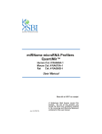
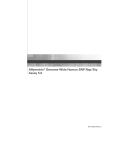
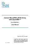
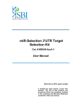
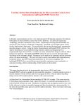
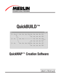
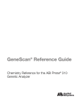
QuantiMir RT Kit
Small RNA Quantitation System
Cat. # RA420A-1
User Manual
Store kit at -20°C on receipt
(ver. 2-070209)
A limited-use label license covers this
product. By use of this product, you
accept the terms and conditions outlined
in the Licensing and Warranty Statement
contained in this user manual.
QuantiMir™ RT Kit
Cat. # RA420A-1
Contents
I. Introduction and Background
A. Overview
B. Importance of MicroRNAs and Other Small RNAs
C. Overview of Protocol
D. List of Components
E. Additional Required Materials
2
2
4
5
5
II. Protocol
A. QuantiMir™ RT Reaction Setup
B. Primer Design Considerations
C. End-point PCR Reactions using QuantiMir™ cDNAs
D. Real-time qPCR Reactions using QuantiMir™ cDNAs
6
7
8
9
III. Quality Control and Sample Data
A. Sensitivity Tests
B. Specificity Tests
C. Sample Data
10
11
12
IV. Troubleshooting
15
V. References
16
VI. Appendix
A. Related Products
B. Technical Support
VII. Licensing and Warranty Statement
888-266-5066 (Toll Free)
650-968-2200 (outside US)
17
18
19
Page 1
System Biosciences (SBI)
User Manual
I. Introduction and Background
A. Overview
This manual provides details and information necessary to use the QuantiMir™ RT Kit to tag
and convert small non-coding RNAs into detectable and quantifiable cDNAs. To ensure
optimal results, please read the entire manual before using the reagents and material supplied
with this kit.
B. Importance of MicroRNAs and Other Small Non-Coding RNAs
The field of non-coding RNAs has gained increasing attention in recent years, particularly due
to the discovery of small interfering RNAs (siRNAs) and micro RNAs (miRNA). These RNAs
are short (typically 19-24 nucleotides) single stranded moieties that regulate the expression of
target genes by interacting with complementary sites within the target mRNAs and either
repressing translation or eliciting target mRNA degradation. miRNAs and siRNAs are
conserved groups of non-coding RNAs with very important regulatory roles.
Mature miRNAs and siRNAs are excised from stem-loop precursors, which are themselves
transcribed as part of longer primary transcripts. These primary miRNAs appear to be first
processed by the RNase Drosha in the nucleus, after which the precursor miRNAs are
exported to the cytoplasm where the RNase Dicer further processes them. These enzymes
are also involved in the generation of mature small inhibitory RNAs (siRNA) from exogenously
transferred double stranded siRNA precursors.
The current, standard method for detecting and quantifying novel miRNA and siRNA molecules
involves Northern blotting with hybridization. Detecting and quantitating known miRNAs can
be done using pre-designed reverse priming and reverse transcription followed by primer sets
built for the specific miRNA for Real-time PCR analysis. These sets require many steps and
can take several hours to complete and trouble-shoot. The QuantiMir™ RT kit provides all the
reagents necessary to anchor-tail and convert small, non-coding RNAs into cDNA starting from
total RNA samples. Once the user performs the reactions on their RNA samples, the cDNAs
are ready to use for either End-point PCR experiments or to perform Real-time qPCR analysis.
The user simply only needs to design the forward primer which corresponds to the miRNA or
siRNA of interest and provide Real-time PCR SYBR green master mix (for Real-time PCR) or
typical PCR mastermix with Taq DNA polymerase for End-point PCR expression profiling.
Recently, previously unknown germline specific classes of miRNA-like molecules were
identified in mouse testes and mouse oocytes, illustrating the need for continued and in-depth
miRNA discovery efforts across a wide range of tissues. These facts taken together
demonstrate an ever-increasing need for simple, robust, and sensitive methods that enable
discovery and quantitation of microRNAs and their precursors.
Page 2
ver. 1-2009
www.systembio.com
QuantiMir™ RT Kit
Cat. # RA420A-1
Fig. 1. Diagram of MicroRNA biogenesis, processing and function.
888-266-5066 (Toll Free)
650-968-2200 (outside US)
Page 3
System Biosciences (SBI)
User Manual
C. Overview of Protocol
Real-time PCR Analysis or
End-point PCR and Gel Analysis
Fig. 2. Workflow schematic for a typical QuantiMir™ RT experiment.
* Trizol-purified total RNA is recommended to ensure small
RNA fraction present.
Page 4
ver. 1-2009
www.systembio.com
QuantiMir™ RT Kit
Cat. # RA420A-1
D. List of Components
Each QuantiMir™ RT Kit contains the following components with enough material to perform
20 reactions as outlined in this manual:
40 μl
5X PolyA Polymerase Buffer
10 μl Poly A Reaction
10 μl
PolyA Polymerase
(enough for 20 reactions)
20 μl
25 mM MnCl2
30 μl
5 mM ATP
10 μl
Oligo dT Adaptor
20 μl RT Reaction
80 μl
5X Reverse Transcriptase Buffer
(enough for 20 reactions)
20 μl
Reverse Transcriptase
30 μl
0.1 M Dithiothreitol (DTT)
50 μl
dNTP Mix
600 μl
3’ Universal Reverse PCR Primer
End-point or qPCR Assay
50 μl
5’ Human U6 Control Forward Primer (10μM)
50 μl
5’ Mouse U6 Control Forward Primer (10μM)
50 μl
5’ Human/Mouse miR-16 Control Forward Primer
(10μM)
1.2 ml
RNase-free Water
Human U6 snRNA Control
Forward Primer Sequence:
Human/Mouse miR-16 Control
Forward Primer Sequence:
5’ –CGCAAGGATGACACGCAAATTC – 3’
Tm = 59°C, amplicon size = 84 bp
5’ – TAGCAGCACGTAAATATTGGCG – 3’
Tm = 58.9°C, amplicon size = 68 bp
Mouse U6 snRNA Control
Forward Primer Sequence:
5’ – TGGCCCCTGCGCAAGGATG – 3’
Tm = 63°C, amplicon size = 97 bp
The kit is shipped on blue ice and should be stored at -20°C upon arrival. Properly stored kits
are stable for 1 year from the date received.
E. Additional Required Materials
•
•
•
•
•
•
Thermocycler (with heated lid)
Thermocycler PCR tubes or plates for end-point reactions
PCR Mastermix, including Taq polymerase for PCR
3.0-3.5% Agarose Gel in Tris-Borate EDTA (TBE) or Tris-Acetate EDTA (TAE) Buffer
DNA Size Ladder with markers from 50 to 2,000 bp (Bio-Rad AmpliSize™ DNA Ladder;
Cat. # 170-8200)
Real-time qPCR Instrument
IMPORTANT:
•
Recommended 2X SYBR Green qPCR Mastermixes:
SBI has tested and recommends SYBR Green Master mix from three vendors: Power
SYBR Master Mix® (Cat. #s 4368577, 4367650, 4367659, 4368706, 4368702, 4368708,
4367660) from Applied Biosystems; SYBR GreenER™ qPCR SuperMix for ABI PRISM®
instrument from Invitrogen (Cat. #s 11760-100, 11760-500, and 11760-02K); and RT2
Real-Time™ SYBR Green / ROX PCR (Cat. #s PA-012 and PA-112) from SuperArray.
888-266-5066 (Toll Free)
650-968-2200 (outside US)
Page 5
System Biosciences (SBI)
User Manual
II. Protocol
A. QuantiMir™ RT Reaction Setup
It is important to start with total RNA that includes
the small RNA fraction.
Start:
In a thin-walled PCR tube or
PCR-compatible plate well, combine:
5 μl
2 μl
+ 1 μl
1.5 μl
0.5 μl
10 μl
STEP 1:
PolyA Tail
Total RNA (10 pg – 10μg)
5X PolyA Buffer
25mM MnCl2
5mM ATP
PolyA Polymerase
Total in tube
Incubate for 30 min. at 37°C
Add: + 0.5 μl Oligo dT Adaptor
STEP 2:
Anneal Anchor
dT Adaptor
STEP 3:
Heat for 5 min. at 60°C
Let cool to room temp for 2 min.
Add:
Synthesize
cDNAs
4 μl
2 μl
+ 1.5 μl
1.5 μl
1 μl
20.5 μl
5X RT Buffer
dNTP mix
0.1M DTT
RNase-free H2O
Reverse Transcriptase
Total in tube
Incubate for 60 min. at 42°C
Heat for 10 min. at 95°C
*The QuantiMir™ cDNAs can be stored at -20°C.
Done!
How much cDNA template per qPCR reaction?
Starting RNA
Page 6
QuantiMir cDNA per qPCR reaction
10pg -100 ng
0.5ul to 1ul
100ng – 1 μg
Dilute 1: 100
> 1 μg
Dilute 1: 1000
ver. 1-2009
www.systembio.com
QuantiMir™ RT Kit
Cat. # RA420A-1
B. Primer Design Considerations for QuantiMir™ cDNA Detection and
Quantitation
The user needs only to design the forward “sense” orientation primer to perform the end-point
or qPCR reactions. MicroRNAs typically range in size from 19 – 24 nt. We recommend using
the exact sequence of the miRNA or siRNA being studied when designing the forward primer.
If the miRNA under study is known and documented, using the miRBase database can be an
easy starting point:
(http://microrna.sanger.ac.uk/sequences/search.shtml).
An example of the known and documented miRNA, Human miR-16, is shown below.
Hsa-miR-16
Simple: Directly use sequence
of mature miRNA as forward
primer in oligo design.
The mature miRNA sequence 5’ – uagcagcacguaaauauuggcg – 3’ can be simply converted to
a DNA sequence and used directly as the forward primer for end-point and qPCR analysis.
Forward primer for hsa-miR-16 (included in kit):
5’ – TAGCAGCACGTAAATATTGGCG – 3’
Tm= 58.9°C, 45% GC and length = 22 bases.
If the user is developing a new assay for a novel miRNA, follow the guidelines of the example
above. Design the primer to have a Tm of at least 50°C and to have a length of at least 18
bases. If the primer being designed for the miRNA to be studied has a Tm below 50°C, lower
the annealing temperature in your cycling conditions for end-point and qPCR instrument
settings. An example of successfully using this approach is demonstrated in Figure 5.
888-266-5066 (Toll Free)
650-968-2200 (outside US)
Page 7
System Biosciences (SBI)
User Manual
C. End-point PCR Reactions
The following PCR reaction conditions are recommended:
+
1 μl
0.5 μl
1 μl
2.5 μl
1 μl
20 μl
25 μl
QuantiMir™ cDNA (diluted, see p.# 7)
Universal Reverse Primer (10 μM)
miRNA-specific Forward Primer
(10 μM, user designed)
10X PCR Buffer with 2.5 mM MgCl2
10 mM dNTPs mix
RNase-free water
Total
Prepare reaction in a suitable PCR tube or plate and thermocycle as follows:
Heat denature at 95°C, 10 min.
Heat denature at 95°C, 15 sec.
Anneal Primers at 60°C, 1 min.
] 30 cycles
Hold at 15°C (optional)
Prepare and pour a 3.5% agarose (1X TAE or 1X TBE) gel with a suitable stain (Ethidium
Bromide, etc.). Add 2.5 μl of 10X Loading dye, mix, and load 10 μl into a well of the gel. Also
run a suitable DNA size marker (50-2,000 bp) along with your samples. Take care to only
electrophorese your gels for 10-15 minutes and then visualize. An example of end-point PCR
primer tests and gel electrophoresis is shown in Figure 3.
New miRNA-like Sequences
Fig. 2. Sequences of adaptors and primers used in this study.
Fig. 3. QuantiMir™ end-point PCR for miR-16 and seven newly
identified miRNA-like sequences by SBI. Numbers on the top of the gel
correspond to cDNA clone number. Letters indicate specific sequence found
in corresponding clone. Include a DNA size ladder with markers in the
range of 50-2,000 bp (e.g., Bio-Rad AmpliSize™ DNA Ladder, Cat. # 1708200). M denotes DNA Marker lanes.
* The size range of the PCR products typically ranges between 50 and 100 bp.
The adaptor segment is 46 bp with the miRNA sequence comprising the
remaining PCR product size.
Page 8
ver. 1-2009
www.systembio.com
QuantiMir™ RT Kit
Cat. # RA420A-1
D. Real-time qPCR Reactions using QuantiMir™ cDNAs
For end-point PCR reactions, please refer to the Section C above, “End-point PCR Reactions
using QuantiMir™ cDNAs”.
1. qPCR Reaction Setup
To determine the expression profile for your miRNA under study, mix the following per
well:
For Single well determination:
+
15 μl
1 μl
0.5 μl
1 μl
12.5 μl
30 μl
2X SYBR Green qPCR Mastermix buffer
User-designed Forward Primer (10 μM)
Universal Reverse Primer (10 μM)
Diluted QuantiMir™ cDNA (see p.# 7)
RNase-free water
Total / well
The Mastermix contents can be scaled up or down depending upon on your experimental
needs. Once reagents are loaded into the wells, cover the plate with the optical adhesive
cover and spin briefly in a centrifuge to bring contents to bottom of wells. Place plate in the
correct orientation (well A1, upper left) into the Real-time qPCR instrument and perform
analysis run.
2. Real-time qPCR Instrument Parameters
Follow the guidelines as detailed for your specific Real-time instrumentation. The
following parameters tested by SBI were performed on an Applied Biosystems 7300 Realtime PCR System but can also apply to an ABI 7500 or an ABI 7900 96-well system. The
details of the thermocycling conditions used in testing at SBI are shown below. A
screenshot from SBI’s instrument set up is shown on the right. Default conditions are
used throughout except for those cases where the Forward Primer’s Tm is below 55°C,
then the annealing temperature in Step 2 of Stage 3 is lowered from 60°C to 50°C.
qPCR cycling and
data accumulation
conditions:
1.
2.
3.
4.
50°C 2 min.
95°C 10 min.
95°C 15 sec.
60°C 1 min.
(40 cycles of steps 3
and 4), data read at
60°C 15 sec. Step
(gold rectangle)
An additional recommendation is to include a melt analysis after the qPCR run to assess
the Tm of the PCR amplicon to verify the specificity of the amplification reaction. Refer to
the User Manual for your specific instrument to conduct the melt analysis and the data
analyses of the amplification plots and Cycle Threshold (Ct) calculations. In general,
Cycle thresholds should be set within the exponential phase of the amplification plots with
software automatic baseline settings.
888-266-5066 (Toll Free)
650-968-2200 (outside US)
Page 9
System Biosciences (SBI)
User Manual
III. Quality Control and Sample Data
A. Sensitivity Tests
The QuantiMir™ cDNAs were synthesized using decreasing amounts of total starting RNA
input from a pool of Human Brain, Heart, Kidney, Placenta, and Testes RNAs. Real-time
quantitative qPCR assays were performed with Forward primers specific for Human miR-16
and Human miR-24 (For procedure, see Section II.D.1, Protocol: Real-time qPCR).
Fig. 4. Real-time qPCR data for Human miR-16 and Human miR-24.
Real-time qPCR amplification plots are shown in the upper inset. Cycle threshold (Ct) values were
determined using the software automatic baseline and Ct settings. The Bar graph depicts the relative
%Signal per RNA input amount for the microRNA. The graph below shows the linear regression analysis
2
with a R value of 0.971 for miR-16 and 0.993 for miR-24. Both microRNAs are readily detectable down
to 200 pg of total starting RNA input.
Page 10
ver. 1-2009
www.systembio.com
QuantiMir™ RT Kit
Cat. # RA420A-1
B. Specificity Tests
To assess the specificity and proper orientation of the miRNA array, oligonucleotide primers
are synthesized both in the “sense” and the “antisense” orientation. An example for the
known, documented miRNA miR-542-3p is detailed below.
Hsa-miR-542-3p
Sequence of mature miRNA
as forward primer in “sense”
oligo design, and then
designed in the “antisense”
oligo as control.
The mature miRNA sequence 5’ – ugugacagauugauaacugaaa – 3’ can be converted to a DNA
sequence along with designing its complement, or “antisense” primer sequence.
Forward “sense” primer for hsa-miR-542-3p:
5’ – TGTGACAGATTGATAACTGAAA – 3’
Forward “antisense” primer for hsa-miR-542-3p:
5’ – TTTCAGTTATCAATCTGTCACA – 3’
Tm= 49.6°C, 32% GC and length = 22 bases.
Fig. 5. Sense and antisense test of the QuantiMir™ cDNA. Dilutions of
the QuantiMir™ cDNA template as well as no template controls (NTC) were
tested with either sense or antisense orientation for the Human miR-542-3p
molecule. Quantitative results are observed for the “sense” orientation of
miR-542-3p. No signals are observed in the “antisense” or no template
controls. The annealing temperature for the qPCR cycling conditions was
lowered to 50°C.
888-266-5066 (Toll Free)
650-968-2200 (outside US)
Page 11
System Biosciences (SBI)
User Manual
C. Sample Data
1. Tissue Expression Pattern Determinations using the QuantiMir™ Kit
The QuantiMir™ cDNA sets were synthesized from 18 separate normal Human tissues
and tested with 2 primers specific for 2 known miRNA molecules: miR-1 (heart and
skeletal muscle-specific) and miR-122a (abundant in liver). The amplification plots and
corresponding expression bar graphs are shown in Figure 6, panels a and b.
a.
b.
Fig. 6. Real-time qPCR data using primers specific for Human miR-1
(Panel a.) and for miR-122a (Panel b.). The amplification plots are shown
on the left with the resulting expression profile bar graphs based on Ct
values is shown on the right. The default qPCR cycling conditions were
used with an annealing temperature of 60°C in Step 2 of Stage 3.
These two known miRNAs, miR-1 and mir-122a, have very specific tissue expression
patterns. Real-time qPCR data confirmed that miR-1 is restricted to skeletal muscle and
heart. The sensitivity of the assays also reveals very low but detectable signals in
additional tissues. miR-122a is known to be highly abundant in liver.
Page 12
ver. 1-2009
www.systembio.com
QuantiMir™ RT Kit
Cat. # RA420A-1
2. Analysis of Tumor and Normal Tissue MicroRNA Expression Levels using
the QuantiMir™ Kit and Real-time qPCR
The QuantiMir™ cDNAs were synthesized from both Normal and Tumor Breast, Lung,
Ovary, Colon, and Lymph node RNAs. MicroRNA forward primers specific for miR-9-1,
miR-155, miR-125, miR-145, miR-7, miR-17-3p, miR-18a, miR-20a and miR-92 were
using to detect the corresponding microRNA species in the tissues detailed in the
expression graph below (Figure 7). The signals were normalized to expression levels of
the U6 snRNA transcript. Fold increases and decreases in Normal vs. Tumor tissues are
graphed below and are consistent with published findings for the particular microRNA in
the specific tumor type.
Fig. 7. Quantitative analysis of MicroRNA expression in tumor and
normal tissue samples. The Bar graph data are grouped by tissue type
with normal tissues in blue bars and tumor tissues in red bars. The specific
MicroRNAs being detected are listed below the bar graphs. The expression
levels are normalized to U6 snRNA transcript levels to control for RNA input.
The MicroRNA expression levels are depicted as ΔCt vales (Y axis). Realtime assays were performed as described in this manual.
888-266-5066 (Toll Free)
650-968-2200 (outside US)
Page 13
System Biosciences (SBI)
User Manual
3. Detection and Quantitation of siRNA Production from Transduced shRNA
Expression Constructs
The QuantiMir™ cDNAs were synthesized from Human T-Rex 293 cells transduced with
equivalent amounts of Lentivirus containing the SBI shRNA expression constructs for the
p53 shRNA driven by the H1 promoter with Tet repressor elements (p53.H1.Tx2L) or
without Tet responsive elements (p53.H1) present in the promoters. The T-Rex 293 cell
line constitutively expresses the Tet repressor protein. The predicted structure of the p53
shRNA and the oligonucleotide primer designed (underlined) to detect the mature siRNA
(boxed in the structure) are shown below in Figure 8a. After transduction, the cells were
left untreated (- DOX) or treated with 1μg/ml Doxycycline (+ DOX) for 48 hours to alleviate
the Tet repressor effects. The quantitative PCR results normalized to the U6 signals are
shown in the bar graphs below in Figure 8b.
Normalized expression levels
Fig. 8. Detection of mature siRNA expression from cell culture
experiments.
Page 14
ver. 1-2009
www.systembio.com
QuantiMir™ RT Kit
Cat. # RA420A-1
Troubleshooting
Problem
Possible Solution
No PCR bands or qPCR
signals using user-designed
Forward Primer with
QuantiMir™ cDNA templates.
Alter thermocycle conditions—lower
annealing temperature. Include the
U6 and/or miR-16 control Forward
primers in separate reactions as PCR
controls.
Too many PCR bands using
user-designed Forward Primer
with QuantiMir™ cDNA
templates.
Alter thermocycle conditions—elevate
annealing temperature. Include the
U6 control Forward primer in a
separate reaction as PCR control.
Signals in qPCR negative
controls.
Dilute your QuantiMir cDNA by 1;100
and try again.
Correct size band and larger
Potential precursor pri-miRNA or prePCR band(s) observed in end- miRNA molecules are being
point PCR tests.
amplified.
888-266-5066 (Toll Free)
650-968-2200 (outside US)
Page 15
System Biosciences (SBI)
User Manual
References
1.
Sonthelmer, E. J., Carthew, R. W. 2005. Silence from within: Endogenous siRNAs and miRNAs. Cell
122:9-12.
2.
Zamore, P.D., Haley, B. 2005. Ribo-gnome: The big world of small RNAs. Science 309: 1519-1524.
3.
Bartel, D. 2004. MicroRNAs: Genomics, Biogenesis, Mechanism, and Function. Cell 116: 281-297.
4.
Kim, Narry V. 2005.Small RNAs: Classification, Biogenesis, and Function. Mol. Cells. 19:1-15.
5.
Valencia-Sanchez, MA., Liu, J., Hannon, GJ., Parker, R., 2006. Control of translation and mRNA
degradation by miRNAs and siRNAs. Genes Dev 20: 515-525.
6.
Lewis B.P, Burge C.B, Bartel, D.P. 2005. Conserved seed pairing, often flanked by adenosines,
indicates that thousands of human genes are microRNA targets. Cell 120: 15-20.
7.
Xie X., Lu J., Kulbokas, E.J., Goulub, T.R., Mooth, V., Lindblad-Toh, K., Lander, E.S. and Kellis, M.
Systematic discovery of regulatory motifs in human promoters and 3’ UTRs by comparison of several
mammals. Nature.434:338-45.
8.
Lagos-Quintana, M., Rauhut, R., Lendeckel, W., Tuschl, T. 2001. Identification of Novel Coding for
Small Expresses RNAs. Science 294: 853-858.
9.
Basyuk, E., Suavet, F., Doglio, A., Bordonne, R., Bertrand, E. 2003. Human let-7 stem-loop precursors
harbor features of RNase III cleavage products. Nucleic Acids Res 31: 6593-6597.
10. Chomczynski P., and Mackey, K. One-hour downward capillary blotting of RNA at neutral pH. 1994,
Anal. Biochem. 221, 303-305.
11. Shi, R., Chiang, V.L., 2005. Facile means for quantifying microRNA expression by real-time PCR.
BioTechniques. 39:519-525.
12. Ding, Y., Chan, C.Y., and Lawrence, C.E. (2005) RNA secondary structure prediction by centroids in a
Boltzmann weighted ensemble. RNA 11, 1157-1166.
13. Griffiths-Jones,S., Grocock, R.J., Van Dongen, S., Bateman, A., Enright, A.J. 2006. miRBase:
microRNA sequences, targets and gene nomenclature. Nucleic Acids Research 34: D140-D144.
14. Shingara, J., Keiger, K., Shelton, J., Laosinchai-Wolf, W., Powers, P., Conrad, R., Brown, D.,
Labourier, E. 2005. An optimized isolation and labeling platform for accurate microRNA expression
profiling. RNA 11:1461-1470.
15. He, L., Thomson, J.M., Hemann, M.T., Hernando-Monge, E., Mu, D., Goodson, S., Powers, S.,
Cordon-Cardo, C., Lowe, S.W., Hannon, G.J., Hammond, S.M. 2005. A microRNA polycistron as a
potential human oncogene. Nature 435: 828-833.
16. Lai, E.C., Wiel, C., Rubin, G.M. 2004. Complementary miRNA pairs suggest a regulatory role for
miRNA:miRNA duplexes. RNA 10:171-175.
17. Ambros, V., Bartel, B., Bartel, D.P., Burge, C.B., Carrington, J.C., Chen, X., Dreyfuss, G., Eddy, S.R.,
Griffiths-Jones, S., Marshall, M., Matzke, M., Ruvkun, G., Tuschl, T. 2003. A uniform system for
microRNA annotation. RNA 9:277-279.
18. Obernosterer, G., Leuschner, P.J.F., Alenius, M., Martinez, J. 2006. Post-transcriptional regulation of
microRNA expression. RNA 12:1-7.
19. Dostie, J., Mourelatos, Z., Yang, M., Sharma, A., Dreyfuss, G. 2003. Numerous microRNPs in neuronal
cells containing novel microRNAs. RNA 9: 180-186.
Page 16
ver. 1-2009
www.systembio.com
QuantiMir™ RT Kit
Cat. # RA420A-1
IV. Appendix
A. Related Products
•
Cancer microRNA qPCR Array (Cat. # RA610A-1)
Pre-formatted 96-well plate containing assays for 95 oncomirs and U6 control. 10
complete profiles plus QuantiMir kit included.
•
Stem Cell microRNA qPCR Array (Cat. # RA620A-1)
Pre-formatted 96-well plate containing assays for 95 different microRNA invloved in stem
cell self-renewal, hematopoiesis, neural development and tissue specificity. Arra also
contains U6 control. 10 complete profiles plus QuantiMir kit included.
•
miRNome Profilers (Cat. # RA660A-1 [Human],
Cat. # RA670A-1 [Mouse])
Profile all known microRNAs (including minor star* forms) for Human or Mouse. Sanger
miRBase 100% updated, 20 complete profiles with QuantiMir kit included.
•
miR-SnaREs (Cat. # RA7xxA-1 – RA8xxA-1)
Epitope-tagged microRNA processing factors to enrich samples for microRNAs and other
small RNAs. Human and Mouse Argonautes, Pasha and Dicer constructs available.
•
Lenti-miRs (Cat. # PMIRHxxA-1)
Over express microRNA precursors using lentivectors.
•
miRZips (Cat. # MZIPxxA-1)
Permanent microRNA knockdown using RNAi lentivectors with anti-sense microRNAs..
•
Global MicroRNA Amplification Kit (Cat. # RA400A-1)
Simple amplification kit allows cDNA amplification for qRT-PCR and microarray studies
from as little as 50 ng of starting total RNA.
•
Full Spectrum™ Complete Transcriptome RNA Amplification Kit (Cat. # RA101A-1)
The Full Spectrum RNA Amplification Kit provides an inexpensive method to amplify
reverse transcribed RNA in a sequence independent, unbiased, and uniform manner with
better representation of 5’ end of mRNA sequences. This approach maintains the relative
levels of each transcript in the starting mRNA samples—even when using starting
amounts of RNA as low as 5ng or when using heavily degraded RNA.
•
Full Spectrum™ MultiStart Primers for T7 IVT (Cat. # RA300A-2)
Extract more data from your RNA than currently available primers in nearly all
commercially-available T7 IVT kits using Full Spectrum™ technology. Just replace the
existing T7 primer with the Full Spectrum™ primers. Compatible with Affymetrix
GeneChip® hybridization.
888-266-5066 (Toll Free)
650-968-2200 (outside US)
Page 17
System Biosciences (SBI)
User Manual
B. Technical Support
For more information about SBI products and to download manuals in PDF format, please visit
our web site:
http://www.systembio.com
For additional information or technical assistance, please call or email us at:
System Biosciences (SBI)
1616 North Shoreline Blvd.
Mountain View, CA 94043
Phone: (650) 968-2200
(888) 266-5066 (Toll Free)
Fax:
(650) 968-2277
E-mail:
General Information:
[email protected]
Technical Support:
[email protected]
Ordering Information:
[email protected]
Page 18
ver. 1-2009
www.systembio.com
QuantiMir™ RT Kit
Cat. # RA420A-1
V. Licensing and Warranty Statement
Limited Use License
Use of the QuantiMir™ RT Kit (i.e., the “Product”) is subject to the following terms and conditions. If the
terms and conditions are not acceptable, return all components of the Product to System Biosciences (SBI)
within 7 calendar days. Purchase and use of any part of the Product constitutes acceptance of the above
terms.
Purchase of the product does not grant any rights or license for use other than those explicitly listed in this
Licensing and Warranty Statement. Use of the Product for any use other than described expressly herein
may be covered by patents or subject to rights other than those mentioned. SBI disclaims any and all
responsibility for injury or damage which may be caused by the failure of the buyer or any other person to
use the Product in accordance with the terms and conditions outlined herein.
SBI has pending patent applications related to the Product. For information concerning licenses for
commercial use, contact SBI.
Limited Warranty
SBI warrants that the Product meets the specifications described in the accompanying Product Analysis
Certificate. If it is proven to the satisfaction of SBI that the Product fails to meet these specifications, SBI will
replace the Product or provide the purchaser with a refund. This limited warranty shall not extend to anyone
other than the original purchaser of the Product. Notice of nonconforming products must be made to SBI
within 30 days of receipt of the Product.
SBI’s liability is expressly limited to replacement of Product or a refund limited to the actual purchase price.
SBI’s liability does not extend to any damages arising from use or improper use of the Product, or losses
associated with the use of additional materials or reagents. This limited warranty is the sole and exclusive
warranty. SBI does not provide any other warranties of any kind, expressed or implied, including the
merchantability or fitness of the Product for a particular purpose.
SBI is committed to providing our customers with high-quality products. If you should have any questions or
concerns about any SBI products, please contact us at (888) 266-5066.
© 2009 System Biosciences (SBI).
888-266-5066 (Toll Free)
650-968-2200 (outside US)
Page 19