1
DRAMIŃSKI 4Vet User Manual
_____________________________________________________________________ ____________________
Table of contentsS5
1.
2.
3.
4.
5.
6.
7.
8.
Introduction .....................................................................................................................................................5
1.1.
Information on the user manual for the device .......................................................................................5
1.2.
Warnings and comments used in this manual .......................................................................................5
1.3.
Brief information on ultrasonography .....................................................................................................5
1.4.
Basic information about the ultrasound scanner ....................................................................................6
Safety of use ...................................................................................................................................................6
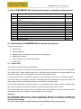
List of DRAMIŃSKI 4Vet ultrasound scanner elements and equipment ........................................................8
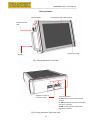
Construction of DRAMIŃSKI 4Vet ultrasound scanner ..................................................................................8
4.1.
Device body ............................................................................................................................................8
4.2.
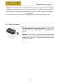
Ultrasonic probe. ..................................................................................................................................11
4.3.
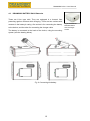
DRAMIŃSKI BATTERY PACK Batteries..............................................................................................12
4.4.
Battery pack charger ............................................................................................................................13
4.5.
Power supply ........................................................................................................................................14
Technical data ...............................................................................................................................................15
Preparing the device for operation and how to finish work. ..........................................................................16
6.1.
General principles of using the touch panel .........................................................................................16
6.2.
Connecting the probe. ..........................................................................................................................16
6.3.
Starting the device in power supply mode ...........................................................................................17
6.4.
Starting the device in battery operation mode .....................................................................................17
6.5.
Finishing work ......................................................................................................................................18
6.6.
Transport ..............................................................................................................................................18
Preparation for diagnostic examination ........................................................................................................18
Description of user panel and its available functions ....................................................................................19
8.1.
Basic panel (B, B+B, B+M modes).......................................................................................................19
8.2.
Active fields ..........................................................................................................................................21
8.3.
Panel in Colour Doppler mode .............................................................................................................22
8.4.
Panel in Power Doppler mode .............................................................................................................23
8.5. Panel in Pulse Wave Doppler mode ........................................................................................................25
9.
Imaging modes..............................................................................................................................................25
9.1.
B mode .................................................................................................................................................25
9.2.
B+B mode.............................................................................................................................................26
9.2.1.
Enabling B+B mode .....................................................................................................................26
9.2.2.
Switching windows in B+B mode.................................................................................................26
9.3.
B+M mode ............................................................................................................................................27
9.3.1.
Enabling B+M mode ....................................................................................................................27
9.4.
Doppler mode .......................................................................................................................................28
10.
Description of ultrasound scanner functions ............................................................................................29
10.1. Image parameters optimisation ............................................................................................................29
10.1.1.
Signal gain control [Gain 1] and [Gain 2].....................................................................................29
10.1.2.
Selecting head frequency: ...........................................................................................................29
10.1.3.
Changing depth of penetration (scanning range): .......................................................................29
10.1.4.
Focusing (the beam):...................................................................................................................29
10.1.5.
Zoom:...........................................................................................................................................30
10.1.6.
Adjusting the Gamma level..........................................................................................................30
10.1.7.
Invert colour: ................................................................................................................................31
10.2. Image freeze: .......................................................................................................................................31
10.3. Dimensioning........................................................................................................................................32
10.3.1.
Grid. .............................................................................................................................................33
10.3.2.
Narrowing ....................................................................................................................................34
10.3.3.
Volume.........................................................................................................................................35
10.3.4.
Length ..........................................................................................................................................36
10.3.5.
Surface area ................................................................................................................................37
10.3.6.
Ellipse surface area .....................................................................................................................38
10.3.7.
Editing measurements .................................................................................................................39
10.3.8.
Clear ............................................................................................................................................39
10.4. Colour Doppler imaging........................................................................................................................40
10.4.1.
Enabling colour Doppler imaging.................................................................................................40
3