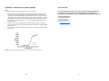
1
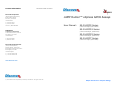
Contact Information DRX UM 95-0040CP cHX 0912V1 DiscoveRx Corporation (World Wide Headquarters) 42501 Albrae Street Fremont, CA 94538 United States t | 510.979.1415 f | 510.979.1650 toll-free | 866.448.4864 cAMP Hunter™ eXpress GPCR Assays User Manual: 95-0040CP2 Series (contains CP2 Reagent, P/N 30-409CP) KINOMEscan A division of DiscoveRx 11180 Roselle Street, Suite D San Diego, CA 92121 United States t | 800.644.5687 f | 858.630.4600 95-0040CP19 Series (contains CP19 Reagent, P/N 30-443CP) 95-0040CP23 Series (contains CP23 Reagent, P/N 30-450CP) 95-0040CP7 Series (contains CP7 reagent, P/N 30-428CP) DiscoveRx Corporation Ltd. (Europe Headquarters) Faraday Wharf, Holt Street Birmingham Science Park Aston Birmingham, B7 4BB United Kingdom t | +44.121.260.6142 f | +44.121.260.6143 www.discoverx.com © 2012 DiscoveRx Corporation, Fremont, CA 94538. All rights reserved. Simple Solutions for Complex Biology CONTENTS APPENDIX B: FREQUENTLY ASKED QUESTIONS LEGAL SECTION PAGE 3 INTENDED USE PAGE 4 TECHNOLOGY PRINCIPLE PAGE 4 KIT CONTENTS AND STORAGE CONDITIONS PAGE 5 MATERIALS PROVIDED PAGE 6 ADDITIONAL MATERIALS REQUIRED PAGE 6 CELL PLATING REAGENT REQUIREMENTS PAGE 7 PREPARATION OF FORSKOLIN SOLUTION PAGE 7 ASSAY PLATE MAPS PAGE 8 THAWING AND PLATING FROZEN CELLS PAGE 10 ASSAY PLATE PREPARATION PAGE 10 Gs-COUPLED RECEPTOR— AGONIST RESPONSE PAGE 11 Gs-COUPLED RECEPTOR— ANTAGONIST RESPONSE PAGE 13 Gi-COUPLED RECEPTOR— AGONIST RESPONSE PAGE 15 Gi-COUPLED RECEPTOR— ANTAGONIST RESPONSE PAGE 17 QUICK START PROCEDURE—AGONIST DOSE RESPONSE PAGE 20 QUICK START PROCEDURE—ANTAGONIST DOSE RESPONSE PAGE 21 APPENDIX A: PREPARATION OF CAMP STANDARD PAGE 22 APPENDIX B: FREQUENTLY ASKED QUESTIONS PAGE 23 Q: A: I did not see a signal with my control agonist. There may be differences in agonist purchased from different vendors. Confirm that the control agonist used is the same ligand used in the dose response shown in the provided cell-specific data sheet. Always use a freshly prepared stock of compounds when possible. Q: I did not see a response with my compound. A1: The concentration of DMSO or Ethanol used for dilution is too high. Maintain concentration of the agonist/antagonist diluent at ≤ 1%. A2: Confirm that the final ligand concentration is correct. Some ligands are “sticky” and difficult to dissolve. A3: Confirm that the cell line responds to the control agonist. A4: Repeat the experiment using a new lot of control agonist. Q: A: My cells arrived thawed. Can I use them? No. Call technical support for a replacement. Q: A: How long is the prepared detection reagent good for? The working detection reagent solution must be used within 24 hours of mixing. Store protected from light at 4°C. Q: A: What instruments can I use to read the plates? Any bench top luminometer will work with the cAMP Hunter™ eXpress Assays. For 96-well analysis, we recommend the LumiLITE™ Microplate Reader from DiscoveRx (Cat. #75-0001). Q: A: How long is the signal stable for? The signal is stable for up to 24 hours following the addition of detection reagent. Q: A: My cells are floating after the 24 hours incubation. The cells are not viable, contact technical support for a replacement. Q: A: Can I switch plates or should I use the plate provided? You can use any clear bottom white or opaque walled plate. For additional information or technical support, contact DiscoveRx or visit www.discoverx.com. 2 23 APPENDIX A: PREPARATION OF CAMP STANDARD NOTE: The cAMP Standard can be prepared before agonist compound addition. 1. Using polypropylene microcentrifuge tubes, dilute the cAMP Standard (2.5 x 10⁻⁴ M) 1:4 with Cell Assay Buffer (1 part cAMP Standard + 3 parts Buffer). This dilution is the highest standard concentration of 5.21 x 10⁻⁶ M in the final 180 µl assay volume (diluted cAMP Standard plus Detection Reagents). Prepare 10 additional 3-fold serial dilutions, with the last dilution yielding the lowest standard concentration of 8.82 x 10⁻¹¹ M. Use Cell Assay Buffer alone for the zero standard. 2. Transfer 15 µL from each cAMP standard dilution (Step 1 above) to duplicate wells on the cAMP Hunter eXpress GPCR Assay plate according to the Plate Maps on p.8 and p.9. 3. Transfer 45 µL of the diluted cAMP Antibody mix to each well and proceed with the protocol. LEGAL SECTION For some products/cell lines, certain 3rd party gene specific patents may be required to use the cell line. It is the purchaser’s responsibility to determine if such patents or other intellectual property rights are required. Licensing Department DiscoveRx Corporation 42501 Albrae Street Fremont, CA 94538 t | 510.979.1415 x 104 e | [email protected] Figure 6. A representative cAMP Standard Curve using the recommended protocol. 22 3 INTENDED USE QUICK-START PROCEDURE: ANTAGONIST DOSE RESPONSE cAMP Hunter™ eXpress GPCR Assays provide a robust, highly sensitive and easyto-use method for monitoring G-protein coupled receptor (GPCR) activation based on cyclic adenosine monophosphate (cyclic AMP or cAMP) detection in live cells. The fully characterized cAMP Hunter cells combined with optimized chemiluminescent detection reagents, cell plating reagents and plates* provide a complete solution for interrogation of GPCRs. The pre-validated, frozen eXpress cells have been manufactured for short term use and are provided in a convenient, ready-to-screen format that saves time and adds convenience. Assays have been designed for 96well plate analysis. ANTAGONIST CURVE STANDARD CURVE Plate 100 μL of cells/well Incubate overnight @ 37°C *Test compounds are not included and must be provided by the researcher. Aspirate media & add 45 μL of diluted cAMP Antibody. Add 7.5 μL of antagonist TECHNOLOGY PRINCIPLE GPCR activation following ligand binding initiates a series of second messenger cascades that result in a cellular response. This signaling involves a membrane bound enzyme called adenylate cyclase (Fig. 1). Gi- and Gs-coupled receptors modulate cAMP by either inhibiting or stimulating adenylate cyclase, respectively. Unlike other force-coupled technologies, cAMP Hunter eXpress cells utilize the natural coupling status of the GPCR to monitor activation of Gi-and Gs-coupled receptors. Following ligand stimulation, the functional status of the GPCR is monitored by measuring cellular cAMP levels using a homogeneous, gain-of-signal competitive immunoassay based on Enzyme Fragment Complementation (EFC). Incubate 15 minutes @ 37°C Add 15 μL of serially diluted cAMP standard to empty wells. Add 7.5 μL of agonist (+forskolin for Gi targets) Add 45 μL of diluted cAMP antibody Incubate 30 minutes @ 37°C Add 60 µL Working Detection Reagents/cAMP Solution D Incubate 1 hr @ RT in the dark Figure 1. GPCR activation results in either an increase (Gs) or decrease (Gi) of intracellular levels of the second messenger cAMP. Add 60 µL cAMP Solution A Incubate 3 hr @ RT in the dark Read Chemiluminescent Signal 4 21 QUICK-START PROCEDURE: AGONIST DOSE RESPONSE AGONIST CURVE STANDARD CURVE KIT CONTENTS AND STORAGE CONDITIONS cAMP HUNTER™ EXPRESS COMPONENTS REQUIRE MULTIPLE STORAGE TEMPERATURES. OPEN BOXES IMMEDIATELY AND STORE CONTENTS AS INSTRUCTED. SHELF LIFE: Use kit within 6 months from the date of receipt under proper storage conditions. Plate 100 μL of cells/well Box 1: Incubate overnight @ 37°C CAMP HUNTER EXPRESS CELLS STORAGE: Short term (2 weeks or less): Store vials at -80°C immediately upon arrival. Long term (greater than 2 weeks): Place vials in the vapor phase of liquid nitrogen (N2). Aspirate media & add 45 μL of diluted cAMP Antibody. Add 15 μL of serially diluted cAMP standard to empty wells. Add 15 μL of agonist (+forskolin for Gi targets) Add 45 μL of diluted cAMP antibody cAMP Hunter eXpress cells arrive frozen on dry ice. Cells are delivered in individual vials containing 3.75 x 10⁶ cells in 200 µL of freezing medium. Each vial contains sufficient cell numbers to generate (1) 96-well microplate prepared at the seeding density described. When removing cryovials from liquid N2 storage, use tongs and place immediately on dry ice in a covered container. Wait at least one minute for any liquid N2 inside the vial to evaporate and proceed with the thawing protocol (p.10). Do not touch the bottom of the tubes at any time to avoid inadvertent thawing of the cells. If cells are not frozen upon arrival, do not proceed. Contact technical support. Incubate 30 minutes @ 37°C BOX 2: Add 60 µL Working Detection Reagents/cAMP Solution D Incubate 1 hr @ RT in the dark CAMP HUNTER EXPRESS DETECTION REAGENTS, CELL PLATING REAGENT: INCLUDING FORSKOLIN AND Once thawed, store the Detection Reagents and Cell Plating (CP) Reagent at 4C. Avoid multiple freeze/thaw cycles. In rare instances, the CP Reagent may be yellow in color after thawing. Although this indicates a slight change in pH, continue with the assay as this does not impact assay performance. BOX 3: 96-WELL TISSUE CULTURE TREATED PLATES: Store at Room Temperature Add 60 µL cAMP Solution A Incubate 3 hr @ RT in the dark Read Chemiluminescent Signal 20 5 MATERIALS PROVIDED Description Contents Box 1: cAMP Hunter™ eXpress Cells (1 GPCR Target) 1 vial 3.75 x 10⁶ cells ea Box 2: cAMP Hunter eXpress Detection Reagents • cAMP Lysis Buffer • cAMP Solution D • cAMP Solution A • cAMP Antibody Reagent • cAMP Standard (250 µM) • Substrate Reagent 1* • Substrate Reagent 2* 3. After completion of the antagonist incubation, transfer 7.5 µL of agonist to all wells in Columns 2-12 of the cAMP eXpress assay plate (refer to Plate Maps on p.9 for more details). DO NOT add agonist compound to well A1. Instead, add 7.5 µL of Cell Assay Buffer plus vehicle. The final screening concentration of vehicle should be identical to the concentration in the presence of agonist. This sample serves as a negative control. 4. Incubate the plate for 30 minutes at 37°C. Storage 2 vials 3.75 x 10⁶ cells ea 10 vials 3.75 x 10⁶ cells ea -80°C (short term) Liq. N2 (long term) C. cAMP STANDARD PREPARATION 3.8 mL 5 mL 8 mL 2.5 mL 1 mL 1 mL 0.2 mL 7.6 mL 10 mL 16 mL 5 mL 1 mL 2 mL 0.4 mL 38 mL 50 mL 80 mL 25 mL 1 mL 10 mL 2 mL Forskolin (dried powder) Cell Assay Buffer Cell Plating Reagent** 1 x 0.25 mg 12.5 mL 1 x 20 mL 2 x 0.25 mg 25 mL 2 x 20 mL 10 x 0.25 mg 125 mL 2 x 100 mL 96-well Tissue Culture Treated Plates 1 plate 2 plates 10 plates -20°C The cAMP Standard can be prepared either before agonist compound addition or during the 30 minute agonist incubation period. Refer to Appendix A, p.22 for more details. D. PREPARATION AND ADDITION OF DETECTION REAGENTS 1. Room Temperature Prepare a 1:1 stock of working cAMP detection reagent and cAMP Solution D. Make working cAMP detection reagent by mixing 19 parts Lysis Buffer, 5 parts Substrate Reagent 1 and 1 part Substrate Reagent 2. Components *Centrifuge vial before opening for maximal recovery. Screw the cap on tightly to prevent evaporation. **Refer to cell-line specific datasheets for optimized cell plating reagent included with each kit. Entire Plate (96 wells) cAMP Lysis Buffer 3.8 mL Substrate Reagent 1 1.0 mL Substrate Reagent 2 0.20 mL cAMP Solution D ADDITIONAL MATERIALS REQUIRED 5.0 mL TOTAL volume The following materials are required but not provided: 10.0 mL This working solution can be stored at 4°C for up to 24 hours. Store protected from light. 1. Pipettes and pipette tips 2. Tissue culture disposables 2. 3. GPCR control agonist as recommended in the cell line specific datasheet. Visit www.discoverx.com/pathway_assays/control_ligands.php for the complete DiscoveRx offering 3. 4. GPCR test compound(s) 4. 5. Disposable reagent reservoir (Thermo Scientific, Cat. #8094 or similar) Add 60 µl of cAMP Solution A to the appropriate wells. DO NOT pipette up and down in the well to mix or vortex/shake plates. 6. V-bottom 96-well compound dilution plates (DiscoveRx, Cat. #92-0011) 5. Incubate for 3 hours at room temperature (23°C), protected from light. 7. Fisherbrand Adhesive Plate Seals (Fisher, Cat. #80-408-240) 8. Multi-mode or luminescence plate reader. For 96-well analysis, we recommend the LumiLITE™ Microplate Reader (DiscoveRx, Cat. #75-0001) 9. IBMX (DiscoveRx, Cat. #92-0007) 6 Following agonist incubation, add 60 µL of working cAMP detection reagents/ cAMP Solution D mixture to the appropriate wells. DO NOT pipette up and down in the well to mix or vortex/shake plates. Incubate for 1 hour at room temperature (23°C), protected from light. 6. Read samples on any standard luminescence plate reader. 7. Use GraphPad Prism® or other comparable program to plot your cAMP response. 19 Example: If the expected IC50 is 10 nM, prepare the highest starting concentration at 4 M. CELL PLATING REAGENT REQUIREMENTS a. In a 15 ml conical tube, prepare a mixture of a 8X concentration of the vehicle of choice in Cell Assay Buffer to a final volume of 2.5 mL. The final screening concentration of vehicle should be identical to the concentration in the presence of antagonist. Each cAMP Hunter™ eXpress GPCR Assay has been validated for optimal assay performance using the specific Cell Plating (CP) Reagent included in the kit. DO NOT substitute Cell Plating Reagent from an alternate kit at any time. b. Add 80 µL of Cell Assay Buffer diluent prepared above to wells A1-A11 of a clean V-bottom compound dilution plate. c. In well A12 of the compound dilution plate, prepare an 8X concentration of the highest dose of antagonist compound in Cell Assay Buffer to a final volume of 120 μL. Mix well by pipetting up and down several times. Discard the pipet tip. d. With a clean pipet tip, remove 40 µL of 8X compound from well A12, add it to well A11 and mix by gently pipetting up and down. Discard the pipet tip. e. Repeat this process 8 more times, preparing serial dilutions from right to left across the plate. f. DO NOT add antagonist compound to wells A1 and A2. Add only Cell Assay Buffer diluent. These samples serve as negative controls and complete the dose curve. g. Repeat this process for each compound tested. h. Set compound dilution plate aside until compounds are ready to be added. If necessary, cover the plate with microseal film to prevent evaporation of compound. 3. Transfer 7.5 µL from wells A1-A12 from the antagonist dilution plate (Step A2.h) to triplicate (or quadruplicate) wells on the cAMP Hunter eXpress GPCR Assay plate according to the plate maps on p.9. 4. Incubate the plate for 15 minutes at 37°C. For catalog numbers ending in CP2: use CP2 Reagent (P/N 30-409CP) CP19: use CP19 Reagent (P/N 30-443CP) CP23: use CP23 Reagent (P/N 30-450CP) CP7: use CP7 Reagent (P/N 30-428CP) PREPARATION OF FORSKOLIN SOLUTION Forskolin activates the enzyme adenylate cyclase and increases intracellular levels of cAMP. For a Gi receptor, agonist binding inhibits intracellular cAMP accumulation induced by Forskolin. In order to measure Gi-coupled receptors, the agonist compound is added in the presence of Forskolin. Activation of the Gi-coupled receptor therefore inhibits the Forskolin-induced production of cAMP. As a result, the dose response curve generated in the presence of agonist plus Forskolin will have a negative slope. NOTE: Please refer to target specific datasheet for recommended concentrations of forskolin. If forskolin is not used immediately, prepare multiple aliquots and store at –20°C. Avoid multiple freeze/thaw cycles. When stored properly, the solution is stable for up to 2 months. Since forskolin treatment results in increased intracellular cAMP levels in all cAMP Hunter™ eXpress cells, it can serve as a positive control for performance of the cells and detection reagents. B. AGONIST COMPOUND PREPARATION AND ADDITION 1. During antagonist incubation, determine the EC80 concentration of the agonist from the agonist dose response curve (described on p.15). Prepare an 8X EC80 concentration of agonist compound as shown below: Example: If the expected EC80 of the agonist compound is 10 nM, prepare a 1.5 mL solution at 80 nM. 2. In a 15 ml conical tube, prepare a mixture of an 8X concentration of agonist compound and an 8X concentration of reconstituted Forskolin in Cell Assay Buffer to a final volume of 1.5 mL. NOTE: Refer to target specific datasheet for recommended final forskolin concentrations. 18 7 ASSAY PROCEDURE — AGONIST DOSE RESPONSE CURVES D. PREPARATION AND ADDITION OF DETECTION REAGENTS The steps outlined below provide the assay volumes and procedure for performing agonist assays using the cAMP Hunter eXpress cells and detection reagents. Although plate layouts and experimental designs may vary, we recommend performing a 12-point dose curve for each compound using at least triplicate wells for each dilution. We also recommend including the cAMP Standard with each experiment. The cAMP Standard can be run on the same plate (Plate Map A) or on a separate plate (Plate Map B). The protocol and volumes described below are designed for a complete 96-well plate. 1. Prepare a 1:1 stock of working cAMP detection reagent and cAMP Solution D. Make working cAMP detection reagent by mixing 19 parts Lysis Buffer, 5 parts Substrate Reagent 1 and 1 part Substrate Reagent 2. Components Entire Plate (96 wells) cAMP Lysis Buffer 3.8 mL Substrate Reagent 1 1.0 mL Substrate Reagent 2 0.20 mL cAMP Solution D 5.0 mL TOTAL volume 10.0 mL This working solution can be stored at 4°C for up to 24 hours. Store protected from light. 2. 3. Figure 2. This illustration shows a 12-point dose curve with 3 data points each; 2 compounds per plate for a total of 4 compounds per eXpress kit. Following agonist incubation, add 60 µL of working cAMP detection reagents/ cAMP Solution D mixture to the appropriate wells. DO NOT pipette up and down in the well to mix or vortex/shake plates. Incubate for 1 hour at room temperature (23°C), protected from light. 4. Add 60 µl of cAMP Solution A to the appropriate wells. DO NOT pipette up and down in the well to mix or vortex/shake plates. 5. Incubate for 3 hours at room temperature (23°C), protected from light. 6. Read samples on any standard luminescence plate reader. 7. Use GraphPad Prism® or other comparable program to plot your cAMP response. Gi-COUPLED RECEPTOR—ANTAGONIST RESPONSE A. ANTAGONIST COMPOUND PREPARATION AND ADDITION 1. Dissolve antagonist compound in the vehicle of choice (DMSO, ethanol, water, or other) at the desired stock concentration. NOTE: Solvents can affect assay performance. cAMP eXpress assays are routinely carried out in the presence of ≤ 1% solvent (i.e. DMSO, ethanol, or other). If other solvents or solvent concentrations are used, optimize the assay conditions accordingly. 2. Figure 3. This illustration shows a 12-point dose curve with 4 data points each; 2 compounds per plate for a total of 4 compounds per eXpress kit. cAMP Standard is run on a separate assay plate. 8 Prepare 3-fold serial dilutions of antagonist compound in Cell Assay Buffer containing the appropriate solvent (DMSO, ethanol, water, or other). The concentration of each dilution should be prepared at 8X of the final screening concentration (i.e. 7.5 µL antagonist + 7.5 µL agonist + 45 µL Cell Assay Buffer/cAMP Antibody Reagent). For each dilution, the final concentration of solvent should remain constant. Preparation of 10-point dose curve serial dilutions of compound: We recommend starting with a concentration that is 50X the expected IC50 value for the compound (400X the final screening concentration). 17 Example: If the expected EC50 is 10 nM, prepare the highest starting concentration at 2 µM. ASSAY PROCEDURE — ANTAGONIST DOSE RESPONSE a. In a 15 ml conical tube, prepare a mixture of a 4X concentration of the vehicle of choice and a 4X concentration of reconstituted forskolin in Cell Assay Buffer to a final volume of 2.5 mL. The final screening concentration of vehicle should be identical to the concentration in of the highest dose of agonist. The steps outlined below provide the assay volumes and procedure for performing antagonist assays using the cAMP Hunter eXpress cells and detection reagents. Although plate layouts and experimental designs may vary, we recommend performing a 10-point dose curve for each compound using at least triplicate wells for each dilution. We also recommend including the cAMP Standard with each experiment. The cAMP Standard can be run on the same plate (Plate Map C) or on a separate plate (Plate Map D). The protocol and volumes described below are designed for a complete 96-well plate. NOTE: Refer to target specific datasheet for recommended final forskolin concentrations. b. Add 80 µL of Cell Assay Buffer diluent prepared above to wells A1-A11 of a clean V-bottom compound dilution plate. c. In well A12 of the compound dilution plate, prepare a mixture of a 4X concentration of the highest dose of agonist compound plus a 4X concentration of reconstituted Forskolin in Cell Assay Buffer to a final volume of 120 µL. Mix well by pipetting up and down several times. Discard the pipet tip. d. With a clean pipet tip, remove 40 µL of 4X compound from well A12, add it to well A11 and mix by gently pipetting up and down. Discard the pipet tip. e. Repeat this process 9 more times, preparing serial dilutions from right to left across the plate. f. DO NOT add agonist compound to well A1. Add only Cell Assay Buffer diluent. This sample serves as the no agonist control and completes the dose curve. g. Repeat this process for each compound tested. h. Set compound dilution plate aside until compounds are ready to be added. If necessary, cover the plate with microseal film to prevent evaporation of compound. Figure 4. This illustration shows a 10-point dose curve with 3 data points each; 2 compounds per plate for a total of 4 compounds per eXpress kit. B. ADDITION OF AGONIST COMPOUND(S) 1. Transfer 15 µL from wells A1-A12 from the compound dilution plate (Step A2.h) to triplicate (or quadruplicate) wells on the cAMP Hunter eXpress GPCR Assay plate according to the Plate Maps on p.8. 2. Incubate the plate for 30 minutes at 37°C. C. cAMP STANDARD PREPARATION The cAMP Standard can be prepared either before agonist compound addition or during the 30 minute agonist incubation period. Refer to Appendix A, p.22 for more details. Figure 5. This illustration shows a 10-point dose curve with 4 data points each; 2 compounds per plate for a total of 4 compounds per eXpress kit. cAMP Standard is run on a separate assay plate. 16 9 DAY 1: THAWING AND PLATING FROZEN CELLS D. PREPARATION AND ADDITION OF DETECTION REAGENTS The following steps outline the procedure for thawing and plating frozen cAMP Hunter eXpress cells from cryogenic vials. Each vial contains sufficient cell numbers to generate (1) 96-well microplate prepared at the seeding density described. 1. Prepare a 1:1 stock of working cAMP detection reagent and cAMP Solution D. Make working cAMP detection reagent by mixing 19 parts Lysis Buffer, 5 parts Substrate Reagent 1 and 1 part Substrate Reagent 2. 1. Pre-warm Cell Plating (CP) Reagent in a 37°C water bath. Components 2. Remove cell vial from –80°C or liquid N2 storage and place immediately on dry ice prior to thawing. DO NOT EXPOSE VIALS TO ROOM TEMPERATURE. cAMP Lysis Buffer NOTE: When removing cryovials from liquid N2, place immediately on dry ice in a covered container. Wait at least one minute before opening in order to allow any liquid N2 inside the vial to evaporate. Place the cell vial(s) briefly (10 seconds to 1 min) in a 37°C water bath until only small ice crystals remain and the cell pellet(s) is almost completely thawed. 4. Add 0.5 mL of pre-warmed CP Reagent to the cell vial. Pipette up and down gently several times to ensure that cells are evenly distributed. 2. 5. Immediately transfer the cells to 12 mL of pre-warmed CP Reagent and pour into a disposable reagent reservoir. Plate 100 µL of cells into each well (30,000 cells/well) of the provided 96-well tissue culture plate. 3. 0.20 mL 5.0 mL TOTAL volume 10.0 mL This working solution can be stored at 4°C for up to 24 hours. Store protected from light. Following agonist incubation, add 60 µL of working cAMP detection reagents/ cAMP Solution D mixture to the appropriate wells. DO NOT pipette up and down in the well to mix or vortex/shake plates. Incubate for 1 hour at room temperature (23°C), protected from light. Add 60 µl of cAMP Solution A to the appropriate wells. DO NOT pipette up and down in the well to mix or vortex/shake plates. 5. Incubate for 3 hours at room temperature (23°C), protected from light. After seeding the cells into the microplate, place it in a 37°C, 5% CO2 in a humidified incubator for 18-24 hours prior to testing. 6. Read samples on any standard luminescence plate reader. 7. Use GraphPad Prism® or other comparable program to plot your cAMP response. Gi-COUPLED RECEPTOR—AGONIST RESPONSE In a 15 ml conical tube, mix Cell Assay Buffer and cAMP Antibody Reagent according to the table below: Entire Plate (96 wells) Cell Assay Buffer 3. 1.0 mL Substrate Reagent 2 4. Components 2. Substrate Reagent 1 NOTE: We recommend including the cAMP Standard on each assay plate. If cAMP Standard is included, DO NOT add eXpress cells to wells in rows G or H (refer to plate map A on p.7 for more details). DAY 2: PREPARATION OF ASSAY PLATE 1. 3.8 mL cAMP Solution D 3. 6. Entire Plate (96 wells) A. COMPOUND PREPARATION 1. NOTE: Solvents can affect assay performance. cAMP eXpress assays are routinely carried out in the presence of ≤ 1% solvent (i.e. DMSO, ethanol, or other). If other solvents or solvent concentrations are used, optimize the assay conditions accordingly. 5 mL cAMP Antibody Reagent 2.5 mL TOTAL volume 7.5 mL Remove cAMP Hunter eXpress cells (previously plated on day 1) from the incubator. Cells should be adherent and greater than 50% confluent at the time of the assay. Carefully aspirate media. NOTE: Residual media left in the wells can decrease assay performance. Be sure to remove as much of the media as possible without disrupting the cell monolayer. 10 Dissolve agonist compound in the vehicle of choice (DMSO, ethanol, water, or other) at the desired stock concentration. 2. Prepare 3-fold serial dilutions of agonist compound in Cell Assay Buffer containing Forskolin and the appropriate solvent (DMSO, ethanol, water, or other). The concentration of each dilution should be prepared at 4X of the final screening concentration (i.e. 15 µL compound + 45 µL Cell Assay Buffer/cAMP Antibody Reagent/Forskolin). For each dilution, the final concentration of solvent should remain constant. Preparation of 11-point dose curve serial dilutions of compound: We recommend starting with a concentration that is 50X the expected EC50 value for the compound (200X the final screening concentration). 15 e. Repeat this process 8 more times, preparing serial dilutions from right to left across the plate. f. DO NOT add antagonist compound to wells A1 and A2. Add only Cell Assay Buffer diluent. These samples serve as negative controls and complete the dose curve. g. Repeat this process for each compound tested. h. Set compound dilution plate aside until antagonist compounds are ready to be added. Cover the plate with microseal film to prevent evaporation of compound. 3. Transfer 7.5 µL from wells A1-A12 from the antagonist dilution plate (Step A2.h) to triplicate (or quadruplicate) wells on the cAMP Hunter eXpress GPCR Assay plate according to the plate maps on p.9. 4. Incubate the plate for 15 minutes at 37°C. 4. Add 45 µl of Cell Assay Buffer/Antibody mixture to all wells designated for compound addition. When running the cAMP Standard, leave rows G and H empty (Refer to Plate Maps A and C, p.8-9). 5. Set plate aside until needed. Reserve the remaining Cell Assay Buffer/Antibody mixture for preparation of cAMP Standard described in Appendix A, p.21). 6. Proceed to the appropriate cAMP Hunter eXpress Protocol based on whether you are testing: Gs target in Agonist mode (p.11) Gs target in Antagonist mode (p.13) Gi target in Agonist mode (p.15) Gi target in Antagonist mode (p.17) Gs-COUPLED RECEPTOR - AGONIST RESPONSE B. AGONIST COMPOUND PREPARATION AND ADDITION A. COMPOUND PREPARATION 1. 1. During the antagonist incubation, determine the EC80 concentration of the agonist from the agonist dose response curve (described on p.11). Prepare an 8X EC80 concentration of agonist compound as shown below: Example: If the expected EC80 of the agonist compound is 10 nM, prepare a 1.5 mL solution at 80 nM. 2. In a 15 ml conical tube, prepare an 8X concentration of agonist compound in Cell Assay Buffer to a final volume of 1.5 mL. 3. After completion of the antagonist incubation, transfer 7.5 µL of agonist to all wells in Columns 2-12 of the cAMP eXpress assay plate (refer to Plate Maps on p.9 for more details). DO NOT add agonist compound to well A1. Instead, add 7.5 µL of Cell Assay Buffer plus vehicle. The final screening concentration of vehicle should be identical to the concentration in the presence of agonist. This sample serves as a negative control. 4. Incubate the plate for 30 minutes at 37°C. C. cAMP STANDARD PREPARATION The cAMP Standard can be prepared either before agonist compound addition or during the 30 minute agonist incubation period. Refer to Appendix A, p.22 for more details. Dissolve agonist compound in the vehicle of choice (DMSO, ethanol, water, or other) at the desired stock concentration. NOTE: Solvents can affect assay performance. cAMP eXpress assays are routinely carried out in the presence of ≤ 1% solvent (i.e. DMSO, ethanol, or other). If other solvents or solvent concentrations are used, optimize the assay conditions accordingly. 2. Prepare 3-fold serial dilutions of agonist compound in Cell Assay Buffer containing the appropriate solvent (DMSO, ethanol, water, or other). The concentration of each dilution should be prepared at 4X of the final screening concentration (i.e. 15 µL compound + 45 µL Cell Assay Buffer/cAMP Antibody Reagent). For each dilution, the final concentration of solvent should remain constant. Preparation of 11-point dose curve serial dilutions of compound: We recommend starting with a concentration that is 50X the expected EC50 value for the compound (200X the final screening concentration). Example: If the expected EC50 is 10 nM, prepare the highest starting concentration at 2 µM. a) In a 15 ml conical tube, prepare a 4X concentration of the vehicle of choice in Cell Assay Buffer to a final volume of 2.5 mL. The final screening concentration of vehicle should be identical to the concentration in the highest dose of agonist. OPTIONAL: IBMX (3-isobutyl-1-methylxanthine) has been shown to be a potent inhibitor of adenosine 3’, 5’-cyclic monophosphate phosphodiesterase (cAMP PDE). For Gs coupled receptor assays, addition of up to 0.5 mM IBMX can result in an increase of intracellular cAMP levels. b) Add 80 µL of the above Cell Assay Buffer diluent to wells A1-A11 of a clean V-bottom compound dilution plate. c) In well A12 of the compound dilution plate, prepare a 4X concentration of the highest dose of agonist compound in Cell Assay Buffer to a final volume of 120 µL. Mix well by pipetting up and down several times. Discard the pipet tip. 14 11 d. With a clean pipet tip, remove 40 µL of 4X compound from well A12, add it to well A11 and mix by gently pipetting up and down. Discard pipet tip. e. Repeat this process 9 more times, preparing serial dilutions from right to left across the plate. f. DO NOT add agonist compound to well A1. Add only Cell Assay Buffer diluent. This sample serves as the no agonist control and completes the dose curve. 3. Incubate for 1 hour at room temperature (23°C), protected from light. 4. Add 60 µl of cAMP Solution A to the appropriate wells. DO NOT pipette up and down in the well to mix or vortex/shake plates. 5. Incubate for 3 hours at room temperature (23°C), protected from light. 6. Read samples on any standard luminescence plate reader. 7. Use GraphPad Prism® or other comparable program to plot your cAMP response. g. Repeat this process for each compound tested. h. Set compound dilution plate aside until antagonist compounds are ready to be added. Cover the plate with microseal film to prevent evaporation of compound. GS-COUPLED RECEPTOR—ANTAGONIST RESPONSE A. ANTAGONIST COMPOUND PREPARATION AND ADDITION B. ADDITION OF AGONIST COMPOUND(S) 1. 1. Transfer 15 µL from wells A1-A12 from the compound dilution plate (Step A2.h) to triplicate (or quadruplicate) wells on the cAMP Hunter eXpress GPCR Assay plate according to the Plate Maps on p.8. 2. Incubate the plate for 30 minutes at 37°C. The cAMP Standard can be prepared either before agonist compound addition or during the 30 minute agonist incubation period. Refer to Appendix A, p.22 for more details. D. PREPARATION AND ADDITION OF DETECTION REAGENTS 1. Prepare a 1:1 stock of working cAMP detection reagent and cAMP Solution D. Make working cAMP detection reagent by mixing 19 parts Lysis Buffer, 5 parts Substrate Reagent 1 and 1 part Substrate Reagent 2. Components Entire Plate (96 wells) cAMP Lysis Buffer 3.8 mL Substrate Reagent 1 1.0 mL Substrate Reagent 2 0.20 mL cAMP Solution D 5.0 mL TOTAL volume 10.0 mL This working solution can be stored at 4°C for up to 24 hours. Store protected from light. 2. Following agonist incubation, add 60 µL of working cAMP detection reagents/ cAMP Solution D mixture to the appropriate wells. Do Not pipette up and down in the well to mix or vortex/shake plates. 12 NOTE: Solvents can affect assay performance. cAMP eXpress assays are routinely carried out in the presence of ≤ 1% solvent (i.e. DMSO, ethanol, or other). If other solvents or solvent concentrations are used, optimize the assay conditions accordingly. 2. C. cAMP STANDARD PREPARATION Dissolve antagonist compound in the vehicle of choice (DMSO, ethanol, water, or other) at the desired stock concentration. Prepare 3-fold serial dilutions of antagonist compound in Cell Assay Buffer containing the appropriate solvent (DMSO, ethanol, water, or other). The concentration of each dilution should be prepared at 8X of the final screening concentration (i.e. 7.5 µL antagonist + 7.5 µL agonist + 45 µL Cell Assay Buffer/cAMP Antibody Reagent). For each dilution, the final concentration of solvent should remain constant. Preparation of 10-point dose curve serial dilutions of compound: We recommend starting with a concentration that is 50X the expected IC50 value for the compound (400X the final screening concentration). Example: If the expected IC50 is 10 nM, prepare the highest starting concentration at 4 µM. a. In a 15 ml conical tube, prepare an 8X concentration of the vehicle of choice in Cell Assay Buffer to a final volume of 2.5 mL. The final screening concentration of vehicle should be identical to the concentration in the highest dose of antagonist. OPTIONAL: IBMX (3-isobutyl-1-methylxanthine) has been shown to be a potent inhibitor of adenosine 3’, 5’-cyclic monophosphate phosphodiesterase (cAMP PDE). For Gs coupled receptor assays, addition of up to 0.5 mM IBMX can result in an increase of intracellular cAMP levels. b. Add 80 µL of the above Cell Assay Buffer diluent to wells A1-A11 of a clean V-bottom compound dilution plate. c. In well A12 of the compound dilution plate, prepare an 8X concentration of the highest dose of antagonist compound in Cell Assay Buffer to a final volume of 120 µL. Mix well by pipetting up and down several times. Discard the pipet tip. d. With a clean pipet tip, remove 40 µL of 8X compound from well A12, add it to well A11 and mix by gently pipetting up and down. Discard pipet tip. 13 d. With a clean pipet tip, remove 40 µL of 4X compound from well A12, add it to well A11 and mix by gently pipetting up and down. Discard pipet tip. e. Repeat this process 9 more times, preparing serial dilutions from right to left across the plate. f. DO NOT add agonist compound to well A1. Add only Cell Assay Buffer diluent. This sample serves as the no agonist control and completes the dose curve. 3. Incubate for 1 hour at room temperature (23°C), protected from light. 4. Add 60 µl of cAMP Solution A to the appropriate wells. DO NOT pipette up and down in the well to mix or vortex/shake plates. 5. Incubate for 3 hours at room temperature (23°C), protected from light. 6. Read samples on any standard luminescence plate reader. 7. Use GraphPad Prism® or other comparable program to plot your cAMP response. g. Repeat this process for each compound tested. h. Set compound dilution plate aside until antagonist compounds are ready to be added. Cover the plate with microseal film to prevent evaporation of compound. GS-COUPLED RECEPTOR—ANTAGONIST RESPONSE A. ANTAGONIST COMPOUND PREPARATION AND ADDITION B. ADDITION OF AGONIST COMPOUND(S) 1. 1. Transfer 15 µL from wells A1-A12 from the compound dilution plate (Step A2.h) to triplicate (or quadruplicate) wells on the cAMP Hunter eXpress GPCR Assay plate according to the Plate Maps on p.8. 2. Incubate the plate for 30 minutes at 37°C. The cAMP Standard can be prepared either before agonist compound addition or during the 30 minute agonist incubation period. Refer to Appendix A, p.22 for more details. D. PREPARATION AND ADDITION OF DETECTION REAGENTS 1. Prepare a 1:1 stock of working cAMP detection reagent and cAMP Solution D. Make working cAMP detection reagent by mixing 19 parts Lysis Buffer, 5 parts Substrate Reagent 1 and 1 part Substrate Reagent 2. Components Entire Plate (96 wells) cAMP Lysis Buffer 3.8 mL Substrate Reagent 1 1.0 mL Substrate Reagent 2 0.20 mL cAMP Solution D 5.0 mL TOTAL volume 10.0 mL This working solution can be stored at 4°C for up to 24 hours. Store protected from light. 2. Following agonist incubation, add 60 µL of working cAMP detection reagents/ cAMP Solution D mixture to the appropriate wells. Do Not pipette up and down in the well to mix or vortex/shake plates. 12 NOTE: Solvents can affect assay performance. cAMP eXpress assays are routinely carried out in the presence of ≤ 1% solvent (i.e. DMSO, ethanol, or other). If other solvents or solvent concentrations are used, optimize the assay conditions accordingly. 2. C. cAMP STANDARD PREPARATION Dissolve antagonist compound in the vehicle of choice (DMSO, ethanol, water, or other) at the desired stock concentration. Prepare 3-fold serial dilutions of antagonist compound in Cell Assay Buffer containing the appropriate solvent (DMSO, ethanol, water, or other). The concentration of each dilution should be prepared at 8X of the final screening concentration (i.e. 7.5 µL antagonist + 7.5 µL agonist + 45 µL Cell Assay Buffer/cAMP Antibody Reagent). For each dilution, the final concentration of solvent should remain constant. Preparation of 10-point dose curve serial dilutions of compound: We recommend starting with a concentration that is 50X the expected IC50 value for the compound (400X the final screening concentration). Example: If the expected IC50 is 10 nM, prepare the highest starting concentration at 4 µM. a. In a 15 ml conical tube, prepare an 8X concentration of the vehicle of choice in Cell Assay Buffer to a final volume of 2.5 mL. The final screening concentration of vehicle should be identical to the concentration in the highest dose of antagonist. OPTIONAL: IBMX (3-isobutyl-1-methylxanthine) has been shown to be a potent inhibitor of adenosine 3’, 5’-cyclic monophosphate phosphodiesterase (cAMP PDE). For Gs coupled receptor assays, addition of up to 0.5 mM IBMX can result in an increase of intracellular cAMP levels. b. Add 80 µL of the above Cell Assay Buffer diluent to wells A1-A11 of a clean V-bottom compound dilution plate. c. In well A12 of the compound dilution plate, prepare an 8X concentration of the highest dose of antagonist compound in Cell Assay Buffer to a final volume of 120 µL. Mix well by pipetting up and down several times. Discard the pipet tip. d. With a clean pipet tip, remove 40 µL of 8X compound from well A12, add it to well A11 and mix by gently pipetting up and down. Discard pipet tip. 13 e. Repeat this process 8 more times, preparing serial dilutions from right to left across the plate. f. DO NOT add antagonist compound to wells A1 and A2. Add only Cell Assay Buffer diluent. These samples serve as negative controls and complete the dose curve. g. Repeat this process for each compound tested. h. Set compound dilution plate aside until antagonist compounds are ready to be added. Cover the plate with microseal film to prevent evaporation of compound. 3. Transfer 7.5 µL from wells A1-A12 from the antagonist dilution plate (Step A2.h) to triplicate (or quadruplicate) wells on the cAMP Hunter eXpress GPCR Assay plate according to the plate maps on p.9. 4. Incubate the plate for 15 minutes at 37°C. 4. Add 45 µl of Cell Assay Buffer/Antibody mixture to all wells designated for compound addition. When running the cAMP Standard, leave rows G and H empty (Refer to Plate Maps A and C, p.8-9). 5. Set plate aside until needed. Reserve the remaining Cell Assay Buffer/Antibody mixture for preparation of cAMP Standard described in Appendix A, p.21). 6. Proceed to the appropriate cAMP Hunter eXpress Protocol based on whether you are testing: Gs target in Agonist mode (p.11) Gs target in Antagonist mode (p.13) Gi target in Agonist mode (p.15) Gi target in Antagonist mode (p.17) Gs-COUPLED RECEPTOR - AGONIST RESPONSE B. AGONIST COMPOUND PREPARATION AND ADDITION A. COMPOUND PREPARATION 1. 1. During the antagonist incubation, determine the EC80 concentration of the agonist from the agonist dose response curve (described on p.11). Prepare an 8X EC80 concentration of agonist compound as shown below: Example: If the expected EC80 of the agonist compound is 10 nM, prepare a 1.5 mL solution at 80 nM. 2. In a 15 ml conical tube, prepare an 8X concentration of agonist compound in Cell Assay Buffer to a final volume of 1.5 mL. 3. After completion of the antagonist incubation, transfer 7.5 µL of agonist to all wells in Columns 2-12 of the cAMP eXpress assay plate (refer to Plate Maps on p.9 for more details). DO NOT add agonist compound to well A1. Instead, add 7.5 µL of Cell Assay Buffer plus vehicle. The final screening concentration of vehicle should be identical to the concentration in the presence of agonist. This sample serves as a negative control. 4. Incubate the plate for 30 minutes at 37°C. C. cAMP STANDARD PREPARATION The cAMP Standard can be prepared either before agonist compound addition or during the 30 minute agonist incubation period. Refer to Appendix A, p.22 for more details. Dissolve agonist compound in the vehicle of choice (DMSO, ethanol, water, or other) at the desired stock concentration. NOTE: Solvents can affect assay performance. cAMP eXpress assays are routinely carried out in the presence of ≤ 1% solvent (i.e. DMSO, ethanol, or other). If other solvents or solvent concentrations are used, optimize the assay conditions accordingly. 2. Prepare 3-fold serial dilutions of agonist compound in Cell Assay Buffer containing the appropriate solvent (DMSO, ethanol, water, or other). The concentration of each dilution should be prepared at 4X of the final screening concentration (i.e. 15 µL compound + 45 µL Cell Assay Buffer/cAMP Antibody Reagent). For each dilution, the final concentration of solvent should remain constant. Preparation of 11-point dose curve serial dilutions of compound: We recommend starting with a concentration that is 50X the expected EC50 value for the compound (200X the final screening concentration). Example: If the expected EC50 is 10 nM, prepare the highest starting concentration at 2 µM. a) In a 15 ml conical tube, prepare a 4X concentration of the vehicle of choice in Cell Assay Buffer to a final volume of 2.5 mL. The final screening concentration of vehicle should be identical to the concentration in the highest dose of agonist. OPTIONAL: IBMX (3-isobutyl-1-methylxanthine) has been shown to be a potent inhibitor of adenosine 3’, 5’-cyclic monophosphate phosphodiesterase (cAMP PDE). For Gs coupled receptor assays, addition of up to 0.5 mM IBMX can result in an increase of intracellular cAMP levels. b) Add 80 µL of the above Cell Assay Buffer diluent to wells A1-A11 of a clean V-bottom compound dilution plate. c) In well A12 of the compound dilution plate, prepare a 4X concentration of the highest dose of agonist compound in Cell Assay Buffer to a final volume of 120 µL. Mix well by pipetting up and down several times. Discard the pipet tip. 14 11 DAY 1: THAWING AND PLATING FROZEN CELLS D. PREPARATION AND ADDITION OF DETECTION REAGENTS The following steps outline the procedure for thawing and plating frozen cAMP Hunter eXpress cells from cryogenic vials. Each vial contains sufficient cell numbers to generate (1) 96-well microplate prepared at the seeding density described. 1. Prepare a 1:1 stock of working cAMP detection reagent and cAMP Solution D. Make working cAMP detection reagent by mixing 19 parts Lysis Buffer, 5 parts Substrate Reagent 1 and 1 part Substrate Reagent 2. 1. Pre-warm Cell Plating (CP) Reagent in a 37°C water bath. Components 2. Remove cell vial from –80°C or liquid N2 storage and place immediately on dry ice prior to thawing. DO NOT EXPOSE VIALS TO ROOM TEMPERATURE. cAMP Lysis Buffer NOTE: When removing cryovials from liquid N2, place immediately on dry ice in a covered container. Wait at least one minute before opening in order to allow any liquid N2 inside the vial to evaporate. Place the cell vial(s) briefly (10 seconds to 1 min) in a 37°C water bath until only small ice crystals remain and the cell pellet(s) is almost completely thawed. 4. Add 0.5 mL of pre-warmed CP Reagent to the cell vial. Pipette up and down gently several times to ensure that cells are evenly distributed. 2. 5. Immediately transfer the cells to 12 mL of pre-warmed CP Reagent and pour into a disposable reagent reservoir. Plate 100 µL of cells into each well (30,000 cells/well) of the provided 96-well tissue culture plate. 3. 0.20 mL 5.0 mL TOTAL volume 10.0 mL This working solution can be stored at 4°C for up to 24 hours. Store protected from light. Following agonist incubation, add 60 µL of working cAMP detection reagents/ cAMP Solution D mixture to the appropriate wells. DO NOT pipette up and down in the well to mix or vortex/shake plates. Incubate for 1 hour at room temperature (23°C), protected from light. Add 60 µl of cAMP Solution A to the appropriate wells. DO NOT pipette up and down in the well to mix or vortex/shake plates. 5. Incubate for 3 hours at room temperature (23°C), protected from light. After seeding the cells into the microplate, place it in a 37°C, 5% CO2 in a humidified incubator for 18-24 hours prior to testing. 6. Read samples on any standard luminescence plate reader. 7. Use GraphPad Prism® or other comparable program to plot your cAMP response. Gi-COUPLED RECEPTOR—AGONIST RESPONSE In a 15 ml conical tube, mix Cell Assay Buffer and cAMP Antibody Reagent according to the table below: Entire Plate (96 wells) Cell Assay Buffer 3. 1.0 mL Substrate Reagent 2 4. Components 2. Substrate Reagent 1 NOTE: We recommend including the cAMP Standard on each assay plate. If cAMP Standard is included, DO NOT add eXpress cells to wells in rows G or H (refer to plate map A on p.7 for more details). DAY 2: PREPARATION OF ASSAY PLATE 1. 3.8 mL cAMP Solution D 3. 6. Entire Plate (96 wells) A. COMPOUND PREPARATION 1. NOTE: Solvents can affect assay performance. cAMP eXpress assays are routinely carried out in the presence of ≤ 1% solvent (i.e. DMSO, ethanol, or other). If other solvents or solvent concentrations are used, optimize the assay conditions accordingly. 5 mL cAMP Antibody Reagent 2.5 mL TOTAL volume 7.5 mL Remove cAMP Hunter eXpress cells (previously plated on day 1) from the incubator. Cells should be adherent and greater than 50% confluent at the time of the assay. Carefully aspirate media. NOTE: Residual media left in the wells can decrease assay performance. Be sure to remove as much of the media as possible without disrupting the cell monolayer. 10 Dissolve agonist compound in the vehicle of choice (DMSO, ethanol, water, or other) at the desired stock concentration. 2. Prepare 3-fold serial dilutions of agonist compound in Cell Assay Buffer containing Forskolin and the appropriate solvent (DMSO, ethanol, water, or other). The concentration of each dilution should be prepared at 4X of the final screening concentration (i.e. 15 µL compound + 45 µL Cell Assay Buffer/cAMP Antibody Reagent/Forskolin). For each dilution, the final concentration of solvent should remain constant. Preparation of 11-point dose curve serial dilutions of compound: We recommend starting with a concentration that is 50X the expected EC50 value for the compound (200X the final screening concentration). 15 Example: If the expected EC50 is 10 nM, prepare the highest starting concentration at 2 µM. ASSAY PROCEDURE — ANTAGONIST DOSE RESPONSE a. In a 15 ml conical tube, prepare a mixture of a 4X concentration of the vehicle of choice and a 4X concentration of reconstituted forskolin in Cell Assay Buffer to a final volume of 2.5 mL. The final screening concentration of vehicle should be identical to the concentration in of the highest dose of agonist. The steps outlined below provide the assay volumes and procedure for performing antagonist assays using the cAMP Hunter eXpress cells and detection reagents. Although plate layouts and experimental designs may vary, we recommend performing a 10-point dose curve for each compound using at least triplicate wells for each dilution. We also recommend including the cAMP Standard with each experiment. The cAMP Standard can be run on the same plate (Plate Map C) or on a separate plate (Plate Map D). The protocol and volumes described below are designed for a complete 96-well plate. NOTE: Refer to target specific datasheet for recommended final forskolin concentrations. b. Add 80 µL of Cell Assay Buffer diluent prepared above to wells A1-A11 of a clean V-bottom compound dilution plate. c. In well A12 of the compound dilution plate, prepare a mixture of a 4X concentration of the highest dose of agonist compound plus a 4X concentration of reconstituted Forskolin in Cell Assay Buffer to a final volume of 120 µL. Mix well by pipetting up and down several times. Discard the pipet tip. d. With a clean pipet tip, remove 40 µL of 4X compound from well A12, add it to well A11 and mix by gently pipetting up and down. Discard the pipet tip. e. Repeat this process 9 more times, preparing serial dilutions from right to left across the plate. f. DO NOT add agonist compound to well A1. Add only Cell Assay Buffer diluent. This sample serves as the no agonist control and completes the dose curve. g. Repeat this process for each compound tested. h. Set compound dilution plate aside until compounds are ready to be added. If necessary, cover the plate with microseal film to prevent evaporation of compound. Figure 4. This illustration shows a 10-point dose curve with 3 data points each; 2 compounds per plate for a total of 4 compounds per eXpress kit. B. ADDITION OF AGONIST COMPOUND(S) 1. Transfer 15 µL from wells A1-A12 from the compound dilution plate (Step A2.h) to triplicate (or quadruplicate) wells on the cAMP Hunter eXpress GPCR Assay plate according to the Plate Maps on p.8. 2. Incubate the plate for 30 minutes at 37°C. C. cAMP STANDARD PREPARATION The cAMP Standard can be prepared either before agonist compound addition or during the 30 minute agonist incubation period. Refer to Appendix A, p.22 for more details. Figure 5. This illustration shows a 10-point dose curve with 4 data points each; 2 compounds per plate for a total of 4 compounds per eXpress kit. cAMP Standard is run on a separate assay plate. 16 9 ASSAY PROCEDURE — AGONIST DOSE RESPONSE CURVES D. PREPARATION AND ADDITION OF DETECTION REAGENTS The steps outlined below provide the assay volumes and procedure for performing agonist assays using the cAMP Hunter eXpress cells and detection reagents. Although plate layouts and experimental designs may vary, we recommend performing a 12-point dose curve for each compound using at least triplicate wells for each dilution. We also recommend including the cAMP Standard with each experiment. The cAMP Standard can be run on the same plate (Plate Map A) or on a separate plate (Plate Map B). The protocol and volumes described below are designed for a complete 96-well plate. 1. Prepare a 1:1 stock of working cAMP detection reagent and cAMP Solution D. Make working cAMP detection reagent by mixing 19 parts Lysis Buffer, 5 parts Substrate Reagent 1 and 1 part Substrate Reagent 2. Components Entire Plate (96 wells) cAMP Lysis Buffer 3.8 mL Substrate Reagent 1 1.0 mL Substrate Reagent 2 0.20 mL cAMP Solution D 5.0 mL TOTAL volume 10.0 mL This working solution can be stored at 4°C for up to 24 hours. Store protected from light. 2. 3. Figure 2. This illustration shows a 12-point dose curve with 3 data points each; 2 compounds per plate for a total of 4 compounds per eXpress kit. Following agonist incubation, add 60 µL of working cAMP detection reagents/ cAMP Solution D mixture to the appropriate wells. DO NOT pipette up and down in the well to mix or vortex/shake plates. Incubate for 1 hour at room temperature (23°C), protected from light. 4. Add 60 µl of cAMP Solution A to the appropriate wells. DO NOT pipette up and down in the well to mix or vortex/shake plates. 5. Incubate for 3 hours at room temperature (23°C), protected from light. 6. Read samples on any standard luminescence plate reader. 7. Use GraphPad Prism® or other comparable program to plot your cAMP response. Gi-COUPLED RECEPTOR—ANTAGONIST RESPONSE A. ANTAGONIST COMPOUND PREPARATION AND ADDITION 1. Dissolve antagonist compound in the vehicle of choice (DMSO, ethanol, water, or other) at the desired stock concentration. NOTE: Solvents can affect assay performance. cAMP eXpress assays are routinely carried out in the presence of ≤ 1% solvent (i.e. DMSO, ethanol, or other). If other solvents or solvent concentrations are used, optimize the assay conditions accordingly. 2. Figure 3. This illustration shows a 12-point dose curve with 4 data points each; 2 compounds per plate for a total of 4 compounds per eXpress kit. cAMP Standard is run on a separate assay plate. 8 Prepare 3-fold serial dilutions of antagonist compound in Cell Assay Buffer containing the appropriate solvent (DMSO, ethanol, water, or other). The concentration of each dilution should be prepared at 8X of the final screening concentration (i.e. 7.5 µL antagonist + 7.5 µL agonist + 45 µL Cell Assay Buffer/cAMP Antibody Reagent). For each dilution, the final concentration of solvent should remain constant. Preparation of 10-point dose curve serial dilutions of compound: We recommend starting with a concentration that is 50X the expected IC50 value for the compound (400X the final screening concentration). 17 Example: If the expected IC50 is 10 nM, prepare the highest starting concentration at 4 M. CELL PLATING REAGENT REQUIREMENTS a. In a 15 ml conical tube, prepare a mixture of a 8X concentration of the vehicle of choice in Cell Assay Buffer to a final volume of 2.5 mL. The final screening concentration of vehicle should be identical to the concentration in the presence of antagonist. Each cAMP Hunter™ eXpress GPCR Assay has been validated for optimal assay performance using the specific Cell Plating (CP) Reagent included in the kit. DO NOT substitute Cell Plating Reagent from an alternate kit at any time. b. Add 80 µL of Cell Assay Buffer diluent prepared above to wells A1-A11 of a clean V-bottom compound dilution plate. c. In well A12 of the compound dilution plate, prepare an 8X concentration of the highest dose of antagonist compound in Cell Assay Buffer to a final volume of 120 μL. Mix well by pipetting up and down several times. Discard the pipet tip. d. With a clean pipet tip, remove 40 µL of 8X compound from well A12, add it to well A11 and mix by gently pipetting up and down. Discard the pipet tip. e. Repeat this process 8 more times, preparing serial dilutions from right to left across the plate. f. DO NOT add antagonist compound to wells A1 and A2. Add only Cell Assay Buffer diluent. These samples serve as negative controls and complete the dose curve. g. Repeat this process for each compound tested. h. Set compound dilution plate aside until compounds are ready to be added. If necessary, cover the plate with microseal film to prevent evaporation of compound. 3. Transfer 7.5 µL from wells A1-A12 from the antagonist dilution plate (Step A2.h) to triplicate (or quadruplicate) wells on the cAMP Hunter eXpress GPCR Assay plate according to the plate maps on p.9. 4. Incubate the plate for 15 minutes at 37°C. For catalog numbers ending in CP2: use CP2 Reagent (P/N 30-409CP) CP19: use CP19 Reagent (P/N 30-443CP) CP23: use CP23 Reagent (P/N 30-450CP) CP7: use CP7 Reagent (P/N 30-428CP) PREPARATION OF FORSKOLIN SOLUTION Forskolin activates the enzyme adenylate cyclase and increases intracellular levels of cAMP. For a Gi receptor, agonist binding inhibits intracellular cAMP accumulation induced by Forskolin. In order to measure Gi-coupled receptors, the agonist compound is added in the presence of Forskolin. Activation of the Gi-coupled receptor therefore inhibits the Forskolin-induced production of cAMP. As a result, the dose response curve generated in the presence of agonist plus Forskolin will have a negative slope. NOTE: Please refer to target specific datasheet for recommended concentrations of forskolin. If forskolin is not used immediately, prepare multiple aliquots and store at –20°C. Avoid multiple freeze/thaw cycles. When stored properly, the solution is stable for up to 2 months. Since forskolin treatment results in increased intracellular cAMP levels in all cAMP Hunter™ eXpress cells, it can serve as a positive control for performance of the cells and detection reagents. B. AGONIST COMPOUND PREPARATION AND ADDITION 1. During antagonist incubation, determine the EC80 concentration of the agonist from the agonist dose response curve (described on p.15). Prepare an 8X EC80 concentration of agonist compound as shown below: Example: If the expected EC80 of the agonist compound is 10 nM, prepare a 1.5 mL solution at 80 nM. 2. In a 15 ml conical tube, prepare a mixture of an 8X concentration of agonist compound and an 8X concentration of reconstituted Forskolin in Cell Assay Buffer to a final volume of 1.5 mL. NOTE: Refer to target specific datasheet for recommended final forskolin concentrations. 18 7 MATERIALS PROVIDED Description Contents Box 1: cAMP Hunter™ eXpress Cells (1 GPCR Target) 1 vial 3.75 x 10⁶ cells ea Box 2: cAMP Hunter eXpress Detection Reagents • cAMP Lysis Buffer • cAMP Solution D • cAMP Solution A • cAMP Antibody Reagent • cAMP Standard (250 µM) • Substrate Reagent 1* • Substrate Reagent 2* 3. After completion of the antagonist incubation, transfer 7.5 µL of agonist to all wells in Columns 2-12 of the cAMP eXpress assay plate (refer to Plate Maps on p.9 for more details). DO NOT add agonist compound to well A1. Instead, add 7.5 µL of Cell Assay Buffer plus vehicle. The final screening concentration of vehicle should be identical to the concentration in the presence of agonist. This sample serves as a negative control. 4. Incubate the plate for 30 minutes at 37°C. Storage 2 vials 3.75 x 10⁶ cells ea 10 vials 3.75 x 10⁶ cells ea -80°C (short term) Liq. N2 (long term) C. cAMP STANDARD PREPARATION 3.8 mL 5 mL 8 mL 2.5 mL 1 mL 1 mL 0.2 mL 7.6 mL 10 mL 16 mL 5 mL 1 mL 2 mL 0.4 mL 38 mL 50 mL 80 mL 25 mL 1 mL 10 mL 2 mL Forskolin (dried powder) Cell Assay Buffer Cell Plating Reagent** 1 x 0.25 mg 12.5 mL 1 x 20 mL 2 x 0.25 mg 25 mL 2 x 20 mL 10 x 0.25 mg 125 mL 2 x 100 mL 96-well Tissue Culture Treated Plates 1 plate 2 plates 10 plates -20°C The cAMP Standard can be prepared either before agonist compound addition or during the 30 minute agonist incubation period. Refer to Appendix A, p.22 for more details. D. PREPARATION AND ADDITION OF DETECTION REAGENTS 1. Room Temperature Prepare a 1:1 stock of working cAMP detection reagent and cAMP Solution D. Make working cAMP detection reagent by mixing 19 parts Lysis Buffer, 5 parts Substrate Reagent 1 and 1 part Substrate Reagent 2. Components *Centrifuge vial before opening for maximal recovery. Screw the cap on tightly to prevent evaporation. **Refer to cell-line specific datasheets for optimized cell plating reagent included with each kit. Entire Plate (96 wells) cAMP Lysis Buffer 3.8 mL Substrate Reagent 1 1.0 mL Substrate Reagent 2 0.20 mL cAMP Solution D ADDITIONAL MATERIALS REQUIRED 5.0 mL TOTAL volume The following materials are required but not provided: 10.0 mL This working solution can be stored at 4°C for up to 24 hours. Store protected from light. 1. Pipettes and pipette tips 2. Tissue culture disposables 2. 3. GPCR control agonist as recommended in the cell line specific datasheet. Visit www.discoverx.com/pathway_assays/control_ligands.php for the complete DiscoveRx offering 3. 4. GPCR test compound(s) 4. 5. Disposable reagent reservoir (Thermo Scientific, Cat. #8094 or similar) Add 60 µl of cAMP Solution A to the appropriate wells. DO NOT pipette up and down in the well to mix or vortex/shake plates. 6. V-bottom 96-well compound dilution plates (DiscoveRx, Cat. #92-0011) 5. Incubate for 3 hours at room temperature (23°C), protected from light. 7. Fisherbrand Adhesive Plate Seals (Fisher, Cat. #80-408-240) 8. Multi-mode or luminescence plate reader. For 96-well analysis, we recommend the LumiLITE™ Microplate Reader (DiscoveRx, Cat. #75-0001) 9. IBMX (DiscoveRx, Cat. #92-0007) 6 Following agonist incubation, add 60 µL of working cAMP detection reagents/ cAMP Solution D mixture to the appropriate wells. DO NOT pipette up and down in the well to mix or vortex/shake plates. Incubate for 1 hour at room temperature (23°C), protected from light. 6. Read samples on any standard luminescence plate reader. 7. Use GraphPad Prism® or other comparable program to plot your cAMP response. 19 QUICK-START PROCEDURE: AGONIST DOSE RESPONSE AGONIST CURVE STANDARD CURVE KIT CONTENTS AND STORAGE CONDITIONS cAMP HUNTER™ EXPRESS COMPONENTS REQUIRE MULTIPLE STORAGE TEMPERATURES. OPEN BOXES IMMEDIATELY AND STORE CONTENTS AS INSTRUCTED. SHELF LIFE: Use kit within 6 months from the date of receipt under proper storage conditions. Plate 100 μL of cells/well Box 1: Incubate overnight @ 37°C CAMP HUNTER EXPRESS CELLS STORAGE: Short term (2 weeks or less): Store vials at -80°C immediately upon arrival. Long term (greater than 2 weeks): Place vials in the vapor phase of liquid nitrogen (N2). Aspirate media & add 45 μL of diluted cAMP Antibody. Add 15 μL of serially diluted cAMP standard to empty wells. Add 15 μL of agonist (+forskolin for Gi targets) Add 45 μL of diluted cAMP antibody cAMP Hunter eXpress cells arrive frozen on dry ice. Cells are delivered in individual vials containing 3.75 x 10⁶ cells in 200 µL of freezing medium. Each vial contains sufficient cell numbers to generate (1) 96-well microplate prepared at the seeding density described. When removing cryovials from liquid N2 storage, use tongs and place immediately on dry ice in a covered container. Wait at least one minute for any liquid N2 inside the vial to evaporate and proceed with the thawing protocol (p.10). Do not touch the bottom of the tubes at any time to avoid inadvertent thawing of the cells. If cells are not frozen upon arrival, do not proceed. Contact technical support. Incubate 30 minutes @ 37°C BOX 2: Add 60 µL Working Detection Reagents/cAMP Solution D Incubate 1 hr @ RT in the dark CAMP HUNTER EXPRESS DETECTION REAGENTS, CELL PLATING REAGENT: INCLUDING FORSKOLIN AND Once thawed, store the Detection Reagents and Cell Plating (CP) Reagent at 4C. Avoid multiple freeze/thaw cycles. In rare instances, the CP Reagent may be yellow in color after thawing. Although this indicates a slight change in pH, continue with the assay as this does not impact assay performance. BOX 3: 96-WELL TISSUE CULTURE TREATED PLATES: Store at Room Temperature Add 60 µL cAMP Solution A Incubate 3 hr @ RT in the dark Read Chemiluminescent Signal 20 5 INTENDED USE QUICK-START PROCEDURE: ANTAGONIST DOSE RESPONSE cAMP Hunter™ eXpress GPCR Assays provide a robust, highly sensitive and easyto-use method for monitoring G-protein coupled receptor (GPCR) activation based on cyclic adenosine monophosphate (cyclic AMP or cAMP) detection in live cells. The fully characterized cAMP Hunter cells combined with optimized chemiluminescent detection reagents, cell plating reagents and plates* provide a complete solution for interrogation of GPCRs. The pre-validated, frozen eXpress cells have been manufactured for short term use and are provided in a convenient, ready-to-screen format that saves time and adds convenience. Assays have been designed for 96well plate analysis. ANTAGONIST CURVE STANDARD CURVE Plate 100 μL of cells/well Incubate overnight @ 37°C *Test compounds are not included and must be provided by the researcher. Aspirate media & add 45 μL of diluted cAMP Antibody. Add 7.5 μL of antagonist TECHNOLOGY PRINCIPLE GPCR activation following ligand binding initiates a series of second messenger cascades that result in a cellular response. This signaling involves a membrane bound enzyme called adenylate cyclase (Fig. 1). Gi- and Gs-coupled receptors modulate cAMP by either inhibiting or stimulating adenylate cyclase, respectively. Unlike other force-coupled technologies, cAMP Hunter eXpress cells utilize the natural coupling status of the GPCR to monitor activation of Gi-and Gs-coupled receptors. Following ligand stimulation, the functional status of the GPCR is monitored by measuring cellular cAMP levels using a homogeneous, gain-of-signal competitive immunoassay based on Enzyme Fragment Complementation (EFC). Incubate 15 minutes @ 37°C Add 15 μL of serially diluted cAMP standard to empty wells. Add 7.5 μL of agonist (+forskolin for Gi targets) Add 45 μL of diluted cAMP antibody Incubate 30 minutes @ 37°C Add 60 µL Working Detection Reagents/cAMP Solution D Incubate 1 hr @ RT in the dark Figure 1. GPCR activation results in either an increase (Gs) or decrease (Gi) of intracellular levels of the second messenger cAMP. Add 60 µL cAMP Solution A Incubate 3 hr @ RT in the dark Read Chemiluminescent Signal 4 21 APPENDIX A: PREPARATION OF CAMP STANDARD NOTE: The cAMP Standard can be prepared before agonist compound addition. 1. Using polypropylene microcentrifuge tubes, dilute the cAMP Standard (2.5 x 10⁻⁴ M) 1:4 with Cell Assay Buffer (1 part cAMP Standard + 3 parts Buffer). This dilution is the highest standard concentration of 5.21 x 10⁻⁶ M in the final 180 µl assay volume (diluted cAMP Standard plus Detection Reagents). Prepare 10 additional 3-fold serial dilutions, with the last dilution yielding the lowest standard concentration of 8.82 x 10⁻¹¹ M. Use Cell Assay Buffer alone for the zero standard. 2. Transfer 15 µL from each cAMP standard dilution (Step 1 above) to duplicate wells on the cAMP Hunter eXpress GPCR Assay plate according to the Plate Maps on p.8 and p.9. 3. Transfer 45 µL of the diluted cAMP Antibody mix to each well and proceed with the protocol. LEGAL SECTION For some products/cell lines, certain 3rd party gene specific patents may be required to use the cell line. It is the purchaser’s responsibility to determine if such patents or other intellectual property rights are required. Licensing Department DiscoveRx Corporation 42501 Albrae Street Fremont, CA 94538 t | 510.979.1415 x 104 e | [email protected] Figure 6. A representative cAMP Standard Curve using the recommended protocol. 22 3 CONTENTS APPENDIX B: FREQUENTLY ASKED QUESTIONS LEGAL SECTION PAGE 3 INTENDED USE PAGE 4 TECHNOLOGY PRINCIPLE PAGE 4 KIT CONTENTS AND STORAGE CONDITIONS PAGE 5 MATERIALS PROVIDED PAGE 6 ADDITIONAL MATERIALS REQUIRED PAGE 6 CELL PLATING REAGENT REQUIREMENTS PAGE 7 PREPARATION OF FORSKOLIN SOLUTION PAGE 7 ASSAY PLATE MAPS PAGE 8 THAWING AND PLATING FROZEN CELLS PAGE 10 ASSAY PLATE PREPARATION PAGE 10 Gs-COUPLED RECEPTOR— AGONIST RESPONSE PAGE 11 Gs-COUPLED RECEPTOR— ANTAGONIST RESPONSE PAGE 13 Gi-COUPLED RECEPTOR— AGONIST RESPONSE PAGE 15 Gi-COUPLED RECEPTOR— ANTAGONIST RESPONSE PAGE 17 QUICK START PROCEDURE—AGONIST DOSE RESPONSE PAGE 20 QUICK START PROCEDURE—ANTAGONIST DOSE RESPONSE PAGE 21 APPENDIX A: PREPARATION OF CAMP STANDARD PAGE 22 APPENDIX B: FREQUENTLY ASKED QUESTIONS PAGE 23 Q: A: I did not see a signal with my control agonist. There may be differences in agonist purchased from different vendors. Confirm that the control agonist used is the same ligand used in the dose response shown in the provided cell-specific data sheet. Always use a freshly prepared stock of compounds when possible. Q: I did not see a response with my compound. A1: The concentration of DMSO or Ethanol used for dilution is too high. Maintain concentration of the agonist/antagonist diluent at ≤ 1%. A2: Confirm that the final ligand concentration is correct. Some ligands are “sticky” and difficult to dissolve. A3: Confirm that the cell line responds to the control agonist. A4: Repeat the experiment using a new lot of control agonist. Q: A: My cells arrived thawed. Can I use them? No. Call technical support for a replacement. Q: A: How long is the prepared detection reagent good for? The working detection reagent solution must be used within 24 hours of mixing. Store protected from light at 4°C. Q: A: What instruments can I use to read the plates? Any bench top luminometer will work with the cAMP Hunter™ eXpress Assays. For 96-well analysis, we recommend the LumiLITE™ Microplate Reader from DiscoveRx (Cat. #75-0001). Q: A: How long is the signal stable for? The signal is stable for up to 24 hours following the addition of detection reagent. Q: A: My cells are floating after the 24 hours incubation. The cells are not viable, contact technical support for a replacement. Q: A: Can I switch plates or should I use the plate provided? You can use any clear bottom white or opaque walled plate. For additional information or technical support, contact DiscoveRx or visit www.discoverx.com. 2 23 Contact Information DRX UM 95-0040CP cHX 0912V1 DiscoveRx Corporation (World Wide Headquarters) 42501 Albrae Street Fremont, CA 94538 United States t | 510.979.1415 f | 510.979.1650 toll-free | 866.448.4864 cAMP Hunter™ eXpress GPCR Assays User Manual: 95-0040CP2 Series (contains CP2 Reagent, P/N 30-409CP) KINOMEscan A division of DiscoveRx 11180 Roselle Street, Suite D San Diego, CA 92121 United States t | 800.644.5687 f | 858.630.4600 95-0040CP19 Series (contains CP19 Reagent, P/N 30-443CP) 95-0040CP23 Series (contains CP23 Reagent, P/N 30-450CP) 95-0040CP7 Series (contains CP7 reagent, P/N 30-428CP) DiscoveRx Corporation Ltd. (Europe Headquarters) Faraday Wharf, Holt Street Birmingham Science Park Aston Birmingham, B7 4BB United Kingdom t | +44.121.260.6142 f | +44.121.260.6143 www.discoverx.com © 2012 DiscoveRx Corporation, Fremont, CA 94538. All rights reserved. Simple Solutions for Complex Biology