1
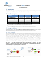
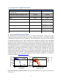
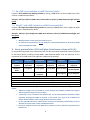
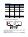
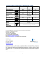
Important Product News Date: February 21, 2011 Product Change Notification Dear Valued Customer, Upon reception of the LANCE Ultra cAMP kit, please store the Eu-cAMP tracer aliquoted and frozen at -20°C. We recommend that you limit the number of freeze-thaw cycles of the tracer. All other kit components should be stored at 4°C. For additional information, please consult the user manual. 1 2 LANCE® Ultra cAMP Kit For Research Use Only 1. Intended use The LANCE® Ultra cAMP kit is intended for the quantitative determination of 3’,5’-cyclic adenosine monophosphate (cAMP) in cell lysate and cellular membrane samples. 2. Provided reagents cAMP standard, 50 µM TRF0262 1,000 points* 1 vial, 1 mL TRF0263 10,000 points* 1 vial, 1 mL TRF0264 50,000 points* 1 vial, 1 mL Eu-cAMP tracer**§ 1 vial, 110 µL 1 vial, 1 mL 5 vials, 1 mL each 1 vial, 37 µL 1 vial, 340 µL 1 vial, 1.68 mL 1 bottle, 25 mL 1 bottle, 250 mL 4 bottles, 250 mL each 1 vial, 1 mL 1 bottle, 10 mL 1 bottle, 50 mL Component ULight™-anti-cAMP ** cAMP Detection Buffer BSA Stabilizer (7.5% solution) * When using the recommended protocols (20-µL assay in 384-well microplates). ** Centrifuge tubes for a few seconds before use to improve recovery of content. § Store the Eu-cAMP tracer aliquoted and frozen at -20°C. Avoid repeated freeze-thaw cycles. 3. Storage conditions Upon receiving the kit, store the Eu-cAMP tracer aliquoted and frozen at -20°C, and all other reagents at 2-8oC protected from light. The expiration date of the kit is indicated on the box label. 4. Assay principle The LANCE Ultra cAMP assay is a homogeneous time-resolved fluorescence resonance energy transfer (TR-FRET) immunoassay designed to measure cAMP produced upon modulation of adenylyl cyclase activity by G-protein coupled receptors (GPCRs). The assay is based on the competition between the europium (Eu) chelate-labeled cAMP tracer and sample cAMP for binding sites on cAMP-specific monoclonal antibodies labeled with the ULight™ dye (Figure 1). When antibodies are bound to the Eulabeled cAMP tracer, light pulse at 320 or 340 nm excites the Eu chelate molecule of the tracer. The energy emitted by the excited Eu chelate is transferred by FRET to ULight molecules on the antibodies, which in turn emit light at 665 nm. Residual energy from the Eu chelate will produce light at 615 nm. In the absence of free cAMP, maximal TR-FRET signal is achieved (Figure 1, left panel). Free cAMP produced by stimulated cells competes with the Eu-cAMP tracer for the binding to the ULight-mAb, causing a decrease in TR-FRET signal (Figure 1, right panel). In the absence of free cAMP Figure 1. LANCE® Ultra cAMP assay principle In the presence of free cAMP 3 5. Reagents not supplied in the kit Recommended source Product no. Invitrogen 14025-092 Invitrogen 15630-080 Forskolin Sigma F6886 IBMX Sigma I7018 OptiPlate-384, white PerkinElmer ProxiPlate-384 Plus, white PerkinElmer OptiPlate-96, white PerkinElmer OptiPlate-1536, white PerkinElmer TopSeal™-A 384 PerkinElmer 6007290 (pack of 50) 6007299 (pack of 200) 6008280 (pack of 50) 6008289 (pack of 200) 6005290 (pack of 50) 6005299 (pack of 200) 6004290 (pack of 50) 6004299 (pack of 200) 6005250 Item Hank’s Balanced Salt Solution (HBSS) (1X) (no phenol red) HEPES Buffer Solution (1 M) pH 7.2 to 7.5 6. Assay optimization guidelines 25,000 20,000 IC10 cAM P 15,000 10,000 5,000 0 IC90 cAM P - -11 -10 -9 -8 -7 -6 Log [cAMP] (M) -5 TR-FRET Signal (665 nm) TR-FRET Signal (665 nm) The following protocol assumes that both the cell number and stimulation conditions have been optimized, as these parameters often vary for each receptor and cell line. It is therefore strongly recommended to generate either forskolin (Gs and Gi receptors) or full agonist (Gs receptors) concentration-response curves in order to determine the optimal cell number per well. We suggest testing from 250 to 5,000 cells per well in a 20-µL assay. The optimal cell number will be the one for which the forskolin or agonist concentration-response curve covers most of the dynamic range of the cAMP standard curve (IC10 – IC90). This typically corresponds to the cell density giving the highest signal to background (S/B) ratio calculated using the maximal signal (untreated cells) and the minimal signal obtained with a saturating concentration of agonist or forskolin (fully activated cells). From the example presented below (Figure 2), the optimal cell concentration selected for subsequent experiments (ex. agonist dose-response curves) would be 1,000 cells/well. Note, however, that at 500 cells per well, the assay window is already acceptable and therefore, the optimal cell density will ultimately depend on your assay needs. Additional assay development guidelines are available on PerkinElmer’s website (www.perkinelmer.com). 25,000 20,000 15,000 Cells/well S/B 500 18 1,000 32 2,000 35 3,000 36 10,000 5,000 0 - -9 -8 -7 -6 -5 -4 -3 Log [Forskolin] (M) Figure 2. Determination of optimal cell density. Left panel: cAMP standard curve; right panel: cell and forskolin cross-titration. 4 7. Reagent preparation 7.1 Stimulation Buffer The recommended Stimulation Buffer for cell-based assays is 1X HBSS, 5 mM HEPES, 0.5 mM IBMX, 0.1% BSA (pH 7.4). Make fresh. To prepare 15 mL of Stimulation Buffer, add the following to a tube: 14 mL of 1X HBSS (Invitrogen, cat. # 14025-092) 75 µL of 1M HEPES (Invitrogen, cat. # 15630-080) 30 µL of 250 mM IBMX dissolved in DMSO (Sigma, cat.# I7018) 200 µL of 7.5% BSA Stabilizer (included in the kit) Adjust pH to 7.4 with 0.1N NaOH and complete volume to 15 mL with 1X HBSS NOTES: Alternative buffers such as cell culture medium containing 10% FBS and phenol red can also be used. For cAMP standard curves, addition of 0.5 mM IBMX to the Stimulation Buffer is optional. For celland membrane-based assays, IBMX could be replaced by another phosphodiesterase inhibitor (e.g., 100 µM RO-201724). Addition of BSA might not be essential for your cellular assay. However, if BSA is used, we strongly recommend the BSA Stabilizer (7.5% solution) included in the kit, as it is a highly purified preparation of BSA, free of europium and heavy metal ion contaminants. 7.2 cAMP standard serial dilutions in Stimulation Buffer Prepare the 4X cAMP standard serial dilutions in Stimulation Buffer from the 50 µM cAMP standard supplied with the kit, as indicated in the table below. Dilution [Final] (M) [4X] (M) Volume of dilution Stimulation Buffer 1 2 3 4 5 6 7 8 9 10 11 12 (ctrl) 1 X 10⁻⁶ 3 X 10⁻⁷ 1 X 10⁻⁷ 3 X 10⁻⁸ 1 X 10⁻⁸ 3 X 10⁻⁹ 1 X 10⁻⁹ 3 X 10⁻¹⁰ 1 X 10⁻¹⁰ 3 X 10-11 1 X 10-11 0 4 X 10⁻⁶ 1.2 X 10⁻⁶ 4 X 10⁻⁷ 1.2 X 10⁻⁷ 4 X 10⁻⁸ 1.2 X 10⁻⁸ 4 X 10⁻⁹ 1.2 X 10⁻⁹ 4 X 10⁻¹⁰ 1.2 X 10⁻¹⁰ 4 X 10-11 0 8 µL of 50 µM cAMP 30 µL of 1 30 µL of 2 30 µL of 3 30 µL of 4 30 µL of 5 30 µL of 6 30 µL of 7 30 µL of 8 30 µL of 9 30 µL of 10 - 92 µL 70 µL 60 µL 70 µL 60 µL 70 µL 60 µL 70 µL 60 µL 70 µL 60 µL 70 µL 5 7.3 Eu-cAMP tracer solution in cAMP Detection Buffer Prepare a 4X Eu-cAMP tracer working solution by making a 1/50 dilution of the Eu-cAMP tracer stock solution in cAMP Detection Buffer. Example: Add 5 µL of the Eu-cAMP tracer stock solution to 245 µL of cAMP Detection Buffer and mix gently. 7.4 ULight™-anti-cAMP solution in cAMP Detection Buffer Prepare a 4X ULight-anti-cAMP working solution by making a 1/150 dilution of the ULight-anti-cAMP stock solution in cAMP Detection Buffer. Example: Add 5 µL of the ULight-anti-cAMP stock solution to 745 µL of cAMP Detection Buffer and mix gently. NOTES: Working solutions can be stored up to 24 hours at 4°C. For optimal assay performance, do not modify the recommended dilutions for both the Eu-cAMP tracer and ULight-anti-cAMP. 8. Assay protocols for a 384-well plate (total assay volume of 20 µL) In the protocols described in the table below, both the cells and tested compounds must be prepared in Stimulation Buffer (including 0.5 mM IBMX). cAMP Detection Buffer must be used only for the preparation of Eu-cAMP tracer and ULight-anti-cAMP working solutions. cAMP standard curve 5 µL cAMP standard 5 µL Stimulation Buffer Gi Agonist Gi Antagonist 5 µL cell suspension Gi Forskolin titration 5 µL cell suspension 5 µL cell suspension 5 µL cell suspension 2.5 µL Agonist 5 µL Forskolin 2.5 µL Forskolin 2.5 µL Forskolin/Agonist Gs Agonist Gs Antagonist 5 µL cell suspension 5 µL Agonist 2.5 µL 2.5 µL Agonist 2.5 µL Antagonist Antagonist Incubate 30 min at room temperature (optional step for cAMP standard curve)* 5 µL 4X Eu-cAMP tracer working solution 5 µL 4X ULight-anti-cAMP working solution Incubate 1 h at room temperature* Read on a TR-FRET microplate reader. Remove microplate seal prior to reading * Cover microplate with a TopSeal™-A film (PerkinElmer, Inc. Cat. # 6005250) or another plate during incubations. NOTES: Additional readings can be performed for at least 24 hours after addition of LANCE Ultra reagents without significant change in assay sensitivity. If preferred, in order to eliminate one addition step, 5 µL of cell suspension in Stimulation Buffer containing 4X ULight-anti-cAMP can be used. In this specific case, 10 µL of 2X Eu-cAMP tracer solution must be added in order to keep the 20-µL total assay volume. For 96- and 1536-well formats, adjust volume of each assay component proportionally in order to maintain the volume ratios used for the 384 plate format. Do not mix reagents from kits with different lot numbers in order to maintain assay performance between lots. - - 6 9. Instrument settings Parameter VICTOR™ EnVision® Lamp/Laser ViewLux®* Flash Energy Area Flash Energy Level Excitation Filter Integrator Cap High 150 320 / 340 3 2X LANCE High Count 615 and 665 (locked protocols) 1) 615 2) 665 50 µs N/A 100% Lamp: 111 (UV2 320) N/A N/A 600,000 DUG11 (UMB, AMC) N/A N/A N/A 1) 203 (Eu 615) 2) 205 (APC 665) 50 µs 1) 618/8 (Eu) 2) 671/8 (LANCE) 50 µs N/A N/A Medium, High and 2x Integrator Level Emission Filter Delay Time Readout Speed, Gain and Binning Lamp: 100 N/A Laser: 20 100 µs 100 µs Window 354 µs (200 µs**) (200 µs**) Lamp: 662, 462 or 412 Mirror Module N/A N/A Laser: 445 or 446 Lamp: 2000 µs Cycle 2000 µs N/A Laser: 16600 µs * Measurement time of 20 seconds recommended for the ViewLux® instrument. ** If signal too low with 100 µs. Number of Flashes N/A 10. Typical LANCE® Ultra cAMP standard curves B TR-FRET Signal (665 nm) TR-FRET Signal (665 nm) A 35,000 EnVision Laser 30,000 EnVision Lamp 25,000 VICTOR 20,000 15,000 10,000 5,000 0 - -11 -10 -9 -8 -7 -6 -5 1,500 ViewLux 1,250 1,000 750 500 250 0 - -11 -10 Log [cAMP] (M) -9 -8 -7 -6 -5 Log [cAMP] (M) EnVision Laser EnVision Lamp VICTOR ViewLux Max counts 24178 16352 30440 1366 Min counts 356 417 638 68 S/B 67.9 39.2 47.7 20.1 IC50 (nM) 1.42 1.42 1.45 1.32 Figure 3. Representative LANCE Ultra cAMP standard curves obtained on different instruments using the recommended settings. A white OptiPlate™-384 microplate with a single cAMP standard curve assay was incubated for 1 hour at room temperature and then read with the (A) EnVision® Multilabel reader (laser and lamp settings), VICTOR™ reader and (B) ViewLux®. NOTE: Depending on the instrument, counts and S/B ratio may vary, but this will not affect significantly assay robustness or sensitivity (IC50). 7 11. Assay volumes recommended for different plate formats ½ AreaPlate™-96 OptiPlate-384 OptiPlate-1536 (Cat. 6005560) (Cat. 6007290) (Cat. 6004290) Total Assay Volume 40 µL 20 µL 8 µL Add cell suspension 10 µL 5 µL 2 µL Add compound (s) 10 µL 5 µL 2 µL Incubate 30 min at RT Add Eu-cAMP tracer 10 µL 5 µL 2 µL Add ULight-anti-cAMP 10 µL 5 µL 2 µL Incubate 1 h at RT Measure TR-FRET signal See instrument settings in Section 9 For technical/application assistance, please contact PerkinElmer technical support: PerkinElmer Life and Analytical Sciences Direct Dial U.S. 800-762-4000 Toll Free Europe 00800-33290000 For Finland please dial 999 800 33 29 0000 E-mail: [email protected] Please visit www.perkinelmer.com for specific country contact details. PerkinElmer, Inc. 940 Winter Street Waltham, MA 02451 USA Phone: (800) 762-4000 or (+1) 203-925-4602 www.perkinelmer.com ©2010 PerkinElmer, Inc. All rights reserved. The PerkinElmer logo and design are registered trademarks of PerkinElmer, Inc. EnVision, ViewLux, and LANCE are registered trademarks of PerkinElmer. Topseal-A, ULight and Victor are trademarks of PerkinElmer. Other trademarks are the property of their respective owners. The ULight dye is claimed in PCT Application No. PCT/US2010/021282 and equivalents. PerkinElmer reserves the right to change this document at any time without notice and disclaims liability for editorial, pictorial or typographical errors. TRF0262-TRF0263-TRF0264-v6 Printed in USA 8