1
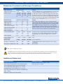
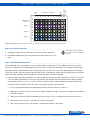
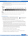
cAMP Hunter™ eXpress GPCR Assay
For chemiluminescent detection of cAMP for GPCR activity
Simple Solutions for Complex Biology
Table of Contents
Overview......................................................................................................................................................................... 1
Technology Principle .................................................................................................................................................... 1
Intended Use.................................................................................................................................................................. 2
Materials Provided and Storage Conditions.............................................................................................................. 3
Additional Materials...................................................................................................................................................... 3
Protocol Schematic ...................................................................................................................................................... 5
Quick Start Procedure................................................................................................................................................ 5
Detailed Protocol (Agonist, 96-well, Gas).................................................................................................................... 6
Step 1: Cell Preparation.............................................................................................................................................. 6
Step 2: Assay Plate Preparation.................................................................................................................................. 7
Step 3: cAMP Standard Preparation........................................................................................................................... 7
Step 4: Agonist Preparation........................................................................................................................................ 8
Step 5: Antibody and Working Solution Addition....................................................................................................... 9
Step 6: Enzyme Acceptor Addition............................................................................................................................. 9
Step 7: Assay Plate Reading....................................................................................................................................... 9
Typical Results............................................................................................................................................................. 10
Appendix....................................................................................................................................................................... 11
Antagonist Protocol for Gas-Coupled Receptors..................................................................................................... 11
Protocol for Gai-Coupled Receptors ....................................................................................................................... 12
PAMs and NAMs .................................................................................................................................................................13
High Throughput Screening ..................................................................................................................................... 13
Crude Biologic Samples .......................................................................................................................................... 13
Adjusted Volumes for 384-well Format..................................................................................................................... 13
FAQs.............................................................................................................................................................................. 14
Limited Use License Agreement................................................................................................................................ 15
Please read entire booklet before proceeding with the assay.
For additional information or Technical Support, contact [email protected]
or visit www.discoverx.com.
© 2015 DiscoveRx Corporation. All Rights Reserved.
DiscoveRx logo and DiscoveRx are registered trademarks of DiscoveRx Corporation. 20370 041615
US: tel | 1.866.448.4864
e | [email protected] EU: tel | +44.121.260.6142
e | [email protected]
www.discoverx.com
cAMP Hunter™ eXpress GPCR Assay User Manual
Overview
The cAMP Hunter eXpress GPCR Assay can be used for small or large molecules. The kits provide a robust, highly
sensitive and easy-to-use assay for monitoring G-protein coupled receptor (GPCR) activation based on 3'-5'-cyclic
adenosine monophosphate (cAMP) production in cells.
Technology Principle
GPCR activation following ligand binding initiates a
series of second messenger cascades that result in a
cellular response. The signaling involves a membrane
bound enzyme called adenylate cyclase. Gai- and Gascoupled receptors modulate cAMP by either inhibiting
or stimulating adenylate cyclase, respectively. With the
cAMP Hunter eXpress GPCR Assay, cells over expressing
GPCRs utilize the natural coupling status of the GPCR
to monitor activation of Gai- and Gas-coupled receptors.
Following ligand stimulation, the functional status of
the GPCR is monitored by measuring the cellular cAMP
levels using a homogeneous (no wash), gain-of-signal
competitive immunoassay based on Enzyme Fragment
Complementation (EFC) technology.
GPCR cAMP Pathway
Ligand
GPCR
Adenylyl
Cyclase
ATP
s
Gαs
cAMP
Gαi
i
The cAMP Hunter eXpress GPCR Assay EFC technology uses a b-galactosidase (b-gal) enzyme split into two
fragments, the Enzyme Donor (ED) and Enzyme Acceptor (EA). Independently these fragments have no β-gal activity;
however, in solution they rapidly complement to form an active b-gal enzyme.
In this assay, cAMP from cells and ED-labeled cAMP (ED-cAMP) compete for anti-cAMP antibody (Ab) binding sites.
Antibody-bound ED-cAMP will not be able to complement with EA, but unbound ED-cAMP is free to complement EA
to form active enzyme, which subsequently produces a luminescent signal. The amount of signal produced is directly
proportional to the amount of cAMP in the cells.
Low Levels of Cellular cAMP
Cellular cAMP
Anti-cAMP Ab
ED-cAMP
EA
High Levels of Cellular cAMP
Substrate
Hydrolysis
Cellular cAMP
US: tel | 1.866.448.4864
Anti-cAMP Ab
ED-cAMP
e | [email protected]
EU: tel | +44.121.260.6142
Active EFC
Enzyme
e | [email protected]
www.discoverx.com
Light
1
cAMP Hunter™ eXpress GPCR Assay User Manual
Intended Use
The cAMP Hunter eXpress GPCR Assay kits provide a robust, highly sensitive and easy-to-use, cell-based functional
assay to study GPCR activity through cAMP production. The pre-validated, frozen eXpress cells have been
manufactured for short term use and are provided in a convenient, ready-to-screen format that saves time and adds
convenience. The kits contain all the reagents needed for the detection of cAMP from eXpress cells expressing Gαi- and
Gαs-coupled receptors induced with a small or large molecule ligand. This flexible assay system has been designed to
work in agonist or antagonist mode for 96- and 384-well plate formats. After plating and stimulation of cells, the user
simply adds the assay reagents to the cells following the homogeneous, easy-to-use protocol provided.
cAMP Hunter eXpress GPCR Assay Schematic Protocol
Plate Cells
Assay
Buffer
Working
ED-cAMP Solution
Ligand
Incubate
US: tel | 1.866.448.4864
e | [email protected]
cAMP
Antibody
Solution
Incubate
EU: tel | +44.121.260.6142
EA
Read signal
Incubate
e | [email protected]
www.discoverx.com
2
cAMP Hunter™ eXpress GPCR Assay User Manual
Materials Provided and Storage Conditions
List of Components
Cell Components
Number of cells
Storage
cAMP Hunter eXpress Cells 1 vial
2 vials
10 vials
Number of Cells
3.75 x 10⁶
cells each
vial in 200
µL
3.75 x 10⁶
cells each
vial in 200
µL
3.75 x 10⁶
cells each
vial in 200
µL
Cells are shipped on dry ice and should arrive in a frozen state.
To ensure maximum cell viability, thaw the vials as soon as
possible upon receipt. If continued storage of the frozen vials
is necessary, store vials at -80°C immediately upon arrival. DO
NOT store at –80°C for >2 weeks. For longer term storage (>2
weeks), store vials in the vapor phase of liquid nitrogen (N2). DO
NOT store vials in liquid phase of N2. (see Safety Warning below)
Kit Components
Volume in each bottle (mL)
Storage
cAMP Lysis Buffer
3.8
7.6
38
cAMP Solution D
5
10
50
cAMP Solution A
8
16
80
cAMP Antibody Reagent
2.5
5
25
cAMP Standard (250 µM)
1
1
1
Substrate Reagent 1
1
2
10
Upon arrival, store reagents at ≤-20°C. The detection kit is
stable until the expiry date indicated on the kit box outer label.
Thaw reagents at room temperature before use. After thawing,
store reagents for up to 1 month at 2-8°C. For longer term
storage, aliquots of all the components may be re-frozen in
opaque containers at ≤-20°C. The reagents can be thawed and
frozen for a total of 3 times without loss in performance.
Substrate Reagent 2
0.2
0.4
2
Forskolin (dried powder)
1 x 0.25
mg
2 x 0.25
mg
10 x 0.25
mg
If Forskolin is not used immediately, prepare multiple aliquots
and store at -20°C. Avoid multiple freeze-thaw cycles. When
stored properly, the solution is stable for up to 2 months.
Cell Assay Buffer
12.5
25
250
-20°C
Assay Complete™ Cell
Plating Reagent
1 x 20 mL
2 x 20 mL
2 x 100 mL -20°C
Plate Format
1 plate
2 plates
10 plates Room Temperature
96-well, No. of data points
100
200
1,000
Included: 96-well white-walled clear-bottom tissue culture
treated plate
384-well, No. of data points
400
800
4,000
Not Included
Bulk cells are available upon request
Safety Warning: A face shield, gloves and lab coat should be worn at all times when handling frozen vials. The manufacturer of the
cryovial recommends storing the vials in the vapor phase above the liquid N2. Upon thawing, if liquid N2 is present in the cryovial, it
rapidly converts back to its gas phase which can result in the explosion of the vial upon its removal from the freezer
Additional Materials
Required Materials
Ordering Information
Disposable Reagent Reservoir
Thermo Scientific, Cat. No. 8094 or similar
Green V-bottom Ligand Dilution Plates (10 plates/pack)
DiscoveRx Cat. No. 92-0011
15 mL polypropylene tubes and 1.5 mL microtubes
Tissue culture disposables (pipettes 1 mL – 25 mL)
Humidified tissue culture incubator (37°C, 5% CO2)
Single and multichannel micro-pipettors and pipette tips
(10 μL – 100 μL)
Multimode or luminescence plate reader
US: tel | 1.866.448.4864
e | [email protected]
EU: tel | +44.121.260.6142
e | [email protected]
www.discoverx.com
3
cAMP Hunter™ eXpress GPCR Assay User Manual
Recommended Materials, Reagents and Equipment
Ordering Information
Control ligands
www.discoverx.com/controlligands
Test Compounds
IBMX, for Gαs assays
DiscoveRx, Cat. No. 92-0007
Instrument Compatibility Chart
Compatible with any Luminometer. Select examples indicated below.
Berthold Technologies: Mithras LB940, CentroLIApc
Molecular Devices: FLIPR, SpectraMax M3/ M4/M5/M5e,
FlexStation 3, SpectraMax L
Biotek: Synergy 2
Perkin Elmer: TopCount, Victor 2 or V, Fusion, LumiCount,
Envision, Micro-beta (Trilux), Viewlux, Northstar, EnSpire
BMG: PheraStar, Cytostar, LumiStar
Promega: GloMax systems
Caliper: LabChip 3000 & EZ Reader
Tecan: Ultra, Evolution
GE: LEAD seeker, Farcyte
Thermo Scientific: Luminoskan Ascent
Hamamatzu: FDS6000, FDSS/RayCatcher
Turner BioSystems: Modulus Microplate
US: tel | 1.866.448.4864
e | [email protected]
EU: tel | +44.121.260.6142
e | [email protected]
www.discoverx.com
4
cAMP Hunter™ eXpress GPCR Assay User Manual
Protocol Schematic
Tip: Use this sheet to note your assay specific
conditions. Post on your bench to use as a quick
reference guide.
Assay Name:___________________________________________________________ Date:________
Product Details:_____________________________________________________________________
Quick-Start Procedure: In a white-walled 96-well tissue culture treated plate perform the following:
Seed cells in 100 µL
Cell Plating Reagent
Agonist
Antagonist
Assay Preperation
Incubate cells overnight
at 37oC, 5% CO2
Remove Cell Plating Reagent
Replace with 30 µL
Cell Assay Buffer
Treat cells with 7.5 μL 6X
Antagonist prepared in
Cell Assay Buffer
Treat cells with 15 μL 3X
Agonist prepared in
Cell Assay Buffer
(Include 3X forskolin for Gαi targets)
Incubate for 15 mins.
at 37oC
Treat cells with 7.5 μL 6X
Agonist prepared in
Cell Assay Buffer
cAMP Hunter eXpress GPCR Detection
(Include 6X forskolin for Gαi targets)
US: tel | 1.866.448.4864
Incubate for _______ mins.
at ________oC
(Specific time & temp. in datasheet)
Add 15 µL Antibody Solution
Add 60 μL cAMP
Working Detection Solution
Incubate for 1 hr. at room
temp. in the dark
Add 60 μL cAMP Solution A
Incubate for 3 hrs. at room
temp. in the dark
Read chemiluminescent signal
e | [email protected]
EU: tel | +44.121.260.6142
e | [email protected]
www.discoverx.com
5
cAMP Hunter™ eXpress GPCR Assay User Manual
Detailed Protocol (Agonist, 96-well, Gas)
The following detailed protocol is specific to detecting cAMP in cells
stimulated by an agonist for Gαs-coupled receptors in a 96-well format plate.
Refer to the appendix for variations
(e.g. agonist/Gai-coupled, antagonist,
HTS, PAMs, NAMs, crude biologics,
and 384-well).
Step 1: Cell Preparation___________________________________________________________________________________
The following steps outline the procedure for thawing and plating frozen
cAMP Hunter eXpress cells from cryogenic vials. Each vial contains
sufficient cell numbers to generate (1) 96-well microplate prepared at the
seeding density described.
a. Remove the cAMP Hunter eXpress Assay cells cryovials from -80°C or
liquid N2 vapor storage and place them immediately on dry ice prior to
thawing.
Do not substitute cell plating from an
alternate kit at any time
Do not expose vials to room
temperature
Safety Warning: A face shield, gloves and lab coat should be worn at all times
when handling frozen vials. The manufacturer of the cryovial recommends storing
the vials in the vapor phase above the liquid N2. Upon thawing, if liquid N2 is
present in the cryovial, it rapidly converts back to its gas phase which can result in
the explosion of the vial upon its removal from the freezer
b. Decontaminate the vial by spraying and wiping with 70% ethanol. All of the procedures from this point on should be
carried out under aseptic conditions in a culture hood.
c. Pre-warm Assay Complete™ Cell Plating Reagent in a 37˚C water bath for 15 mins.
d. Add 0.5 mL of pre-warmed AssayComplete Cell Plating Reagent to the
cell vial to thaw the cells. Pipette up and down gently several times to
ensure that cells are evenly distributed.
Do not thaw vials in 37°C water bath
or centrifuge
e. Immediately transfer the cells to 11.5 mL of pre-warmed AssayComplete Cell Plating Reagent. Mix and pour into a
disposable reagent reservoir.
f. Prepare the 96-well tissue culture treated assay plate using the plate map figure on the next page. Leave the first 2
rows empty (with no cells) to allow for an 11-point cAMP standard curve in duplicate. For the remaining wells, plate
100 ul of cells into each well. For rows C through H, plate 3 different 11 point agonist curves in duplicate. Optionally,
samples can be run on different plates, in triplicates or other variations.
g. Place the seeded plate in a 37°C, 5% CO2 humidified incubator for 18-24 hours prior to testing (refer to the targetspecific datasheet for the recommended incubation time).
US: tel | 1.866.448.4864
e | [email protected]
EU: tel | +44.121.260.6142
e | [email protected]
www.discoverx.com
6
High
1
cAMP std. curve
(no cells)
agonist 1
serial dilutions of agonist
2
3
4
5
6
7
8
Low
No agonist
(control)
cAMP Hunter™ eXpress GPCR Assay User Manual
9 10 11 12
A
B
C
D
agonist 2
E
F
agonist 3
G
H
Agonist assay plate map: Create 11-point curves for cAMP standard curve and 3 different agonists in duplicate.
Step 2: Assay Plate Preparation ___________________________________________________________________________
a. Completely remove the cell media from assay plate wells by aspiration
Removing the media completely
is crucial for reducing variability of
replicates.
b. Immediately add 30 μL of Cell Assay Buffer to all empty wells in the assay
plate.
Step 3: cAMP Standard Preparation _______________________________________________________________________
When optimizing the assay conditions, always include a cAMP standard curve. The standard curve not only verifies
that the kit components are working properly, but also serves as a detection limit guide. If the amount of cAMP being
detected exceeds the detection limit of the cAMP detection kit, the EC50 will start to shift (depending on the coupling
status of Gas or Gai, the shift will be towards the right or left). To avoid this situation, the cell number per well should be
optimized. cAMP standard should be prepared fresh before agonist compound addition.
a. Prepare cAMP standard serial dilutions in a separate plate by diluting the cAMP standard (2.5 x 10 -4M) in a 1:9 ratio
(so 1 part cAMP standard plus 8 parts Cell Assay Buffer). This dilution ratio corresponds to the highest standard
concentration (well No. 1) at 2.31 x 10 -6M in an assay volume of 180 μL. Make 10 additional 3-fold serial dilutions,
using the Cell Assay Buffer, with the last well as the negative control as shown below.
1. Using a separate dilution plate (or polypropylene tubes), label the wells No. 1 to No. 12.
2. Add 160 μL of Cell Assay Buffer and 20 μL of cAMP standard to well No. 1 (this will be the highest concentration
of cAMP standard).
3. Add 80 μL of Cell Assay Buffer to dilution wells No. 2 to No. 12. This is enough volume for more than 4 rows.
4. Remove 40 μL from well No. 1 and add it to well No. 2. Mix gently.
5. With clean tip, remove 40 μL from well No. 2 and add it to well No. 3. Mix gently.
US: tel | 1.866.448.4864
e | [email protected]
EU: tel | +44.121.260.6142
e | [email protected]
www.discoverx.com
7
cAMP Hunter™ eXpress GPCR Assay User Manual
6. Repeat this process until well No. 11. Do not add cAMP standard to well No. 12 since this is the negative control
well (containing only Cell Assay Buffer).
b. Add 15 μL of cAMP standard dilutions in duplicate to the first two rows (rows A and B) of the assay plate as shown
in the previously described assay plate map.
cAMP
Standard
20 μL
40 μL
Cell
Assay
Buffer
40 μL
40 μL
40 μL
40 μL
40 μL
40 μL
40 μL
40 μL
40 μL
Negative
Control
160 μL
80 μL
80 μL
80 μL
80 μL
80 μL
80 μL
80 μL
80 μL
80 μL
80 μL
1
2
3
4
5
6
7
8
9
10
11
12
Final Volume:
140 μL
80 μL
80 μL
80 μL
80 μL
80 μL
80 μL
80 μL
80 μL
80 μL
120 μL
80 μL
Sequential
Dilution Ratio:
1:9
1:3
1:3
1:3
1:3
1:3
1:3
1:3
1:3
1:3
1:3
7.70 x10⁻⁷
2.57 x10⁻⁷
8.56 x10⁻⁸
2.85 x10⁻⁸
9.51 x10⁻⁹
3.17 x10⁻⁹
1.06 x10⁻⁹
~Final Assay
Concentration (M): 2.31x10⁻⁶
80 μL
3.52 x10⁻¹⁰ 1.17 x10⁻¹⁰ 3.91 x10⁻¹¹
cAMP Standard serial dilutions: Make 11 three fold serial dilutions of cAMP standard solution in a separate plate.
Step 4: Agonist Preparation _______________________________________________________________________________
a. Prepare agonist serial dilutions in a separate dilution plate in a 11-point series
of 3X (3-fold) dilutions of agonist in Cell Assay Buffer or appropriate agonist
buffer (as specified on the datasheet). The final concentration of each dilution
should be prepared at 3X of the final screening concentration.
For Gai specific targets or other
ligand variations (e.g. antagonists),
refer to the appendix section.
1. For each agonist, label the wells of a separate dilution plate (or polypropylene tubes) No. 1 to No. 12.
2. Similar to the cAMP standard serial dilutions, add 80 μL of Cell Assay Buffer (or agonist specific buffer) to
dilution wells No. 2 to No. 12. This is enough volume for over 4 rows, and the dilution volume may be adjusted
according to the number of wells desired.
3. Prepare the highest concentration of Agonist in Cell Assay Buffer (or agonist specific buffer). We recommend
preparing a final screening concentration that is 250X the expected EC50 of the agonist. Therefore, prepare a
working concentration that is 750X the expected EC50 per well. E.g. for an expected EC50 of 1 nM, prepare the
highest working concentration at 750 nM. This is 3X the screening or final top concentration of 250 nM, and the
expected EC50 concentration will lie near the center of the dose response curve.
4. Add 120 μL of the highest concentration of Agonist/Cell Assay Buffer to well No. 1.
5. Remove 40 μL from well No. 1 and add it to well No. 2. Mix gently.
6. With clean tip, remove 40 μL from well No. 2 and add it to well No. 3. Mix gently.
7. Repeat process until well No. 11. Do not add agonist to well No. 12 since this is the negative control well.
8. Set up additional serial dilutions for additional agonists in a similar manner.
US: tel | 1.866.448.4864
e | [email protected]
EU: tel | +44.121.260.6142
e | [email protected]
www.discoverx.com
8
cAMP Hunter™ eXpress GPCR Assay User Manual
b. Add 15 μL of each 3X agonist serial dilution in duplicate to the designated agonist rows (e.g. agonist 1 in rows C
and D; agonist 2 in rows E and F; and agonist 3 in rows G and H) of the assay plate as indicated on the previously
described assay plate map.
c. Incubate assay plate at the indicated time and temperature for the specific cell line (please refer to the specific cell
line datasheet for conditions). For most cell lines, incubate for 30 minutes at 37°C. For the best results, the optional
incubation time should be empirically determined.
Step 5: Antibody and Working Solution Addition____________________________________________________________
a. Following agonist incubation, add 15 μL of cAMP
Antibody Reagent to all wells.
b. Prepare a stock of cAMP Working Detection Solution
in a separate 15 ml polypropylene tube, by mixing
19 parts of cAMP Lysis Buffer, 5 parts of Substrate
Reagent 1, 1 part Substrate Reagent 2, and 25 parts
of cAMP Solution D.
c. Add 60 μL of cAMP Working Detection Solution to all
wells of the assay plate. Do not pipette up and down
in the vial to mix or vortex plates.
Make stock within 8 hours of use.
cAMP Working Detection Solution
Components
Volume ratio
Volume per plate
cAMP Lysis Buffer
19
3.8 ml
Substrate Reagent 1
5
1.0 mL
Substrate Reagent 2
1
0.2 mL
cAMP Solution D
25
5.0 mL
Total Volume
d. Incubate assay plate for 1 hour at room temperature
in the dark for the immunocompetition reaction to
occur.
10 mL
cAMP Working Detection Solution is light sensitive, thus
incubation in the dark is necessary.
Step 6: Enzyme Acceptor Addition_________________________________________________________________________
a. Add 60 μL of cAMP Solution A to all wells of the assay plate. Do not pipette up and down in the vial to mix or vortex
plates
b. Incubate assay plate for 3 hours at room temperature in
the dark.
cAMP Working Detection Solution is light sensitive, thus
incubation in the dark is necessary.
Step 7: Assay Plate Reading_______________________________________________________________________________
a. Read samples on a standard luminescence plate reader at 0.1 to 1 second/well for photomultiplier tube readers or
5-10 seconds for imager. The plate may be incubated overnight and the signal may be measured the next day. In
general, the signal continues to increase and reaches a maximum approximately 3-6 hours after the last incubation
step. The actual signal characteristics over time are affected by lab conditions, such as temperature, and the user
should establish an optimal read time. Luminescence readout usually collects signal from all wavelengths. Some
instrument manufacturer may include a cutoff filter at high wavelengths, but usually no wavelength setting is needed
for luminescence readout.
b. Data analysis can be performed using your choice of statistical analysis software (e.g. GraphPad Prism, Molecular
Devices Softmax Pro, Biotek Instruments Gen5, Microsoft Excel, etc.).
US: tel | 1.866.448.4864
e | [email protected]
EU: tel | +44.121.260.6142
e | [email protected]
www.discoverx.com
9
cAMP Hunter™ eXpress GPCR Assay User Manual
Typical Results
Typical results shown below of the cAMP standard curve analysis (top left) and cAMP Hunter eXpress GPCR Assay
using the cAMP Hunter™ dopamine receptor D2 (short isoform) Gai cells (top right) and glucagon-like peptide receptor
1 Gas cells (bottom). Note for the cAMP standard curve graph, the signal continues to increase over time and plates can
be read the following day.
cAMP Standard Curve (96 well)
1hr read
2hr read
3hr read
18 hr read
RLU
15000
10000
5000
10000
Dopamine Receptor D2
(short isoform), Gαi Cells
8000
RLU
20000
6000
4000
2000
0
10 -11
10 -10
10 -9
10 -8
10 -7
10 -6
10 -5
0
10 -13 10 -12
10 -10
10 -8
10 -6
Dopamine [M]
cAMP [M]
1 hr. read
2 hr. read
3 hr. read
18 hr. read
EC50
1.40x10 -9
EC50
1.96x10 -8
2.47x10 -8
2.53x10 -8
1.82x10 -8
S/B
7.1
S/B
37.6
42.2
43.1
50.4
Glucagon-like Peptide Receptor 1, Gαs Cells
8000
RLU
6000
4000
2000
0
10 -13 10 -12 10 -11 10 -10 10 -9 10 -8 10 -7
GLP1(7-36) [M]
EC50
2.21x10 -11
S/B
18.0
US: tel | 1.866.448.4864
e | [email protected]
EU: tel | +44.121.260.6142
e | [email protected]
www.discoverx.com
10
cAMP Hunter™ eXpress GPCR Assay User Manual
Appendix
Antagonist Protocol for Gas Receptors_____________________________________________________________________
Protocol Modification
High
1
cAMP std. curve
(no cells)
antagonist 1
serial dilutions of antagonist
2
3
4
5
6
7
8
Low
+ agonist
- antagonist
- agonist
- antagonist
1. Antagonist serial dilution: Similar to the agonist step 4a, prepare a 10-point series of 3-fold dilutions of antagonist in
Cell Assay Buffer. The concentration of each dilution should be prepared at 6X of the final screening concentration.
See plate map below for an antagonist set-up example.
9 10 11 12
A
B
C
D
antagonist 2
E
F
antagonist 3
G
H
Antagonist assay plate map: Create 10-point curves with cAMP standard curve and 3 different antagonists in duplicate.
2. Antagonist/Agonist addition: Replace step 4b of the agonist detailed protocol with the following directions.
a. Add 7.5 μl of each 6X antagonist serial dilution to the designated antagonist rows of the assay plate.
b. Incubate assay plate for 15 minutes at 37°C. For best results, the optimal incubation time should be empirically
determined.
c. For the agonist challenge, add 7.5 μL of agonist at 6X the EC80.
We recommend running the agonist dose response curve to determine the
EC80. The specific concentration of agonist to be used can vary from EC75
to EC100, based on the target and assay conditions. For Gai targets, instead
of the agonist alone, add 7.5 μL of 6X forskolin plus agonist solution at this time.
For Gai targets, only add the
forskolin during the agonist
challenge step.
d. Continue with step 4c (incubation) of the agonist detailed protocol.
US: tel | 1.866.448.4864
e | [email protected]
EU: tel | +44.121.260.6142
e | [email protected]
www.discoverx.com
11
cAMP Hunter™ eXpress GPCR Assay User Manual
Protocol for Gai-Coupled Receptors_______________________________________________________________________
In order to measure Gai-coupled receptors (targets), the agonist sample is added in the presence of forskolin. Forskolin
activates adenylate cyclase and increases intracellular levels of cAMP. For a Gai receptor, agonist binding or activation
inhibits the intracellular cAMP accumulation induced by forskolin. As a result, the dose response curve generated in the
presence of agonist plus forskolin will have a negative slope. Since forskolin treatment results in increased intracellular
cAMP levels in all cAMP Hunter™ cells, it can serve as a positive control for the performance of the cells and detection
reagents
Protocol Modification
1. Replace step 4a of the agonist detailed protocol with the following steps to
prepare a Gai–coupled receptor ligand plus forskolin 11-point serial dilution.
a. Prepare a 3X (for agonist mode) or 6X (for antagonist mode) final screening
concentration of forskolin. Refer to the target-specific datasheet for
recommended concentrations of forskolin. A general recommendation
for the final forskolin concentration is 10-15 μM for CHO-K1 cells and
15-20 μM for HEK 293. For example, if the forskolin final in-well screening
concentration is recommended to start at 15 μM, prepare a 45 μM forskolin
solution (which is 3X).
For antagonist Gai-coupled
receptors, forskolin is added to
the agonist challenge solution.
For forskolin, avoid multiple
freeze/thaw cycles. Store as
multiple aliquots at -20ºC for
up to 2 months.
b. Prepare the highest concentration, 3X (for agonist mode) or 6X (for antagonist mode), of Gai agonist in the
forskolin solution prepared above.
c. Add 120 μL of the highest concentration of agonist plus forskolin to well No. 1.
d. Remove 40 μL from well No. 1 and add it to well No. 2. Mix gently.
e. With clean tip, remove 40 μL from well No. 2 and add it to well No. 3. Mix gently.
f. Repeat process until well No. 11. Do not add agonist to well No. 12 since this is the negative control well.
g. Set up additional serial dilutions for additional agonists in a similar manner.
2. Replace step 4b of the agonist detailed protocol. Add 15 μL (for agonist mode) or 7.5 μL (for antagonist mode) of
the Gai agonist plus forskolin serial dilutions in duplicate to the designated agonist rows (e.g. agonist 1 in rows C
and D; agonist 2 in rows E and F; and agonist 3 in rows G and H) of the assay plate as indicated on the previously
described assay plate map.
3. Continue with step 4c (incubation) of the agonist detailed protocol.
US: tel | 1.866.448.4864
e | [email protected]
EU: tel | +44.121.260.6142
e | [email protected]
www.discoverx.com
12
cAMP Hunter™ eXpress GPCR Assay User Manual
PAMs and NAMs__________________________________________________________________________________________
For positive allosteric modulators (PAMs), refer to the antagonist protocol above, but change the agonist challenge
concentration to EC20 instead of EC80.
For negative allosteric modulators (NAMs), refer to the antagonist protocol above with no changes.
High Throughput Screening_______________________________________________________________________________
For high throughput screening applications, the cAMP Antibody Reagent can be added before ligand introduction to
save an addition step. Briefly, pre-dilute the cAMP Antibody Reagent from the kit in a 1:2 ratio with Cell Assay Buffer (so
1 part cAMP Antibody Reagent to 2 parts Cell Assay Buffer). Replace step 2a of the agonist detailed protocol by adding
45 μl of diluted antibody mixture to each well. Replace step 4b by adding 15 μl of 4X agonist to each well. Incubate the
assay plate as indicated in step 4c and skip to step 5b of the standard protocol.
Crude Biologic Samples __________________________________________________________________________________
The cAMP Hunter eXpress GPCR kits can be run in the presence of high levels of serum or plasma without significantly
impacting assay performance. Therefore, standard curves and samples can be prepared in neat serum or plasma and
added directly to cells without further dilution. For the best results, the optimized minimum required dilution (MRD) of
crude samples should be empirically determined.
Adjusted Volumes for 384-well Format _____________________________________________________________________
Use the following table to adjust the component volumes per well for 384-well plates.
384-well Format
Assay Reagents
96 plate (per well)
384 plate (per well)
No. of Cells
30,000 *
10,000 *
AssayComplete Cell Plating Reagent
100 μL **
20 μL **
Cell Assay Buffer
30 μL
10 μL
Ligand (e.g. Agonist)
15 μL
5 μL
cAMP Antibody Reagent
15 μL
5 μL
cAMP Working Detection Solution
60 μL
20 μL
cAMP Solution A
60 μL
20 μL
Final Assay Volume **
180 μL
60 μL
* The number of cells is dependent on the cell line.
** The AssayComplete Cell Plating Reagent volume is used to plate cells and this volume is removed during first step.
US: tel | 1.866.448.4864
e | [email protected]
EU: tel | +44.121.260.6142
e | [email protected]
www.discoverx.com
13
cAMP Hunter™ eXpress GPCR Assay User Manual
FAQs
Is IBMX necessary in the DiscoveRx cAMP kits?
• IBMX is not necessary and we do not use it for our internal testing; however, the kit is compatible with the use of
IBMX. If IBMX is to be used, cell number per well and IBMX concentration needs to be optimized so that the amount
of cAMP generated stays within the optimal detection of the assay kit.
How do I use suspension cells?
• Harvest and resuspend suspension cells in Cell Assay Buffer media (either 1X HBSS + 10mM HEPES or PBS) at
the optimal cell density. Typical suspension cell density is approximately 20,000 cells per well in a standard 96-well
plate with a cell viability > 90%.
What if there is no or low signal?
• If plated on clear-bottom assay plates, visually inspect the cells before and after running the assay to ensure that
they are healthy. Make sure proper cell plating procedure is followed to ensure assay performance.
• Make sure detection reagents are stored and prepared properly.
• Make sure forskolin is included for Gai coupled receptors. Please refer to the Appendix for Gai coupled receptor
assay protocol.
• Make sure the proper assay mode is used (agonist mode or antagonist mode).
• Make sure the plate reader is setup properly to read luminescence.
• High levels of protein in the wells may interfere with enzyme fragment complementation. If high levels are present, a
media exchange step could be performed just prior to the detection reagent addition.
What if the response is lower than expected (lower than expected S:B)?
• Make sure that the ligand is prepared properly, take extra care to observe ligand solubility.
• Make sure DMSO and other solvents are not too high and not more than 1% final concentration.
• When testing ligands at high concentrations, make sure the vehicle is compatible.
• Make sure ligand is incubated for the designated time and at designated temperature.
• Make sure plates are protected from light during incubation.
What if the EC50 does not match reported values?
• Include a cAMP standard curve in the assay to ensure that the ligand dose curve is in the dynamic range of the
cAMP detection kit. Moving out of the dynamic range may shift the EC50 of ligands.
• Make sure ligands are incubated at the proper temperature.
• The source of a ligand, particularly for biologics, can have a large effect on the observed pharmacology.
• Changing tips during serial dilutions can help to avoid carryover.
• Receptor expression level may cause receptor reserve issues in ligand testing. Select a cell line that has medium to
low expression of receptors.
What if the variability between replicates is high?
• Make sure dispensing equipment is properly calibrated, and proper pipetting technique is used.
What if my compound is in media containing high concentrations of serum, can I use it as is or will the serum
interfere with the assay?
• Our assays are highly tolerant to high serum content (as high as 80% serum). We recommend that you aspirate the
high serum media prior to adding detection reagents.
For additional information or Technical Support, contact [email protected] or visit www.discoverx.com.
US: tel | 1.866.448.4864
e | [email protected]
EU: tel | +44.121.260.6142
e | [email protected]
www.discoverx.com
14
cAMP Hunter™ eXpress GPCR Assay User Manual
Limited Use License Agreement
DiscoveRx name, logo, and cAMP Hunter are trademarks of DiscoveRx Corporation. DiscoveRx grants to the purchaser
of this product an end-user license under United States and foreign patents owned or licensed exclusively to DiscoveRx
(including US patent numbers, 4708929, 4956274, 5244785, 5444161, 5604091, 5643734 and equivalent foreign
patents) and non-exclusively licensed (including US patent numbers 4931569, 4978614, 5145772, 5326882, 5538847,
and 5851771 and equivalent foreign patents) to use this product solely in the fields of drug discovery and regulatory
qualification of therapeutic drugs and not for any other uses, including but not limited to, other research uses, or use in
human, clinical, animal, or food diagnostics.
If the purchaser has any further questions regarding the rights conferred with purchase of the Materials, please contact:
Licensing Department
DiscoveRx Corporation
42501 Albrae Street
Fremont, CA 94538
t | 510.979.1415 x 104
e | [email protected]
cAMP Hunter eXpress GPCR Assay User Manual 70-234 Rev_8
US: tel | 1.866.448.4864
e | [email protected]
EU: tel | +44.121.260.6142
e | [email protected]
www.discoverx.com
15