1
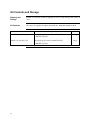
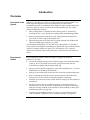
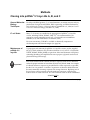
pcDNA™3.1/myc-His A, B, and C Catalog no. V800-20 Rev. Date: 28 October 2010 Manual part no. 28-0137 MAN0000641 Corporate Headquarters Invitrogen Corporation 1600 Faraday Avenue Carlsbad, CA 92008 T: 1 760 603 7200 F: 1 760 602 6500 E: [email protected] For country-specific contact information visit our web site at www.invitrogen.com User Manual ii Table of Contents Kit Contents and Storage .....................................................................................................................iv Introduction ................................................................................................................... 1 Overview ................................................................................................................................................. 1 Methods ......................................................................................................................... 2 Cloning into pcDNA™3.1/myc-His A, B, and C................................................................................. 2 Transformation and Transfection ........................................................................................................ 5 Appendix........................................................................................................................ 7 pcDNA™3.1/myc-His A, B, and C........................................................................................................ 7 pcDNA™3.1/myc-His/lacZ ................................................................................................................... 9 Accessory Products.............................................................................................................................. 10 Technical Support ................................................................................................................................ 11 Purchaser Notification......................................................................................................................... 12 References.............................................................................................................................................. 13 iii Kit Contents and Storage Shipping and Storage pcDNA™3.1/myc-His vectors are shipped on wet ice. Upon receipt, store vectors at –20°C. Kit Contents All vectors are supplied in aliquot detailed below. Store the vectors at –20°C. Vector Composition Amount pcDNA 3.1/myc-His A, B, and C 40 l of 0.5 g/μl vector in 10 mM Tris-HCl, 1 mM EDTA, pH 8.0 20 g pcDNA™3.1/myc-His/lacZ 40 l of 0.5 g/μl vector in 10 mM Tris-HCl, 1 mM EDTA, pH 8.0 20 g ™ iv Introduction Overview Description of the System pcDNA™3.1/myc-His A, B, and C are 5.5-kb vectors derived from pcDNA™3.1(+) and designed for high-level expression, purification, and detection of recombinant proteins in mammalian hosts. High-level stable and non-replicative transient expression can be carried out in most mammalian cells. The vectors contain the following elements: Three reading frames to facilitate in frame cloning with a C-terminal tag encoding the myc (c-myc) epitope and a polyhistidine metal-binding peptide Human cytomegalovirus immediate-early (CMV) promoter for high-level expression in a wide range of mammalian cells Episomal replication in cell lines that are latently infected with SV40 or that express the SV40 large T antigen (e.g., COS7) The control plasmid, pcDNA™3.1/myc-His/lacZ, is the pcDNA™3.1/myc-His C vector with a 3.2-kb fragment containing the -galactosidase gene cloned in frame with the C-terminal peptide (see page 9). It is included for use as a positive control for transfection, expression, purification, and detection in the cell line of choice. Experimental Outline Use the following outline to clone and express your gene of interest in pcDNA™3.1/myc-His: Consult the multiple cloning sites described on pages 3-4 to determine which vector (A, B, or C) should be used to clone your gene in frame with the C-terminal myc epitope and the polyhistidine tag. Ligate your insert into the appropriate vector and transform into E. coli. Select transformants on 50–100 g/ml ampicillin. Analyze your transformants for the presence of insert by restriction digestion. Select a transformant with the correct restriction pattern and confirm that your gene is in frame with the C-terminal peptide by sequencing. Transfect your construct into the cell line of choice using your own method of transfection. Test for expression of your recombinant gene by western blot analysis or functional assay. If you do not have an antibody to your protein, Invitrogen offers the Anti-myc antibody or the Anti-His(C-term) antibody to detect your recombinant protein (see page 10). To purify your recombinant protein, you may use a metal-chelating resin such as ProBond™. ProBond™ resin is available separately (see page 10). 1 Methods Cloning into pcDNA™3.1/myc-His A, B, and C General Molecular Biology Techniques For help with DNA ligations, E. coli transformations, restriction enzyme analysis, purification of single-stranded DNA, DNA sequencing, and DNA biochemistry, refer to Molecular Cloning: A Laboratory Manual (Sambrook et al., 1989) or Current Protocols in Molecular Biology (Ausubel et al., 1994). E. coli Strain Many E. coli strains are suitable for the propagation of pcDNA™3.1/myc-His vectors, including TOP10, TOP10F´, DH5™-T1R. We recommend that you propagate vectors containing inserts in E. coli strains that are recombinant deficient (recA) and endonuclease A-deficient (endA). For your convenience, TOP10F’ is available as chemically competent or electrocompetent cells from Invitrogen (see page 10). Maintenance of pcDNA™3.1/ myc-His Important To propagate and maintain the pcDNA™3.1/myc-His vectors, use the supplied 0.5 g/l stock solution in TE, pH 8.0 to transform a recA, endA E. coli strain like TOP10, TOP10F’, DH5, JM109, or equivalent. Select transformants on LB plates containing 50–100 g/ml ampicillin. Be sure to prepare a glycerol stock of each plasmid for long-term storage. Your insert should contain a Kozak consensus sequence with an ATG initiation codon for proper initiation of translation (Kozak, 1987; Kozak 1990). An example of a Kozak consensus sequence is provided below. Other sequences are possible, but the G or A at position –3 and the G at position +4 (shown in bold) illustrates the most commonly occurring sequence with strong consensus. Replacing one of the two bases at these positions provides moderate consensus, while having neither results in weak consensus. The ATG initiation codon is shown underlined. (G/A)NNATGG Continued on next page 2 Cloning into pcDNA™3.1/myc-His A, B, and C, Continued Multiple Cloning Site of Version A Below is the multiple cloning site for pcDNA™3.1/myc-His A. Restriction sites are labeled to indicate the cleavage site. Note that there is a stop codon between the BamH I site and the BstX I site. The boxed nucleotides indicate the variable region. The multiple cloning site has been confirmed by sequencing and functional testing. The vector sequence of pcDNA™3.1/myc-His A is available for downloading from our website at www.invitrogen.com or from Technical Support (see page 11). T7 promoter/priming site Hind III Kpn I BamH I 861 ATTAATACGA CTCACTATAG GGAGACCCAA GCTGGCTAGT TAA GCT TGG TAC CGA GCT CGG Ala Trp Tyr Arg Ala Arg BstX I EcoR I EcoR V BstX I Not I 922 ATC CAC TAG TCC AGT GTG GTG GAA TTC TGC AGA TAT CCA GCA CAG TGG CGG CCG Ile His *** Ser Ser Val Val Glu Phe Cys Arg Tyr Pro Ala Gln Trp Arg Pro Xho I Xba I Apa I myc epitope Sfu I 976 CTC GAG TCT AGA GGG CCC TTC GAA CAA AAA CTC ATC TCA GAA GAG GAT CTG AAT Leu Glu Ser Arg Gly Pro Phe Glu Gln Lys Leu Ile Ser Glu Glu Asp Leu Asn Polyhistidine tag Age I PmeI 1030 ATG CAT ACC GGT CAT CAT CAC CAT CAC CAT TGA GTTTAAACCC GCTGATCAGC Met His Thr Glu His His His His His His *** BGH Reverse priming site 1083 CTCGACTGTG CCTTCTAG Multiple Cloning Site of Version B Below is the multiple cloning site for pcDNA™3.1/myc-His B. Restriction sites are labeled to indicate the cleavage site. The boxed nucleotides indicate the variable region. The multiple cloning site has been confirmed by sequencing and functional testing. The vector sequence of pcDNA™3.1/myc-His B is available for downloading from our website at www.invitrogen.com or from Technical Support (see page 11). T7 promoter/priming site Hind III Kpn I BamH I 861 ATTAATACGA CTCACTATAG GGAGACCCAA GCTGGCTAGT TAAG CTT GGT ACC GAG CTC GGA Leu Gly Thr Glu Leu Gly BstX I EcoR I EcoR V BstX I Not I 923 TCC ACT AGT CCA GTG TGG TGG AAT TCT GCA GAT ATC CAG CAC AGT GGC GGC CGC Ser Thr Ser Pro Val Trp Trp Asn Ser Ala Asp Ile Gln His Ser Gly Gly Arg Xho I XbaI Apa I Sac II Sfu I myc epitope 977 TCG AGT CTA GAG GGC CCG CGG TTC GAA CAA AAA CTC ATC TCA GAA GAG GAT Ser Ser Leu Glu Gly Pro Arg Phe Glu Gln Lys Leu Ile Ser Glu Glu Asp Age I Polyhistidine tag Pme I 1028 CTG AAT ATG CAT ACC GGT CAT CAT CAC CAT CAC CAT TGA GTTT AAACCCGCTG Leu Asn Met His Thr Gly His His His His His His *** BGH Reverse priming site 1081 ATCAGCCTCG ACTGTGCCTT CTAGTTGCCA Continued on next page 3 Cloning into pcDNA™3.1/myc-His A, B, and C, Continued Multiple Cloning Site of Version C Below is the multiple cloning site for pcDNA™3.1/myc-His C. Restriction sites are labeled to indicate the cleavage site. The boxed nucleotides indicate the variable region. The multiple cloning site has been confirmed by sequencing and functional testing. The vector sequence of pcDNA™3.1/myc-His C is available for downloading from our website at www.invitrogen.com or from Technical Support (see page 11). T7 promoter/priming site Hind III Kpn I 861 ATTAATACGA CTCACTATAG GGAGACCCAA GCTGGCTAGT TA AGC TTG GTA CCG AGC Ser Leu Val Pro Ser BamH I BstX I EcoR I EcoR V BstX I 918 TCG GAT CCA CTA GTC CAG TGT GGT GGA ATT CTG CAG ATA TCC AGC ACA GTG Ser Asp Pro Leu Val Gln Cys Gly Gly Ile Leu Gln Ile Ser Ser Thr Val Not I Xho I BstE II Sfu I myc epitope 969 GCG GCC GCT CGA GGT CAC CCA TTC GAA CAA AAA CTC ATC TCA GAA GAG GAT Ala Ala Ala Arg Gly His Pro Phe Glu Gln Lys Leu Ile Ser Glu Glu Asp Age I Polyhistidine tag Pme I 1020 CTG AAT ATG CAT ACC GGT CAT CAT CAC CAT CAC CAT TGA GTTTAAACCC Leu Asn Met His Thr Gly His His His His His His *** BGH Reverse priming site 1069 GCTGATCAGC CTCGACTGTG CCTTCTAGTT GC 4 Transformation and Transfection If you need more details about the techniques discussed, refer to Molecular Cloning: A Laboratory Manual (Sambrook et al., 1989) or Current Protocols in Molecular Biology (Ausubel et al., 1994). Method of Transformation Transform your ligation mixtures into a competent recA, endA E. coli strain (e.g. TOP10, TOP10F´, DH5) and select on LB plates containing 50–100 g/ml ampicillin. Select 10–20 clones and analyze for the presence and orientation of your insert. For your convenience, TOP10F’ is available as chemically competent or electrocompetent cells from Invitrogen (see page 10). MEND ION AT RECOM Introduction We recommend that you sequence your construct to confirm that your gene is fused in frame with the myc epitope and the C-terminal polyhistidine tag. We suggest using the T7 Promoter and BGH Reverse primer sequences. Refer to the diagrams on pages 3–4 for the sequence and location of the primer binding sites. For your convenience, Invitrogen offers a custom primer synthesis service. For more information, visit www.invitrogen.com or contact Technical Support (page 11). Plasmid Preparation Plasmid DNA for transfection into eukaryotic cells must be very clean and free from phenol and sodium chloride. Contaminants will kill the cells and salt will interfere with lipid complexing, decreasing transfection efficiency. We recommend isolating plasmid DNA using the PureLink™ HiPure Miniprep Kit or the PureLink™ HiPure Midiprep Kit (see page 10 for ordering information). Methods of Transfection For established cell lines (e.g., HeLa), consult original references or the supplier of your cell line for the optimal method of transfection. It is recommended that you follow exactly the protocol for your cell line. Pay particular attention to medium requirements, when to pass the cells, and at what dilution to split the cells. Further information is provided in Current Protocols in Molecular Biology. Methods of transfection include calcium phosphate (Chen & Okayama, 1987; Wigler et al., 1977), lipid-mediated (Felgner et al., 1989; Felgner & Ringold, 1989) and electroporation (Chu et al., 1987; Shigekawa & Dower, 1988). For high efficiency transfection in a broad range of mammalian cells, we recommend using Lipofectamine™ 2000 Reagent available from Invitrogen. For more information on Lipofectamine™ 2000 and other transfection reagents available, visit our website at www.invitrogen.com or contact Technical Support (page 11). Positive Control pcDNA™3.1/myc-His/lacZ is provided as a positive control vector for mammalian transfection and expression (see page 9). It may be used to optimize transfection conditions for your cell line. The gene encoding -galactosidase is expressed in mammalian cells under the CMV promoter. A successful transfection will result in -galactosidase expression that can be easily assayed (see below). Detection of Fusion Proteins A number of antibodies are available from Invitrogen (see page 10) that can be used to detect expression of your fusion protein from pcDNA™3.1/myc-His. Continued on next page 5 Transformation and Transfection, Continued Assay for galactosidase Activity Transform your ligation mixtures into a competent recA, endA E. coli strain (e.g., TOP10, TOP10F´, DH5) and select on LB plates containing 50–100 g/ml ampicillin. Select 10–20 clones and analyze for the presence and orientation of your insert. Geneticin® Selection Guidelines Geneticin® is available separately from Invitrogen. Geneticin® blocks protein synthesis in mammalian cells by interfering with ribosomal function. It is an aminoglycoside, similar in structure to neomycin, gentamycin, and kanamycin. Expression of the bacterial aminoglycoside phosphotransferase gene (APH), derived from Tn5, in mammalian cells results in detoxification of Geneticin® (Southern and Berg, 1982). Use as follows: Prepare Geneticin® in a buffered solution (e.g., 100 mM HEPES, pH 7.3). Use 100–1000 g/ml of Geneticin® in complete medium. Calculate concentration based on the amount of active drug (check lot label). Test varying concentrations of Geneticin® on your cell line to determine the concentration that kills your cells (kill curve). Cells differ in their susceptibility to Geneticin®. Cells will divide once or twice in the presence of lethal doses of Geneticin®, so the effects of the drug take several days to become apparent. Complete selection can take from 3 to 6 weeks of growth in selective medium. Preparing Cells for Use the procedure below to prepare cells for lysis prior to purification of your protein on ProBond™. You will need 5 × 10⁶ to 1 × 10⁷ cells for purification of your Lysis protein on a 2-ml ProBond™ column (see the ProBond™ Purification manual). 1. Seed cells in five T-75 flasks or two to three T-175 flasks. 2. Grow the cells in selective medium until they are 80–90% confluent. 3. Harvest the cells by treating with trypsin-EDTA for 2–5 minutes or by scraping the cells in PBS. 4. Inactivate the trypsin by diluting with fresh medium (if necessary) and transfer the cells to a sterile microcentrifuge tube. 5. Centrifuge the cells at 1,500 rpm for 5 minutes. You may lyse the cells immediately or freeze in liquid nitrogen and store at –70°C until needed. Lysis of Cells If you are using ProBond™ resin, refer to the ProBond™ Purification manual for details about sample preparation for chromatography. If you are using another resin, refer to the manufacturer's instruction for recommendations on sample preparation. The C-terminal peptide containing the myc epitope and polyhistidine tag will add approximately 3 kDa to the size of your protein. The size of the lacZ/myc-His fusion protein is approximately 121 kDa. 6 Appendix pcDNA™3.1/myc-His A, B, and C T7 Hind III Kpn I BamH I BstX I EcoR I EcoR V BstX I Not I Xho I Xba I* Apa I** Sfu I myc epitope Age I The figure below summarizes the features of the pcDNA™3.1/myc-His vectors. The nucleotide sequence for pcDNA™3.1/myc-His A is available for downloading from www.invitrogen.com or from Technical Support (page 11). Details of the multiple cloning sites for pcDNA™3.1/myc-His A, B, and C are shown on pages 3–4. V P CM BGH pA 6xHis Term Pme I Map of pcDNA™3.1/mycHis f1 Neomy cin n A, B, C 5.5 kb 0 SV4 A m p i ci l li pcDNA3.1/ myc-His 40 SV Comments for pcDNA3.1/myc-His A 5493 nucleotides p UC * There is a unique BstE II site, but no Xba I or Apa I sites in version C. CMV promoter: bases 209-863 ** There is a unique Sac II site T7 promoter/priming site: bases 863-882 between the Apa I site Multiple cloning site: bases 902-999 and the Sfu I site in version B only. myc epitope: bases 997-1026 Polyhistidine tag: bases 1042-1059 BGH reverse priming site: bases 1082-1099 BGH polyadenylation signal: bases 1081-1295 f1 origin of replication: bases 1358-1771 SV40 promoter and origin: bases 1836-2160 Neomycin resistance gene: bases 2196-2990 SV40 polyadenylation signal: bases 3166-3296 pUC origin: bases 3679-4352 Ampicillin resistance gene: bases 4497-5357 (complementary strand) 7 pcDNA™3.1/myc-His A, B, and C, Continued Features of pcDNA™3.1/mycHis pcDNA™3.1/myc-His A (5,493 bp), pcDNA™3.1/myc-His B (5,497 bp), and pcDNA™3.1/myc-His C (5,489 bp) contain the following elements. All features have been functionally tested. Feature 8 Benefit Human cytomegalovirus (CMV) immediate-early promoter/enhancer Allows efficient, high-level expression of your recombinant protein (Andersson et al., 1989; Boshart et al., 1985; Nelson et al., 1987). T7 promoter/priming site Allows for in vitro transcription in the sense orientation and sequencing through the insert. Multiple cloning site in three reading frames Allows insertion of your gene and facilitates cloning in frame with the myc epitope and polyhistidine C-terminal tag. myc epitope (c-myc) (Glu-Gln-Lys-Leu-Ile-Ser-Glu-GluAsp-Leu) Allows detection of your recombinant protein with the Anti-myc Antibody or Anti-myc-HRP Antibody (Evan et al., 1985). C-terminal polyhistidine tag Allows purification of your recombinant protein on metal-chelating resin such as ProBond™. In addition, the C-terminal polyhistidine tag is the epitope for the Anti-His (C-term) Antibody and the Anti-His (C-term)-HRP Antibody. BGH reverse priming site Allows sequencing through the insert. Bovine growth hormone (BGH) polyadenylation signal Efficient transcription termination and polyadenylation of mRNA (Goodwin and Rottman, 1992). f1 origin Allows rescue of single-stranded DNA SV40 early promoter and origin Allows efficient, high-level expression of the neomycin resistance gene and episomal replication in cells expressing the SV40 large T antigen. Neomycin (Geneticin®) resistance gene Selection of stable transfectants in mammalian cells (Southern and Berg, 1982). SV40 polyadenylation signal Efficient transcription termination and polyadenylation of mRNA. pUC origin High-copy number replication and growth in E. coli. Ampicillin resistance gene (-lactamase) Selection of vector in E. coli. pcDNA™3.1/myc-His/lacZ Map of Control Vector pcDNA™3.1/myc-His/lacZ is a 8,540-bp control vector containing the gene for -galactosidase. pcDNA™3.1/myc-His C was digested with EcoR V and Not I. A 3.2-kb blunt-Not I fragment containing the -galactosidase gene was then ligated into pcDNA™3.1/myc-His C in frame with the C-terminal peptide. V P CM BGH pA Term f1 0 SV4 n Neomy cin A m p i ci l li pcDNA3.1/ myc-His/lacZ 8.5 kb Comments for pcDNA3.1/myc-His/lacZ 8540 nucleotides 6xHis Pme I lacZ Not I Xho I BstE II Sfu I T7 Hind III BamH I myc epitope Age I The figure below summarizes the features of the pcDNA™3.1/myc-His/lacZ vector. The nucleotide sequence for pcDNA™3.1/myc-His/lacZ is available by downloading it from our website (www.invitrogen.com) or by contacting Technical Support (page 11). 40 SV p UC CMV promoter: bases 209-863 T7 promoter/priming site: bases 863-882 lacZ with C-terminal tag: 963-4106 lacZ ORF: bases 963-4019 myc epitope: bases 4044-4073 Polyhistidine tag: bases 4089-4106 BGH reverse priming site: bases 4129-4146 BGH polyadenylation signal: bases 4128-4342 f1 origin of replication: bases 4405-4818 SV40 promoter and origin: bases 4883-5207 Neomycin resistance gene: bases 5243-6037 SV40 polyadenylation signal: bases 6213-63473 pUC origin: bases 67267-7399 Ampicillin resistance gene: bases 7544-8404 (complementary strand) 9 Accessory Products Introduction The following additional products may be used with the pcDNA™3.1/myc-His vectors. For more information, www.invitrogen.com or contact Technical Support (see page 11). Amount Catalog no. 6 purifications K850-01 50 ml R801-01 150 ml R801-15 100 preps K2100-03 25 preps K2100-04 Electrocomp TOP10F’ 2 × 20 rxns 6 × 20 rxns C665-11 C665-24 One Shot™ TOP10F’ (chemically competent cells) 21 × 50 l C3030-03 Product ™ ProBond Purification System ProBond™ Resin ™ PureLink HiPure Plasmid Miniprep Kit ™ PureLink HiPure Plasmid Midiprep Kit ™ Detection of Fusion Proteins A number of antibodies are available from Invitrogen that can be used to detect expression of your fusion protein from pcDNA™3.1/myc-His. The table below describes the antibodies available and ordering information. The amount supplied is sufficient for 25 Westerns. Product 10 Purpose Catalog no. Anti-myc Detects 10 amino acid epitope derived from c-myc R950-25 Anti-myc-HRP See above. Provided as an HRP conjugate for time-saving detection. R951-25 Anti-His(C-term) Detects the C-terminal polyhistidine tag (requires the free carboxyl group for detection) R930-25 Anti-His(C-term)-HRP See above. Provided as an HRP conjugate for time-saving detection. R931-25 Technical Support Web Resources Contact Us Visit the Invitrogen website at www.invitrogen.com for: Technical resources, including manuals, vector maps and sequences, application notes, MSDSs, FAQs, formulations, citations, handbooks, etc. Complete technical support contact information Access to the Invitrogen Online Catalog Additional product information and special offers For more information or technical assistance, call, write, fax, or email. Additional international offices are listed on our website (www.invitrogen.com). Corporate Headquarters: 5791 Van Allen Way Carlsbad, CA 92008 USA Tel: 1 760 603 7200 Tel (Toll Free): 1 800 955 6288 Fax: 1 760 602 6500 E-mail: [email protected] Japanese Headquarters: LOOP-X Bldg. 6F 3-9-15, Kaigan Minato-ku, Tokyo 108-0022 Tel: 81 3 5730 6509 Fax: 81 3 5730 6519 E-mail: [email protected] European Headquarters: Inchinnan Business Park 3 Fountain Drive Paisley PA4 9RF, UK Tel: +44 (0) 141 814 6100 Tech Fax: +44 (0) 141 814 6117 E-mail: [email protected] MSDS Material Safety Data Sheets (MSDSs) are available on our website at www.invitrogen.com/msds. Certificate of Analysis The Certificate of Analysis provides detailed quality control and product qualification information for each product. Certificates of Analysis are available on our website. Go to www.invitrogen.com/support and search for the Certificate of Analysis by product lot number, which is printed on the box. Limited Warranty Invitrogen (a part of Life Technologies Corporation) is committed to providing our customers with high-quality goods and services. Our goal is to ensure that every customer is 100% satisfied with our products and our service. If you should have any questions or concerns about an Invitrogen product or service, contact our Technical Support Representatives. All Invitrogen products are warranted to perform according to specifications stated on the certificate of analysis. The Company will replace, free of charge, any product that does not meet those specifications. This warranty limits the Company’s liability to only the price of the product. No warranty is granted for products beyond their listed expiration date. No warranty is applicable unless all product components are stored in accordance with instructions. The Company reserves the right to select the method(s) used to analyze a product unless the Company agrees to a specified method in writing prior to acceptance of the order. Invitrogen makes every effort to ensure the accuracy of its publications, but realizes that the occasional typographical or other error is inevitable. Therefore the Company makes no warranty of any kind regarding the contents of any publications or documentation. If you discover an error in any of our publications, please report it to our Technical Support Representatives. Life Technologies Corporation shall have no responsibility or liability for any special, incidental, indirect or consequential loss or damage whatsoever. The above limited warranty is sole and exclusive. No other warranty is made, whether expressed or implied, including any warranty of merchantability or fitness for a particular purpose. 11 Purchaser Notification Limited Use Label License No. 22: Vectors and Clones Encoding Histidine Hexamer 12 This product is licensed under U.S. Patent Nos. 5,284,933 and 5,310,663 and foreign equivalents from Hoffmann-LaRoche, Inc., Nutley, NJ and/or Hoffmann-LaRoche Ltd., Basel, Switzerland and is provided only for use in research. Information about licenses for commercial use is available from QIAGEN GmbH, Max-Volmer-Str. 4, D-40724 Hilden, Germany. References Andersson, S., Davis, D. L., Dahlbäck, H., Jörnvall, H., and Russell, D. W. (1989). Cloning, Structure, and Expression of the Mitochondrial Cytochrome P-450 Sterol 26-Hydroxylase, a Bile Acid Biosynthetic Enzyme. J. Biol. Chem. 264, 8222-8229. Ausubel, F. M., Brent, R., Kingston, R. E., Moore, D. D., Seidman, J. G., Smith, J. A., and Struhl, K. (1994). Current Protocols in Molecular Biology (New York: Greene Publishing Associates and WileyInterscience). Boshart, M., Weber, F., Jahn, G., Dorsch-Häsler, K., Fleckenstein, B., and Schaffner, W. (1985). A Very Strong Enhancer is Located Upstream of an Immediate Early Gene of Human Cytomegalovirus. Cell 41, 521-530. Chen, C., and Okayama, H. (1987). High-Efficiency Transformation of Mammalian Cells by Plasmid DNA. Molec. Cell. Biol. 7, 2745-2752. Chu, G., Hayakawa, H., and Berg, P. (1987). Electroporation for the Efficient Transfection of Mammalian Cells with DNA. Nucleic Acids Res. 15, 1311-1326. Evan, G. I., Lewis, G. K., Ramsay, G., and Bishop, V. M. (1985). Isolation of Monoclonal Antibodies Specific for c-myc Proto-oncogene Product. Mol. Cell. Biol. 5, 3610-3616. Felgner, P. L., Holm, M., and Chan, H. (1989). Cationic Liposome Mediated Transfection. Proc. West. Pharmacol. Soc. 32, 115-121. Felgner, P. L., and Ringold, G. M. (1989). Cationic Liposome-Mediated Transfection. Nature 337, 387-388. Goodwin, E. C., and Rottman, F. M. (1992). The 3´-Flanking Sequence of the Bovine Growth Hormone Gene Contains Novel Elements Required for Efficient and Accurate Polyadenylation. J. Biol. Chem. 267, 16330-16334. Kozak, M. (1987). An Analysis of 5´-Noncoding Sequences from 699 Vertebrate Messenger RNAs. Nucleic Acids Res. 15, 8125-8148. Kozak, M. (1990). Downstream Secondary Structure Facilitates Recognition of Initiator Codons by Eukaryotic Ribosomes. Proc. Natl. Acad. Sci. USA 87, 8301-8305. Miller, J. H. (1972). Experiments in Molecular Genetics (Cold Spring Harbor, New York: Cold Spring Harbor Laboratory). Nelson, J. A., Reynolds-Kohler, C., and Smith, B. A. (1987). Negative and Positive Regulation by a Short Segment in the 5´-Flanking Region of the Human Cytomegalovirus Major Immediate-Early Gene. Molec. Cell. Biol. 7, 4125-4129. Sambrook, J., Fritsch, E. F., and Maniatis, T. (1989). Molecular Cloning: A Laboratory Manual, Second Edition (Plainview, New York: Cold Spring Harbor Laboratory Press). Shigekawa, K., and Dower, W. J. (1988). Electroporation of Eukaryotes and Prokaryotes: A General Approach to the Introduction of Macromolecules into Cells. BioTechniques 6, 742-751. Southern, P. J., and Berg, P. (1982). Transformation of Mammalian Cells to Antibiotic Resistance with a Bacterial Gene Under Control of the SV40 Early Region Promoter. J. Molec. Appl. Gen. 1, 327-339. Wigler, M., Silverstein, S., Lee, L.-S., Pellicer, A., Cheng, Y.-C., and Axel, R. (1977). Transfer of Purified Herpes Virus Thymidine Kinase Gene to Cultured Mouse Cells. Cell 11, 223-232. ©2009, 2010 Life Technologies Corporation. All rights reserved. For research use only. Not intended for any animal or human therapeutic or diagnostic use. 13 Notes 14 Corporate Headquarters Invitrogen Corporation 5791 Van Allen Way Carlsbad, CA 92008 T: 1 760 603 7200 F: 1 760 602 6500 E: [email protected] For country-specific contact information, visit our web site at www.invitrogen.com User Manual