1
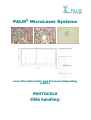
®
PALM®
MicroLaser Systems
L
Ca
atta
ap
pu
ullttiin
ng
g
La
asse
err M
Miiccrro
od
diisssse
eccttiio
on
na
an
nd
dP
Prre
essssu
urre
eC
((L
LM
MP
PC
C))
PROTOCOLS
-DNA handling-
PALM® MicroLaser Systems
Protocols
Preparation of Slides
Remarks to different objectives:
When working with low magnifying objectives like 5x, 10x or 20x, regular 1 mm thick glass
slides and 0.17 mm glass slides can be used. To keep this flexibility for higher magnifications
(40x or 63x) P.A.L.M. recommends using long distance objectives.
With those you have the possibility to adapt the working distance to the different glass slides
by moving the correction ring on the objective.
Due to the short working distance of the 100x magnifying objectives only 0.17 mm thin cover
glass slides can be used.
Samples on glass slides
With the PALM® MicroBeam almost every kind of biological material can be microdissected and
catapulted directly from glass slides. Even archival pathological sections can be used after
removing the cover slip and the mounting medium without any additional steps.
To facilitate easy catapulting additional adhesive substances or “Superfrost + charged slides”
should only be applied when necessary for the attachment of poorly adhering special material
(e.g. some brain sections or blood vessel rings).
Samples on PALM® MembraneSlides
(#1440-1000, 1 mm or #1440-1500, 0.17 mm)
MembraneSlides are special slides covered with a membrane on one side. This membrane is
easily cut together with the sample and acts as a stabilizing backbone during catapulting.
Therefore even large areas are catapulted by a single laser impulse without affecting the
morphological integrity. Use of membrane is especially important for isolated single cells,
chromosomes and also living cells or small organisms.
P.A.L.M. offers slides with PEN membrane. This polyethylene naphthalate membrane is
highly absorptive in the UV-A range, which facilitates laser cutting. The PEN-membrane can be
used for all applications. For special procedures (e.g., ablation of chromosome parts prior to
LMPC) a second membrane type (polyester, POL) is also available. (#1440-1550, POL, 0.17
mm)
• UV treatment
To overcome the hydrophobic nature of the membrane it is advisable to irradiate with UV light
at 254 nm for 30 minutes (e.g., in a cell culture hood). The membrane gets more hydrophilic,
therefore the sections (paraffin and cryosections) adhere better. Positive side effects are
sterilization and destruction of potentially contaminating nucleic acids.
• Poly-L-Lysine treatment
Additional coating with poly-L-Lysine (0.1 % w/v) only will be necessary for poorly adhering
materials and should be performed by distributing a drop of the solution on the membrane
(after UV treatment). Let it air dry at room temperature for 30 minutes. Avoid any leakage
underneath the membrane, as this might result in impairment of Laser Pressure Catapulting.
DNA handling - 0205
2/11
PALM® MicroLaser Systems
Protocols
Mounting frozen sections onto slides
Sections are mounted onto MembraneSlides the same way as routinely done using glass slides.
For cutting and catapulting a coverslip or standard mounting medium must not be applied.
Freeze supporting substances like OCT or similar may be used but should be kept to a
minimum.
Fixation
After mounting the sections there are many possibilities to fix the material. Prepare your
sections onto the slide as you do routinely. Let shortly air dry. Dehydrate in 70% ice-cold
ethanol for 1-5 minutes (acetone, methanol or other are possible as well depending on the
later applications). Subsequently let air dry for some minutes. The slides can now be used at
once (even if they are still somewhat wet), stained or deep-frozen at –80 °C.
Removing the freeze supporting substance
If OCT or another tissue freezing medium is used, it is important to remove that before Laser
Microdissection, because these media will interfere with laser efficiency. Removing of the
medium is easily done by gently washing the slide for about 1 minute in water. If the sections
will be stained, the supporting substance is normally removed “automatically” in the aqueous
staining solutions or the diluted ethanols.
Mounting paraffin embedded sections onto slides
Sections are mounted onto MembraneSlides the same way as routinely done using glass slides.
Floating the section on warm water or hot plate techniques can both be applied.
After mounting let dry overnight in a drying oven at 37 °C up to 56 °C. For cutting and
catapulting a coverslip or standard mounting medium must not be applied.
Deparaffination
Paraffin will reduce the efficiency of the laser, sometimes completely inhibiting cutting and
catapulting. If you are working with unstained sections it is therefore very important not to
forget removing of the paraffin before laser cutting and laser pressure catapulting. If applying
normal staining procedures deparaffination is routinely included in any protocols.
Minimal procedure:
Xylene
Ethanol 100 %
Ethanol 96 %
Ethanol 70 %
rinse with water
2
1
1
1
times for 2 minutes
minute
minute
minute
With the 1 mm PALM® MembraneSlides (#1440-1000) it is possible to extend the time of
Xylene treatment up to 2x15 minutes. Also acetone or isopropanol can be applied with those.
ATTENTION: The thin (0.17 mm) membrane slides are not so resistant against organic
solvents and should be handled according to the Minimal Procedure (see above).
DNA handling - 0205
3/11
PALM® MicroLaser Systems
Protocols
Cytospins
Cytospins can be prepared on glass slides or on MembraneSlides. After centrifugation with a
cytocentrifuge let the cells air-dry. Then fix for 5 minutes in 100 % methanol. Allow the
cytospins to dry at room temperature before staining.
Blood and tissue smear
Distribute a drop of (peripheral) blood or material of a swab smear over the slide. Be careful to
avoid injuries in the membrane, which would lead to leakage during fixation or washing and
therefore impair the laser pressure catapulting.
Let smears air-dry shortly and fix them for 2 up to 5 minutes in 70 % ethanol.
DNA handling - 0205
4/11
PALM® MicroLaser Systems
Protocols
Staining of the Sections
To our experience almost any standard histological staining (like H&E, Methyl Green, etc.) can
be used when you are interested in DNA.
Because of the easy and short protocol P.A.L.M. recommends also the Cresyl Violet staining for
DNA (see below), which was originally developed for RNA.
P.A.L.M.’s recommended staining procedure – Cresyl Violet
1.
2.
3.
4.
5.
dip slide for 20 seconds in 1% cresyl violet acetate solution (*).
remove excess stain on absorbent surface
dip into 70% EtOH
dip into 100% EtOH
air dry shortly (1-2 minutes)
Slides can be used immediately or stored at -80°C before LMPC.
To avoid excess condensation of moisture during thawing, the slides should be frozen and
reemerged in a tightly sealed container (e.g., 50ml Falcon-tube)
(*) Dissolve solid cresyl violet acetate (e.g., Aldrich cat #86,098-0, Fluka #61123 or
similar) at a concentration of 1% (w/v) in 100% EtOH at room temperature with
agitation/stirring for several hours to overnight. Filter the staining solution through a 0.2 m
filter unit before use.
Alternative staining procedures
• Hematoxylin/Eosin (H&E, HE)
HE-staining is used routinely in most histological laboratories and does not interfere with DNA
preparation. The nuclei are stained blue, the cytoplasm pink/red.
Procedure:
directly from distilled water
3 minutes Mayer’s Hematoxylin solution (e.g. SIGMA, #MHS-32)
3 minutes rinsing in tap water or blueing solution
30 seconds up to 3 minutes Eosin Y (e.g. SIGMA, #HT110-2-32)
quick increasing ethanol series
let air-dry at room temperature
• Methyl Green
The nuclei are stained dark green, the cytoplasm light green.
Procedure:
directly from distilled water
5 minutes Methyl Green solution (DAKO, #S1962)
rinse in distilled water
let air-dry at room temperature
DNA handling - 0205
5/11
PALM® MicroLaser Systems
Protocols
• Methylene Blue
The nuclei are stained dark blue.
Procedure:
directly from distilled water
5-10 minutes Methylene Blue solution (0.05 % in water; SIGMA, #31911-2)
rinse in distilled water
let air-dry at room temperature
• Toluidine Blue
The nuclei are stained dark blue. Cytoplasm lighter blue
Procedure:
directly from distilled water
30 seconds Toluidine Blue solution (0.1 % in water; SIGMA, #T-0394)
rinse in distilled water
quick increasing ethanol series
let air-dry at room temperature
• Nuclear Fast Red
The nuclei are stained dark red, the cytoplasm lighter red.
Procedure:
directly from distilled water
5 to 10 minutes Nuclear Fast Red solution (DAKO, #S1963)
rinse in distilled water
let air-dry at room temperature
DNA handling - 0205
6/11
PALM® MicroLaser Systems
Protocols
Laser Microdissection and Pressure Catapulting
(LMPC) Procedures
Please have a look also in the PALM® MicroBeam User Manual.
Tips for improvement of the morphology
For LMPC (Laser Microdissection and Pressure Catapulting) embedding and glass covering of
the specimen is inapplicable. Thus, the rough open surface of the section/material often results
in impaired view of morphology.
• PALM® Adhesive Caps opaque (#1440-0240 and -0250)
The white/opaque filling of PALM® AdhesiveCaps opaque can improve the visual appearance of
the tissue section by enhancing contrast and color similar to the characteristic view of routinely
embedded and glass-covered histological sections.
The opaque filling diffuses the incident microscope light, which smoothens the harshness of
contrast and, depending on material and staining, even minute details as e.g., nuclei and cell
boundaries show up. Even slight differences in color become visible. Two different
microfugetube sizes with these white filled caps are available from P.A.L.M. (200l: #14400240 or 500l: #1440-0250).
• PALM® LiquidCover Glass N (#1440-0600)
The polymeric and low viscose PALM® LiquidCover Glass N completely embeds the tissue and
smoothens the rough tissue surface, resulting in enhanced morphology. The coverslip-like
surface, which is formed, not only improves the optical characteristics of the specimen, but
also protects it against environmental influences (e.g. moisture and associated RNase activity).
For more details and handling please see the PALM® LiquidCover Glass N instruction manual.
• Ethanol
Go forward to search an interesting area on the section and pipette about 5 l of ethanol onto
this area. You may use 70 % or 100 % ethanol. When using absolute ethanol a little destaining
of the section may happen, but the drying is much quicker.
Observe the area on the screen. The depiction of the cells is improved immediately after
having contact with the ethanol. Now mark the cells or cell area of interest with the software
tool. Since ethanol is evaporating rapidly you can soon cut and catapult again.
Different collection devices
PALM® AdhesiveCaps (opaque or clear)
The intention of PALM® AdhesiveCaps is to allow LMPC (Laser Microdissection and Pressure
Catapulting) without applying any capturing liquid into the caps prior to LMPC.
For more details and handling please see also the PALM® AdhesiveCaps Application Manual.
DNA handling - 0205
7/11
PALM® MicroLaser Systems
Protocols
•
Collection procedures
The sticky filling substance occupies most of the volume in PALM® AdhesiveCaps. Hence, the
catapulting distance is decreased to a minimum enabling sample collection at low laser energy
and highest recovering success.
We suggest using a spare cap to avoid cross-contamination if selected ablation of unwanted
material is carried out prior to the relevant LMPC procedure.
Glass-mounted specimen
PALM® AdhesiveCaps are especially recommended for catapulting from glass-mounted
specimen (e.g., by AutoLPC).
LPC from glass-mounted tissue sections usually results in small tissue fragments, which will
tightly adhere to the adhesive filling until lysis. The catapulted fragments remain visible in the
cap until the extraction procedure is performed.
After lysis, digestion or denaturation occasionally structures may still be encountered within
the cap. These are cellular residues retained after complete extraction of the target molecules
and do not indicate incomplete recovery of DNA, RNA or protein.
Membrane-mounted specimen
PALM® AdhesiveCaps can also be used with membrane-mounted specimen. The membrane
serves as a backbone preserving the morphology of the selected tissue area during the LMPC
procedure, i.e. LMPC from membrane-mounted tissue sections usually result in morphologically
beautifully preserved tissue within the cap. Sometimes, the membrane-support remains within
the cap after the extraction procedure. However, in most cases the target compounds have
been entirely dissolved, resulting in a faint appearance of the catapulted tissue piece.
Sometimes it may also be helpful to dissect and catapult smaller sample pieces. The PALM®
AdhesiveCaps can also be used with capturing-liquid as done with regular LPC-Microfugetubes
(see below).
• Extraction procedure for AdhesiveCaps
Depending on the required buffer volume for the extraction step, we suggest the following
protocols after LMPC (please see also the PALM® AdhesiveCaps Application Manual):
Buffer volume less than 20 l
Please add the buffer directly into the cap onto the catapulted specimen. Perform lysis or
digestion in an up-side-down position prior to centrifugation. Centrifugation can be performed
with any standard benchtop centrifuge for 5 min up to 13400 rcf (e.g., Eppendorf 5415D:
12000 rpm)
Buffer volume up to 500 l
Please add the lysis buffer into the centrifuge tube, close the tube with the loaded cap and
process by intensive mixing (vortexing) in an up-side-down position of the tube. We suggest
adding a minimum of 100 l of buffer solution.
Capturing liquid
With capturing liquid applied inside the cap prior to LMPC, immediate centrifugation is possible.
For more detailed information please refer to the PALM® AdhesiveCaps Application Manual.
DNA handling - 0205
8/11
PALM® MicroLaser Systems
Protocols
LPC-Microfugetubes
• Catapulting into the cap
Please have a look also in the PALM® MicroBeam User Manual.
Pipette 3 to 15 l bidistilled water or buffer into the inner ring of the cap. By using 0.5% Igepal
CA-630 (SIGMA #I-3021) it is possible to smear out a small amount of liquid in the cap. Now
the whole cap is covered with liquid without the rebuilding of a droplet.
The catapulted cells or cell areas will stick onto the wet inner surface of the cap and will not
fall down after the catapulting procedure. For single cells or very small areas spinning down
from mineral oil (as recommended in the beginnings of LMPC) may be difficult and therefore
aqueous solutions should be preferred. Be aware that aqueous solutions will dry out after a
while.
When using membrane-mounted samples the dissected membrane acts as a backbone for the
selected area/cell and can therefore be catapulted with a single laser shot from a remaining
“bridge” at the border. The morphological integrity is completely preserved with this procedure
and can be viewed and documented nicely in the cap.
When using glass mounted samples it is advisory to put more liquid (up to 40 l) into the cap
since the smaller “flakes” produced by multiple LPC points cannot be catapulted so straight to
the centre of the cap as areas on membrane. A reduced distance of the capturing surface will
improve the efficiency of capturing the samples.
•
Extraction procedure for regular LPC-caps
After microdissection and catapulting - depending on the desired volume - digestion/lysis is
either performed “upside down” in the cap or lysis buffer is added to the tube and after closure
mixed by inversion. Then the lysate is spun down in a benchtop centrifuge (5 minutes, 13400
rcf; e.g., Eppendorf 5415D: 12000 rpm) and samples can then be stored for later use.
(For more details please see page 8 “Extraction procedure for AdhesiveCaps”)
•
Looking into the cap to see the catapulted samples
To control efficiency of catapulting it is possible to look into the collection device
(e.g., PALM® AdhesiveCaps or LPC-caps) with the 5x, 10x, LD20x, LD40x and LD63x
objectives. By using the software function “go to checkpoint” the slide is moved out of the light
path and the inside of the cap can be viewed with the respective objective. Normally most
catapulted areas/cells can be found within the small inner ring of the caps.
DNA handling - 0205
9/11
PALM® MicroLaser Systems
Protocols
Preparation of DNA from catapulted samples
For DNA-preparation from paraffin sections we usually use a Proteinase K containing Catapult
Buffer. This step is mostly not necessary for frozen sections. Then you can proceed directly to
normal DNA extraction procedures (see below).
Catapult Buffer: (20 mM Tris, 0.1 mM EDTA, 0.5% Igepal, 1% Proteinase K fresh
from Stock)
Formula to prepare 10 ml:
0.05 M EDTA pH 8.0
1 M Tris pH 8.0
Igepal CA-630 (SIGMA #I-3021)
(Proteinase K Stock: 20 mg/ml)
mol.biol. grade H2O
20 l
200 l
50 l
(100 l)
fill up to 10 ml
Proteinase K Solution 20 mg/ml
(Qiagen GmbH, Hilden, Germany; Catalog #19131)
Always prepare a fresh mixture of Catapult Buffer and Proteinase K!
•
Proteinase K digestion procedures
Digest for 2 - 18 hours at 55°C(*) followed by a heating step at 90°C for 10 min to inactivate
Proteinase K. At best use a thermal cycler with a heating lid for the standard digestion.
Recommendable for AutoLPC and small samples: Catapult directly into 20 l Catapult Buffer
containing Proteinase K. Digest upside down in the cap for 2 - 18 hours at 55°C(*) followed by
a heating step at 90°C for 10 min to inactivate Proteinase K.
If not proceeding immediately, store the samples in the refrigerator at 4 °C.
(*)
The time necessary for complete digestion depends on the kind and on the number of
catapulted cells.
•
DNA extraction
After the Proteinase K digest (or directly with frozen tissue without fixation) any suitable DNA
extraction procedure can be performed e.g., Qiagen QiaAmp DNA micro.kit (Qiagen, #56304),
CST Genomic DNA Purification kit Micro tissue (DRI Ltd., Kent, UK, www.chargeswitch.com,
#11203), CST Forensic DNA Purification kit (DRI Ltd., Kent, UK, #11200), Phenol/Chloroform
extraction etc.
Even use of the crude lysate directly in PCR may be possible with small samples
Do not forget to inactivate the proteinase K!
DNA handling - 0205
10/11
PALM® MicroLaser Systems
Protocols
Chromosome preparation
Protocols on request
Isolation of living cells
Protocols on request
Please note: There is also a review brochure available dealing with laser micromanipulation
of live cells (P.A.L.M. Scientific Edition No. 11, ISBN No. 3-9808893-0-0).
For questions, remarks or protocol requests please contact
P.A.L.M. Service & ApplicationLaboratory
Email: [email protected]
Service Line: +49 (0) 81 58 99 71-300
DNA handling - 0205
11/11