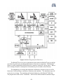
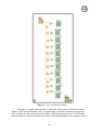
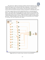
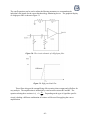
1
Final Design
Expert Anesthesiology
Monitoring System
Team 2:
Timothy Morin
Nathan White
Kane Killelea
Sponsored by:
Joseph H. McIsaac and John D. Enderle
March 23, 2007
Client Contact:
Dr. John D. Enderle
Joseph McIsaac
Editor-in-Chief, EMB Magazine
Biomedical Engineering Book Series Editor
for Morgan and Claypool Publishers
Program Director & Professor for Biomedical
Engineering
University of Connecticut
Bronwell Building, Room 217C
260 Glenbrook Road
Storrs, Connecticut 06269-2247
(860) 486-5521
[email protected]
Chief, Trauma Anesthesia
Hartford Hospital
Suite JB300,
Department of Anesthesiology
80 Seymour St.
Hartford, CT 06102
(860)-545-2117
[email protected]
TABLE OF CONTENTS
SECTION
PAGE NUMBER
Abstract
1 Introduction
1.1 Background (client and disability)
1.2 Purpose of the project
1.3 Previous work done by others
1.3.1 Products
1.3.2 Patent search results
1.4 Map for the rest of the report
2 Project Design
2.1 Design Alternatives
2.1.1 Design 1
2.1.1.1 Objective
2.1.1.2 LabVIEWTM
2.1.1.3 Block Diagram
2.1.1.4 Vital Signals
2.1.1.5 Patients Prior Information
2.1.2 Design 2
2.1.2.1 Objective
2.1.2.2 Circuitry and Filtering
2.1.2.3 LabVIEWTM Front Panel
2.1.2.4 Testing
2.1.2.4.1 LabVIEWTM Testing
2.1.2.4.2 Circuit Testing
2.1.2.5 Integration
2.1.3 Design 3
2.1.3.1 Objective
2.1.3.2 Subunits
2.2 Optimal Design
2.2.1 Objective
2.2.2 Subunits
2.2.2.1 LabVIEWTM Program
2.2.2.1.1 Front panel
2.2.2.2.2 Block Diagram
2.2.2.2 Connectors and their testing
2.2.2.3 GE Marquette and
ADI Blackfin® Processor
2.2.2.3.1 GE Marquette
2.2.2.3.1.1 Hardware Connections….
2.2.2.3.1.2 Acquiring Data
2.2.2.3.1.3 Data Communications
2.2.2.3.1.4 Data Parsing
-1-
5
5-9
6-7
7
7-9
7-8
8-9
9
9-74
10-21
10-13
10
10-11
12
12
13
13-17
13
13-15
15-16
16-18
16
17
18
18-21
18-19
19-21
22-74
23
24-74
24-29
24-26
26-29
29-35
35-42
35-37
35
36
36-37
37
2.2.2.3.2 Blackfin®
2.2.2.4 Patient’s Prior Information
2.2.2.5 Material Requirements
2.2.2.5.1 Case
2.2.2.5.2 Interface Screen
2.2.2.6 Key Pad
2.2.2.7 Cooling Fan
2.2.2.8 Power Source
2.2.2.9 Electrical Circuits
2.2.2.9.1 Low-Pass Filter
2.2.2.9.2 High-Pass Filter
2.2.2.9.3 Band-pass Filter
2.2.2.9.4 Leakage Current
2.2.2.10 Real Time Processing
2.2.2.11 Digital Signal Processing
2.2.2.11.1 Fixed Point vs. Floating Point systems
2.2.2.11.2 Programming languages
2.2.2.12 Testing
2.2.2.12.1 LabVIEWTM Testing
2.2.2.12.2 Circuit Testing
2.2.2.12.3 Total Testing
2.2.2.13 Integration
2.2.2.14 Safety
3 Realistic Constraints
4 Safety Issues
5 Impact of Engineering Solutions
6 Life-Long Learning
7 Budget and Timeline
7.1 Budget
7.2 Timeline
8 Team Members Contributions to the project
9 Conclusion
10 References
11 Acknowledgements
12 Appendix
12.1 Updated Specifications
12.2 Purchase Requisitions and FAX quotes
-2-
38-42
42-47
48-54
48-51
51-54
54-56
56-57
57-60
60-64
60-61
61-62
63
63-64
64-65
65-68
65-66
67-68
68-70
68
68-69
69-70
71
71
71-74
74-75
75-76
77
78-82
78-79
80-82
82-83
83-84
85-86
86
87-88
87
88
Figures:
Figure
PAGE NUMBER
Figure 1: Example front panel of the anesthesia monitoring system.
Figure 2: The circuit schematic and Bode Plot of a low-pass filter.
Figure 3: The circuit schematic and Bode Plot of a high-pass filter.
Figure 4: Example front panel of the anesthesia monitoring system.
Figure 5: function generator and oscilloscope setup
Figure 6: Optimal Design flow chart.
Figure 7: Example front panel of the anesthesia monitoring system
Figure 8: BISTM Vista monitor
Figure 9: Example of a well designed block diagram
Figure 10: Female port for BISTM Monitoring system.
Figure11: 9-pin D connector.
Figure 12: 25-pin D connector.
Figure 13: Physical characteristics of the port.
Figure 14: Code to alarm
Figure 15: Code for alarm heightening.
Figure 16: Code for Noise.
Figure 17: Code for error messages.
Figure 18: Code to find version number and updates of software.
Figure 19: Code to activate recording of events.
Figure 20: Block Diagram explaining outline of the parameter update packet
Figure 21: Code for a data array.
Figure 22: Architecture Core
Figure 23: Blackfin® insignia.
Figure 24: Block Diagram of the Dual-Core
Figure 25: EZ-KIT lite from Analog Devices, Inc.
Figure 26: Blackfin® EZ-extender
Figure 27: Age calculator for dosage.
Figure 28: Weight calculations using LabVIEWTM
Figure 29: Sex calculation using LabVIEWTM
Figure 30: Physical condition dosage calculation using LabVIEWTM
Figure 31: Example case for the monitor.
Figure 32: Front of the case
Figure 33: Rear of the case
Figure 34: 10.4” LCD monitor
Figure 35: Block diagram of LCD display
Figure 36: Specifications for the LCD screen
Figure 37: Possible button layout
Figure 38: Circuit symbol and example of a push-to-make switch
Figure 39: Surface mount tactile switch
Figure 40: Switch dimensions
Figure 41: Cooling fan
Figure 42: Double-Fused Three Function Power Entry Module
Figure 43: Schematic of Power Supply
-3-
11
14
14
16
17
22
25
26
28
30
30
31
31
32
32
32
33
33
33
36
37
38
39
40
41
42
44
45
45
46
49
50
51
52
53
54
55
55
56
56
57
58
58
Figure 44: Specifications for Power Supply
Figure 45: Power Cord
Figure 46: Power Cord Schematic
Figure 47: Power Cord Specification
Figure 48: The circuit schematic of a Low-pass filter
Figure 49: Low-pass Bode Plot
Figure 50: The circuit schematic of a High-pass filter
Figure 51: High-pass Bode Plot
Figure 52: The circuit schematic of a Band-pass filter
Figure 53: Circular buffer operation. This shows an example of how a circular
buffer will look at one instant (a) and the following instant (b).
Figure 54: Floating and Fixed point trade offs
Figure 55: Programming trade-offs.
Figure 56: Example Loop code
Figure 57: function generator and oscilloscope setup
Figure 58: Biopac electrodes
Figure 59: Biopac clip leads
Table
59
59
60
60
61
61
62
62
63
65
66
67
67
69
70
70
PAGE NUMBER
Table 1: EEG and ECG filtering parameters.
Table 2: Important Code.
Table 3: processed EEG Data From EEG snippet code.
Table 4: Header information for EEG snippet code.
Table 5: processed variables and spectra sent to the host once every second code.
Table 6: Raw EEG data code.
Table 7: contains BIS history data records code.
Table 8: code to indicate an event was marked.
Table 9: EEG and ECG filtering parameters
-4-
15
33
34
34
34
34
35
35
61
Abstract:
This anesthesia monitoring system is a well designed device that will read, interpret and
display values by retrieving inputs from the aspect medical BISTM EEG monitor and the GEMarquette anesthesia monitor. This will be done by using a transducer to pass the data from the
two monitors to a Blackfin® chip, which will be programmed with a LabVIEWTM. These
transducers will have to be able to attach to both the monitors and then alternatively have to
connect to the Blackfin® chip within the monitor. This information will then be processed and
exported to a LCD screen that will display the level of consciousness of the patient, using their
prior information, and the two monitors. Then this information will be used to determine a
dosage of anesthesia that should be applied to the patient. This project is necessary to create a
program that can adequately interpret the level of consciousness of the patient under anesthesia,
time stamp the data, and then give the best possible dosage to the anesthesiologist to maintain the
proper level amnesia, analgesia and immobility. This device is different from other past devices
because it will allow the use of two of the top monitoring systems which will then be analyzed
and used cooperatively to give the anesthesiologist a number to use for the administration of
anesthesia and the value can then be used to support the anesthesiologist if something goes
wrong. All the information will be time stamped and rectified by the program, giving a hard
copy of the surgery numerically for scrutiny.
This device is going to have to be using the Blackfin® chip to run the LabVIEWTM
program and process the information without a computer. This is possible and will be run using
only the GE-Marquette anesthesia monitor and the aspect medical BISTM EEG monitor for data
input. The information will then be used to calculate the dosage that should be applied and
displayed on the LCD screen which will be attached directly to the Blackfin® chip using the
display outputs.
1
Introduction:
The following proposal is an in depth explanation of how a specialized unit for
determining the consciousness level of a patient to help the anesthesiologist maintain vigilance
can be designed. Chief of Trauma Anesthesiology at Hartford hospital, Dr. Joseph McIsaac, is
the main client and his needs will be considered for all aspects of this design. The principles of
engineering will be followed in determining the best cost efficient design encompassing all of the
needs of the client. An informative description of the project’s goals are outlined which includes
the device requirements and its overall abilities. Possible programs and components will be
highlighted for use as well.
This device is going to use measurements of 3 signals to determine the level of
consciousness of a patient under anesthesia and find if more anesthetic needs to be applied.
Mainly the monitor will focus on the data that is received from the GE-Marquette anesthesia
monitor, the aspect medical BISTM EEG monitor, and will have to include time stamping.
During the process of anesthesia the patient will have data that will be incorporated into the
LabVIEWTM program, analyzed and the data in graphical form and then recommend an applied
dosage of anesthesia.
-5-
The development of new software and hardware has given anesthesiologists hope for
additional improvements on the current anesthesiology monitoring systems. LabVIEWTM is the
new data acquisition software that will be used in this project to create measurements and a
calculated level of consciousness on a clear front panel. This device should be reliable and
easily used throughout a surgical endeavor. The settings should be easily managed allowing any
level of experience technician to update them. The front panel will display clear graphs with the
corresponding numerical values and appropriate labels. The device should be able to withstand a
great deal of time in the surgical rooms and be durable enough to withstand everyday use. The
device should have a clean and purposeful appearance in the fact that there should not be any
wasted space as the device will be in close proximity of patients while maintaining accessibility
without compromising the surgery.
The device will then be in such a way which minimizes noise through using analog
filtering and amplifications built and after the signal manipulations the data will be sent to the
LabVIEWTM program. In the final product the program will be transferred to a Blackfin® chip,
which will allow the surgeon to control the functions of the LabVIEWTM program without a
personal computer which optimizes the device’s functionality in the surgical environment. The
components necessary for this device consist of a screen, mother board, microprocessor, and a
box to contain the internals. Figure 1 shows a flow chart of the operations of the anesthesia
monitoring system.
This design is slightly different then the last because the imported signals will be data
received from the GE-MarquetteTM anesthesia monitor and the Aspect Medical BISTM EEG
monitor and displaying it on a LabVIEWTM program that will be able to manipulate the received
data, time stamp it, and correlate it to a dosage of anesthesia for the patient.
1.1 Background (Client and disability):
Doctor Joseph McIsaac suggested that he was in need of a device that could import
information from the GE-MarquetteTM anesthesia monitor and the Aspect Medical BISTM EEG
monitor’s, time stamp them and then use that information to recommend a dosage of anesthesia
to maintain a proper level of consciousness for the patient. Our client is interested in the
education of students and the advancement of his ability to become a better anesthesiologist.
This project fills is a chance for the client to support students in learning and simultaneously
create a safer environment in the operating room for patient and surgeon alike.
As an anesthesiologist is judging the level of consciousness of each individual patient
they are using differential equations and precise calculations of fuzzy logic as described by
Joseph McIsaac. He explained the procedure as an educated guess and check that has to be
monitored and then reconsidered through the full time that the patient is on anesthesiology. The
anesthesiologist must also consider each individual patient based on their prior resistance, acute
and chronic disease states, age, weight, gender, exercise tolerance, medication usage, habits such
as smoking, drug and alcohol use. The doses that are applied take time to affect the patient so as
more medicine is added to the patient one must wait till they are sure the “poison” has taken its
effects on the consciousness of the patient.
-6-
Doctor McIsaac suggested that the BIS monitoring system by GE-MarquetteTM anesthesia
monitor should be considered as the basis for our project. He supplied the project with a manual
and some background information that may help direct the project more efficiently.
1.2 Purpose of the project:
This project’s main objective is to develop a device that will act as an “Automatic
Anesthesia Expert System”. With this device the anesthesiologist should be able to see all of the
patient’s relevant information, vital signs and their level of consciousness. The main function is
to help the anesthesiologist perform with the most information possible about the patient’s level
of consciousness through examination of the information provided by the GE-MarquetteTM
anesthesia monitor and the Aspect Medical BISTM EEG monitor. This information will include
the patient’s prior information and give a diagnostic evaluation of consciousness and the proper
quantity of anesthesia that the patient should receive to be properly sedated. This will all be time
stamped to allow for reconstruction of the events in the surgical room during the surgery.
Since new software and hardware has been designed recently it is expected that these new
influential materials will help to advance the anesthesiology monitoring systems. LabVIEWTM is
the new software that will be used in this project to calculate a level of consciousness on a clear
front panel, using the GE-MarquetteTM anesthesia monitor and the Aspect Medical BISTM EEG
monitor. This device should be reliable and easily used throughout a surgical endeavor. The
settings should be easily changed and the front panel should display clear graphs and needed
numerical data with clear labels. The device should be able to withstand a great deal of time in
the surgical rooms and be durable enough to withstand every day use. The device should have a
clean and purposeful appearance in the fact that there should not be any wasted space, as the
device will have to be close to the patient and easily accessed without being in the way of the
surgeons.
1.3 Previous Work Done by Others:
1.3.1 Products
Considering that there are previously designed anesthesiology monitoring systems on the
medical market already, there must be a reason to use this new design. The client has pointed
out that the BIS monitoring system is available and is the model that is being used as an
example. The SNAPP II is another anesthesiology monitoring system that is also on the market
that has been researched.
The main model that will be examined and used as a guide is the Aspect Medical Systems
BIS VISTA or GE anesthesia machine the BISTM
monitor. The BISTM monitor relies on an Electroencephalogram (EEG) to determine the level of
consciousness of a patient and then displays a number between 1 and 100 that corresponds to
their consciousness. The BISTM monitor also highlights and acceptable region to which the
patient is within the correct level of consciousness for the surgery to continue without amnesia,
analgesia, and immobility becoming a factor. There is also an alarm that will sound if the patient
leaves the range at which would be considered approaching brain damage or a wakening state.
TM
-7-
Another model that is available on the market is Everest Biomedical Instruments
anesthesia monitoring system called the SNAP II. This devise is similar to the BISTM
monitor in that it mainly focuses on the level of consciousness of the patient relevant to their
electroencephalogram wave and supplies a level of consciousness numerical value between 1 and
100. The SNAP also has alarm limit bars that will show exactly weather the patient is within
acceptable range of consciousness or not. There is also an elapsed time clock that shows exactly
how long the patient has been under anesthesia. There is also a battery symbol which shows
exactly how much longer the devise will work before it dies.
1.3.2 Patent Search Results
Thoroughly searching for United States patents is an essential part of beginning a project
design. If there are any similar designs that may be infringed upon the design team must know
about them and make sure to either find their own method or honor the patent. Intellectual
property rights are formed by a patent which gives exclusive rights by the state to a patentee for a
certain period of time in exchange for the right to regulate public disclosure of certain details of a
devise or method or composition of matter, which is new, inventive, and useful for industrial
applications.
The first patent was the Vital signs monitoring system patent number 4,705,048 in
November 10, 1987. This patent includes the first sensor unit including a microphone for
mounting on the patient’s chest for picking up breath and heart sounds with a filter and automatic
gain control circuit. There was also a second sensor unit which positioned a microphone beneath
the blood pressure cuff for picking up the blood flow sounds to determine blood pressure. There
were also earphones for monitoring the selected sounds by the physician on hand.
The next patent is a continuation of the first one with a number of 5,010,890 in April 30,
1991. This one has one altercation that allows for continuation of the patient. The patent now
includes a switch selector which allows the physician to move freely throughout the operating
room while still maintaining the portable receiver that allows the physician to hear the
monitoring system.
The next patent consists of an anesthesia machine with a head worn display which
includes the gas delivery system control and a patient monitor system in full cooperation. This
patent is the anesthesia machine with head worn display which was passed on July 9, 2002. The
measured values of the sensor can be displayed on the head worn display devise which has
stereoscopic capabilities. The monitor includes communication ports for selectively monitoring
the sensors of a similar anesthesia machine which can be remotely positioned and the other port
can be saved for downloading patients medical records from a hospital medical record computer.
This entire system is wireless and can facilitate the anesthesiologists movement throughout the
operating room.
The forth patent that was found is closer to the design that will be used to suit the needs
of this monitoring system. This monitor patent is an EEG operative and post-operative patient
monitoring method on May 23, 2000 and was designed mainly to focus on the
-8-
electrocephalograph (EEG) of the patient involved in the surgery. This patient suggests that by
modeling the brain waves, both ongoing and evoked by stimuli, are amplified, digitized and
recorded. The brain waves of the patient prior to the surgery are used and compared to the brain
waves during the procedure to maintain vigilance over the patient’s consciousness. This method
focuses mainly on the relative power in the theta band which indicates blood flow, and
prolongations of the latency periods under brain stem stimuli as indicated by the patient’s ability
to feel pain.
The last patent that was found to be relevant was the EEG operative and post-operative
patient monitoring system and method passed on December 23, 1997. This method focused on
the electrocephalograph (EEG) system monitors patients during and after medical operations to
make sure that sufficient anesthetics are being used to attain the desired plan of anesthesia. This
devise functions by examining the brain waves of a patient and determining if more or less
anesthesia is required.
1.4 Map for the rest of the report
For the duration of this final report the design, budget and other engineering
considerations will be considered and discussed. Directly following this will be the alternative
designs and the optimal design of the expert anesthesiology system. They are however different
from the final design and the optimal design due to conflicts between the client, advisor’s, and
engineers involved in the development of this device. These changes will be discussed as well
and the complete final design will be discussed thoroughly. By examining the alternative
designs, it becomes evident the direction at which the project went and where things had to be
reconsidered. Following these designs will be the realistic constraints, safety issues, and impact
of engineering solutions. While the device was being designed the realistic constraints, safety
issues and impact of engineering solutions must be considered and well addressed. By
considering the impact of engineering solutions, the engineers must consider the possible effects
of this device on different areas in society, such as environment, economy and on the rest of the
world. After this life-long learning is discussed and was developed through out this designing
process. Through out the development of this device, there have been many lessons learned and
many hurdles missed. The end of the report contains a timeline for construction of the device,
the budget, and the team member’s contributions to the report and the design of the device. Then
lastly the acknowledgments, references, and the appendix is attached which displays the final
specifications.
2 Project Design:
This project has been a process of research and revision that has been changed week by
week to obtain the best possible device to meet the needs of the client and advisor. This section
displays the different designs that were considered and researched to create the expert
anesthesiology monitor. Throughout the alternative designs many changes were made, reasons
for the changes and a full explanation of the changes are discussed. The optimal design was
chosen to be the most effective to meet the application purposes because it is the only one that
truly meets the total requirements of the device which has been changed and re-evaluated in
-9-
recent weeks. This design seems to be capable of performing all the functions necessary to meet
the requirements of this projects final purpose.
2.1 Design Alternatives:
2.1.1 Design 1:
2.1.1.1 Objective:
The first design for the expert anesthesia monitoring system focused on obtaining vital
signals from a patient under anesthesia. The main focus of the design was to make a clear
display panel that would give the anesthesiologist an easy view of all the signals at hand. This
device was designed to use measurements of a few vital signs to determine the level of
consciousness of a patient under anesthesia. Mainly the monitor will focus on the patient’s
physical condition, medical conditions and prior exposures. During the process of anesthesia the
patients ECG, volumetric capnography, blood pressure and their EEG signal would have been
taken and used to develop a level of consciousness of a patient.
The device would then be built to specifications that would support the LabVIEWTM
program. The program would have been transferred to a data chip, which will allow the chip to
control the functions of the LabVIEWTM program without a personal computer. The components
necessary for this device consist of a screen, mother board, microprocessor, and a box to contain
the internals.
2.1.1.2 LabVIEWTM
The front panel for this design has a few preliminary aspects that need to be addressed.
The main focus of the front panel is to have a clear graphical and numerical display of the most
important data. The graphs will display as close to real time as possible the patients ECG,
volumetric capnography, blood pressure and their EEG signal. Each graph will have separate
alert levels that will set off the visual and audio alarms to warn the anesthesiologist that the
patient is becoming conscious or beginning to fall into an unrecoverable state. These graphs will
be colorful in a way that will allow for color segregation between graphs. The numerical values
that will be displayed to show the level of consciousness of the patients will be large and clearly
displayed next to their corresponding graph. Figure 1 shows a rough estimate of the visual
appeal of the front panel.
- 10 -
Figure 1: Example front panel of the anesthesia monitoring system.
The patient’s information must be easily manipulated on the front panel by the
anesthesiologist. These factors consist of the patient preliminary statistics based on their prior
resistance, acute and chronic disease states, age, weight, gender, exercise tolerance, medication
usage, and habits such as smoking, drug and alcohol use. These will all have to be configured
neatly and clearly on the front panel so that the anesthesiologist can alter these during the
procedure if necessary. There is a chance that a patient may lie to the doctors and the
information given maybe wrong for one reason or another. This is why it is necessary to make
the preliminary statistics easily manipulated, and ensure the program is not completely
dependent on these factors.
The main model for the front panel is being compared to the Aspect Medical Systems
BISTM VISTA or GE anesthesia machine the BIS monitor as displayed in figure 3. The BISTM
monitor relies on a clear graph of an Electroencephalogram (EEG) to determine the level of
consciousness of a patient at a moments notice by the anesthesiologist and then displays a
number between 1 and 100 that corresponds to their consciousness. The BISTM monitor also
highlights an acceptable region to which the patient is within the correct level of consciousness
for the surgery to continue without amnesia, analgesia, and immobility becoming a factor. There
is also an alarm that will sound if the patient exceeds the previously determined range to alert the
anesthesiologist of the patient approaching brain damage or a wakening state.
- 11 -
2.1.1.3 Block Diagram
The block diagram for the program will be aimed to meet the code followed by national
instruments programmers, which consists of common rules to pursuing a proper program. The
program should be clear and easily manipulated. The titles of all the different parts of the
program are necessary when working with the program and problem shooting. The DAQ must
be used in a manner that allows the program to take different inputs and apply them all to one
block diagram.
The block diagram is going to have a case structure that will allow for the calculations of
each of the vital signs and then the calculation of the level of consciousness produced from them.
Each vital sign will have its own portion of the block diagram. The case structures will allow the
anesthesiologist to use individual portions of the program and disconnect others if the surgery is
interfered with by the sensors.
The block diagram will include global variables that will be placed outside the main
loops. This will allow for the numbers to be manipulated at any point in the device’s process.
Each of these separate loops can be recalled and used in multiple places in the program making
the information easy to work with.
An example of a block diagram that fits the requirements that we are looking to pursue is
shown as figure four. This diagram is clear, well labeled, and organized in an easily followed
manner. This is extremely important in creating a good environment for fixing and accessing
problems that may arise during testing or even during later uses. There are flaws that tend to
escape the grasp of the testing atmosphere and are only discovered when the device is used in the
actually application it was designed for. These errors will have to then be examined and
corrected after the device has been cleared for use. This might mean someone unfamiliar with
the design may have to navigate the block diagram, find the error, and reprogram the section. If
the block diagram is impossible to follow this job becomes immensely more difficult.
2.1.1.4 Vital Signals:
This design focused on four man vital signals to determine the level of consciousness of
the patient. The first signal was the ECG which would help to determine the level of
consciousness by determining how fast the heart was beating, which in turn would give a
parameter from which a comparison could be made from one state to the next. When the heart
rate drops the patient is in a more relaxed state and this can be used to judge the level of
consciousness. The next signal that was used was the EEG or electroencephalograph. The delta
and theta waves are more prevalent when the patient is unconscious and the alpha wave is more
prevalent when the patient is awake and fully functional. Then the blood pressure of the patient
could be used to determine consciousness as well. The blood pressure automatically drops when
the patient is unconscious and the degree of change can be used to determine the level of
consciousness of the patient. The last vital sign was volumetric capnography, which would be
used to determine the amount of respiratory carbon dioxide leaving the patient. This in turn will
help to find how much oxygen the person is using and exactly how conscious they are.
- 12 -
2.1.1.5 Patients Prior Information:
While considering prior resistance, acute and chronic disease states, age, weight, gender,
exercise tolerance, medication usage, and habits such as smoking, drug and alcohol use a patient
has had prior to the anesthesia the average range for vital signs will be much more readily
available. These estimates will increase or decrease the alarm levels of the patient by
determining the amount that the person’s vital signs will be affected by these prior experiences.
For example if the patient is between a certain age range the upper limit will be lowered because
the anesthesia drugs will have a stronger effect on them. Some of the personal information used
will have a stronger effect then others, such as weight and prior drug use. The main problem
with using information given by the patient is that the patient may lie about some information
due to a fear of punishment. If a patient has prior experiences with pain killers they may be more
likely to need more drugs to keep them at the correct consciousness level but they may not tell
the anesthesiologist for fear of persecution. When the anesthesiologist realizes that the drugs are
not causing the amount of anesthesia that they are supposed to, it maybe appropriate to change
the prior drug usage to a higher level to account for this resistance during the procedure.
2.1.2 Design 2
2.1.2.1 Objective
The primary goal for this design was to improve and expand on the basic outline created
in design 1. Like design one, two still included the four vital signals and their corresponding
transducers for EEG, ECG, volumetric capnography, and blood pressure. The design, like
before, takes the signals obtained from the four transducers and determines the level of
consciousness of the patient who’s under the anesthesia through the use of the data acquisition
software LabVIEWTM. The improvements and expansions in this designed were in filtering, the
presentation of the data, testing, integration of the system into the final product.
The filtering has been specified to be done completely in the analog domain. This
method of using all hardware for filtering and amplifying is commonly used for applications that
require a high degree of accuracy. The front panel in the first design was changed from
displaying all of the signals to a simpler design. This design explained the method for testing the
program and device for design two. The section provides guidelines for testing the program and
the circuitry individually and then provides a way to test the device after all of the components
have been integrated into one unit. Lastly, integration methodology was investigated, which in
design one was neglected.
2.1.2.2 Circuitry and Filtering
Analog filtering and amplification is more effective than digital filtering and
amplification because in the analog domain can eliminate noise and amplify the signals with
more accuracy. The proposed circuitry that will be used in design two are low-pass and highpass filters. The purpose of the low-pass filter is to effectively attenuate all of the undesirable
high frequency noises. Figure 14 is an example of a second order low-pass filter. The cutoff
- 13 -
1
. The distinguishing features that make
2πR1C1
it second order filter is the second set of resistor/capacitor combination connected to the positive
terminal. The low-pass filter is shown in figure 2 through the graphical interpretation of using
the bode plot method.
frequency is determined with this equation: f OL =
Figure 2: The circuit schematic and Bode Plot of a low-pass filter.
The high-pass filter is created with a similar setup to the low-pass filter. However, the
C1, C2 and R1, R2 positions are switched. Opposed to the low-pass filter this setup attenuates
low frequencies reducing any noise with a frequency below the cutoff frequency to be
eliminated. Figure 16 displays the High-pass filter. The cutoff frequency is determined in the
1
.
same way as the low-pass. The equation relating the filter with the circuit is f OH =
2πR1C1
The cutoff equations can be used to adjust the filtering parameters to accommodate the
individual vital signals for the expert anesthesiology monitoring device. The graphical display of
a high-pass filter is shown in figure 3.
Figure 3: The circuit schematic and Bode Plot of a high-pass filter.
These filters also provide an amplifying effect creating clearer output and will allow for
easy analysis. The amplification is influenced by ratio between resistors RG and RF. The
- 14 -
RF
. Depending on the type of signal the specific
RG
circuit is dealing a different combination of resistors will be used for applying the correct
amplification. Placing these two filters in series provides filtering effects which are called band
pass and band stop filter depending on the order which they are placed. A band pass filter is a
high and low pass filter, relatively; the band stop filter is in the opposite order which is used for
eliminating certain frequencies within the valuable data. The frequencies for which these filters
have to be adapted to are displayed in Table 1.
equation relating these resistors is Av = 1 +
EEG waves
Alpha
Beta
Detla
Theta
EEG Signal
f OH
0.0833Hz
0.0333Hz
0.143Hz
ECG Signal
f OH
0.05Hz
f OL
0.125Hz
0.077Hz
0.25Hz
f OL
40/100/150Hz
Table 1: EEG and ECG filtering parameters.
2.1.2.3 LabVIEWTM Front Panel
The front panel like previous designs still displays clear graphical and numerical values,
which represent information that the anesthesiologist must be aware of. However this design
was developed in a way that would make the front panel simpler by making the four vital signs
selectable through the use of a drop down menu. The reasoning behind this idea is, currently
anesthesiologists primarily use the EEG to determine the patient’s level of consciousness with a
high degree of accuracy. Therefore additional vital signals would aid in pin pointing the reason
for the patient’s fluctuating level of consciousness but are not necessary in determining their
level. The default vital signal would be set to the EEG because this is the anesthesiologist
primary signal. Below the selectable graph would be the overall level of consciousness display,
like in design one, would incorporate all of the transducer signals to produce the values. Figure
4 is the example front panel for design two.
- 15 -
Figure 4: Example front panel of the anesthesia monitoring system.
2.1.2.4 Testing
2.1.2.4.1 LabVIEWTM Testing
The testing of LabVIEWTM will involve a break down of the program into smaller
portions to make sure that all the parts work independently before combining them into one
program. The program will mostly be broken down into each individual component. After each
of the signals are working independently and the errors or deciphered and fixed, the program will
be consolidated into and will be run through one Data Acquisition or DAQ assistant. This is
where most of the problems in the program should be experienced. When combining multiple
working signals into one, there is an error rate that has to be accounted for. The chance that
multiple programs can be combined without interference is implausible.
The testing without the circuits attached can be done with programmer installed data.
Instead of using the circuits to input the data from the patient, signals can be simulated and
applied to the program to make sure that the program can process and display the data correctly
without error. At this point the filters can be tweaked and the noise will be separated from the
signal and can be filtered out. This will allow for the most precise measurements. Knowing the
signal that should be outputted will allow for a proper diagnosis of noise and error in the
outputted signal and graph.
- 16 -
2.1.2.4.2 Circuit Testing
Testing the electrical hardware will be done in two ways. First using PSpice the circuit
build and simulated with the calculated resistive and capacitive components. Once the board has
gone through extensive virtual trouble shooting it will be translated into electronic hardware.
This hardware will be required to undergo additional tests to check whether or not there were
errors incurred during fabrication and soldering of the components.
Each of the signals will require specialized components to meet their amplification and
filtering needs. First the circuitry will be verified by testing the amplification and filtering.
Given that each signal requires a unique set of filtering and amplifying parameters the circuitry
components will be customized set their requirements.
For example the EEG amplitudes and frequency components vary from 10-100 μV and
from 0.5-30Hz respectively and within these general parameters there are sub parameters
correlating to the different periodicity’s which compose the EEG signal. Each of these
periodicities is characterized by a more specific frequency and amplitude as well as the
conditions which they are most apparent. The filtering of this design will be completed solely in
analog domain.
The approach to verify the functionality of the circuit will be to connect a function
generator to the input of the circuit board and to channel 1 of an oscilloscope and connect
channel two of the oscilloscope to the output. This will provide an easy way to compare the
inputted signal to the outputted signal. The input signal will be attenuated by the same scale that
that output signal is amplified by which will reproduce the un attenuated signal. To manually
test the filtering of the circuit board the frequency of the created signal will be move around each
cutoff frequency. As the frequency exceeds a cutoff frequency the signals should be attenuated
to about zero. Figure 5 displays a general setup for a function generator and oscilloscope.
Figure 5: function generator and oscilloscope setup
- 17 -
2.1.2.5 Integration
The total monitor will have to integrate the LabVIEWTM program to the circuits and to
the sensors. The sensors should input the data from the patient or test subject to circuit, where it
will be amplified and filtered and then it will be sent to the LabVIEWTM program to clearly
display the patient’s current status through the use of graphs and numeric values. The circuit
board will include all of the filtering and amplification. The advantages to using all electrical
hardware for the signal manipulations once the circuit is build it is more robust and analog
filtering and amplification provides better results. However the downfall to manipulating in the
analog domain is that that all the filtering and amplification parameters are fixed, which
eliminates any possibility of adjusting the parameters to optimize the displayed signal. The data
is then converted to the digital domain where the signals are compiled into one DAQ assistant
allowing the program to have one collaborative final output and will be able to analyze each
signal simultaneously. The signal transducers will be implemented into circuit board where the
data will be manipulated to reduce the noise. After the acquisition stage the input will then be
ready for the LabVIEWTM program that is installed onto a Blackfin® chip. Then the display
screen will have to be attached to the circuit board to allow for a clear presentation of the signals.
The LabVIEWTM program will have to be either applied to a PIC chip or a Blackfin® chip to
control the function of the monitoring system. The Blackfin® chip will allow for a more
complex display, which will allow for more alterations on the site. The PIC chip will be more
difficult to use due to its less capable abilities.
2.1.3 Design 3
2.1.3.1 Objective
The objective of this project is to create a monitor that will accurately calculate and
display the consciousness level of the patient under anesthesia. Chief of Trauma Anesthesiology
at Hartford hospital, Dr. Joseph McIsaac, is the main client and his needs will be considered for
all aspects of this design.
This device is going to use measurements of four vital signs to determine the level of
consciousness of a patient under anesthesia. Mainly the monitor will focus on the patient’s
physical condition, medical conditions and prior exposures. During the process of anesthesia the
patient’s ECG, volumetric capnography/pulse oximeter, blood pressure and their EEG signal will
be recorded and analyzed. These four signals will be combined to create an accurate depiction of
the patient’s level of consciousness.
The subunits of the device will be considered in a high level of detail as with the original
two designs. There are a variety of different components that will go into this device, and all
should be carefully selected and manufactured. Perhaps the most important aspect of this device
is the LabVIEWTM program that interprets the four different signals. Other subunits are
important as well such as the case design and the individual transducers that will be used to test
the subject’s vital signs.
- 18 -
Instead of volumetric capnography a pulse oximeter will be used yielding essentially the
same results. The LabVIEWTM block diagram has been reverted back to the original design.
The design of the case has been refined to include materials choice, dimensions, and other
features. In general the methods of testing and the transducers being used have been elaborated
on. In the second alternative design, we discussed leakage current, and in this design we tell how
to test for it and fix it if a leakage current is detected.
2.1.3.2 Subunits
The third version of the design also uses LabVIEWTM as the primary program for
integration of the different physiological signals obtained from the patient. In this version of the
design, the LabVIEWTM program incorporates the filtering of the transducer signals into the
program itself as in the second design. In our first design, the filters were constructed as
physical circuits outside of the LabVIEWTM program. Although using external filters would
create less noise then those implemented in LabVIEWTM, the LabVIEWTM filters are easily
created and used, and can be toggled in order to keep the noise to a minimum.
In this version of the front panel, the previous drop down menu has been eliminated and
replaced by individual graphs for each of the different signals as in the first alternative design.
The drop down menu presented too much of a potential for complications and confusion in
interpreting the graphs. If the anesthesiologist wanted to view one of the signals during surgery,
he or she would have to click the drop down menu, and select the appropriate graph. In place of
the drop-down menu we have included four separate graphs for the four different physiological
signals, and one graph for the overall level of consciousness. The latter of the graphs will be
made more prominent seeing as that is the most important of the five graphs. The individual
graphs are straightforward and can be viewed easily and all at the same time on the screen. This
is useful if the anesthesiologist would like to view a specific signal while still monitoring all the
others including the total level of consciousness.
There are some changes made to the transducers between the previous design and this
one. The main change is that the volumetric capnography signal will no longer be used in our
design. In its place we have included a pulse oximeter, which essentially completes the same
task. In volumetric capnography, the level of oxygen respired is used to infer the level of oxygen
in the blood, and doubles as a method of measuring the volume of air respired and breathing rate.
However, this method is very invasive and eliminates the Pulse oximetry used two different
wavelengths of light from light emitting diodes to indirectly measure the level of oxygen in the
bloodstream. This method of measuring blood oxygen saturation is non-invasive, and
inexpensive. The breathing rate and volume of respired air is no longer measured, but the ease
of measurement of the blood oxygen level outweighs the loss of the volumetric capnography
signal.
The specific operations of the individual transducers and the testing of each respective
transducer have also been refined in this updated version of the design. Biopac sensors will be
used to test the device and ensure that each of the different signals is operating correctly and to
- 19 -
the degree of accuracy required by the client. After the transducers have been tested with
Biopac, they can be integrated with LabVIEWTM and tested for final operation.
For the electrocardiograph (ECG) signal, there will be three electrodes placed on the
body to measure the signal. One electrode must go on one of the subject’s legs, and one on each
of the subject’s wrists. The electrode leads are then attached to the electrodes, with the ground
on the leg. The ECG signal will be tested and calibrated using the Biopac software. Once the
electrodes are receiving usable signals, the transducers can be applied to the LabVIEWTM
program for final testing and implementation.
The electroencephalograph (EEG) signals will also be recorded using the Biopac
software. The Biopac program will be used to test the transducer before it is applied to the
LabVIEWTM program. The electrodes will be placed on the patient’s head in order to measure
the electric potential across the skull to determine brain activity. A third electrode must be
placed on the earlobe in order to ground the patient. In order to test the patent, he/she must
remain in the supine position with the head resting comfortably tilted to one side. This
positioning will yield the best and most accurate results. After the electrodes are attached to the
previously specified locations it must be calibrated, and after it is calibrated the transducer can be
attached to the LabVIEWTM program by using the DAQ assistant. Channel one will display the
raw EEG wave, channel two alpha, channel three beta, channel four delta, and channel five theta.
Once the DAQ assistant gets the signals from the transducer the program can use these signals to
help determine the level of consciousness.
The pulse oximeter transducer will be calibrated in a similar way. The pulse oximeter
probe will be placed on the tip of the subject’s finger and calibrated through Biopac. Once all
the transducers have been sufficiently tested through the Biopac software they can be integrated
into the LabVIEWTM program for the final device testing. The pulse oximeter was chosen
instead of volumetric capnography because it is a less complicated process. To measure the
levels in the blood, a pulse oximeter uses two different wavelengths of light to measure the
oxygen concentration in the blood. Volumetric capnography uses a similar method, but
measures the carbon dioxide level in respired air. Volumetric capnography is not particularly
invasive to the patient, but if the surgeon needs to perform surgery on the head or face of the
patient this method cannot be used. For this reason we chose to use a pulse oximeter because it
is simply a clip that is placed on one of the fingers on either hand, so it will never get in the way
of a surgeon or his/her tasks.
The case has been modified from the previous version to include a much more
streamlined design. This version of the case will be molded from a polymer, specifically ABS
plastic because it has good shock absorption properties and is reasonably inexpensive. The case
should be appropriately sized so as to fit all the components with minimal extra internal space. It
should have sufficient ventilation and fans if necessary to keep the internal components cool and
the exterior should be functional and aesthetically pleasing. This particular case has an opening
for the monitor to be mounted, and an area to the side of the monitor that is appropriate for
mounting buttons for navigating through the device’s menus. The monitor and buttons should be
mounted behind a plastic film to ensure ease of cleaning, and reduce the risk of failure from any
liquids or foreign materials that might come in contact with the front of the case.
- 20 -
Testing for leakage current is imperative in the development of a safe and effective
electronic device. If leakage current is present it can result in extremely dangerous and
potentially fatal current running through the metallic components of the case. If someone
touches any of the exterior metal components of the case the current will be transferred to them.
The second design addressed leakage current, but not how to actually test for any sort of leakage
or how to stop leakage current if it is present. This version of the design addresses the specific
methods involved in testing for leakage current. A leakage current tester must be either
purchased from a vendor or borrowed from another institution. The tester must be used to test
the external metal components and the power cord. This is done by touching a probe to all metal
components, and if any current is measured there is a current leak. To test the power cord, the
plug is physically plugged into the leakage current tester and the cord is rotated around the plug.
The tester will give a reading when this is done if there is any leakage current. If any leakage
current is found, the current must be traced back to the source. The current must then be fixed
by grounding the components of the device causing the leakage.
- 21 -
2.2 Optimal Design
Figure 6: Optimal Design flow chart.
- 22 -
2.2.1 Objective
The objective of this device is to create a program that can take the information from the
two other monitors and analyze them in a way to apply that information with the patient’s prior
information in a way to diagnose a proper dosage to keep the patient under anesthesia. The focus
of this new monitor is to allow for a more sufficient measurement and time analysis of the
patients surgery. This should satisfy the needs of both the client and our advisor.
The new design started when the client told us that our design was not in the direction he
had originally anticipated. Then the advisor decided that the client’s requests were not adequate
for the senior design class. After reinterpreting what both the client and the advisor thought was
adequate for the design, the design was given a total overhaul. The new design was mostly
directed by the advisor of the project, Dr. Enderle. The new object of this project had become to
meet the standards set by our advisor to involve three types of engineering. The first type of
engineering used is computer programming, which will be done to process the information
imported from the other devices and the anesthesiologist. The next type of engineering is
electrical circuits which will be done when building transducers to connect them, connecting the
LCD screen, and the buttons that will be used to insert the patient’s prior information which will
have to be inserted manually.
This device is going to use measurements of 3 signals to determine the level of
consciousness of a patient under anesthesia and find if more anesthetic needs to be applied.
Mainly the monitor will focus on the data that is received from the GE-Marquette anesthesia
monitor, the aspect medical BISTM EEG monitor, and will have to include time stamping.
During the process of anesthesia the patient will have data that will be incorporated into the
LabVIEWTM program, analyzed and the data in graphical form and then recommend an applied
dosage of anesthesia.
This device should be reliable and easily used throughout a surgical endeavor. The
settings should be easily managed allowing any level of experience technician to update them.
The front panel will display clear graphs with the corresponding numerical values and
appropriate labels. The device should be able to withstand a great deal of time in the surgical
rooms and be durable enough to withstand everyday use. The device should have a clean and
purposeful appearance in the fact that there should not be any wasted space as the device will be
in close proximity of patients while maintaining accessibility without compromising the surgery.
These devices have to be able to function in the hospital setting without exploding. The Aspect
Medical BISTM EEG monitor has an explosion hazard which states that it should not be subjected
to a flammable atmosphere or put in an area where concentrations of flammable anesthetics may
be. It is also not designed for an MRI environment. These restrictions will continue to the new
expert anesthesiology monitoring system.
This design is slightly different then the last because the imported signals will be data
received from the GE-MarquetteTM anesthesia monitor and the Aspect Medical BISTM EEG
monitor and displaying it on a LabVIEWTM program that will be able to manipulate the received
data, time stamp it, and correlate it to a dosage of anesthesia for the patient.
- 23 -
2.2.2.1 LabVIEWTM Program
2.2.2.1.1 Front panel
The front panel for this design has a few preliminary aspects that need to be addressed.
The main focus of the front panel is to have a clear graphical and numerical display of the most
important data. The graphs will display a time stamped version of the data that will be imported
from the different devices that are required. The graphs will then be used to analyze the
incoming data and then applied to a diagnostic analysis of patient’s consciousness. The patient’s
information will be applied to the level of consciousness and then used to help determine the
correct dosage of the anesthetics. The graphs will be colorful in a way that will allow for color
segregation between graphs which will allow for the doctor to quickly look up and know exactly
where the patient is and will require little to no additional adjustments during a time of
emergency. The numerical values that will be displayed to show the level of consciousness of
the patients will be large and clearly displayed next to their corresponding graph. The level of
consciousness will be determined using a level of fuzzy logic that will synthesize and
analytically presented for the anesthesiologist. Figure 7 shows a rough estimate of the visual
appeal of the front panel.
The patient’s information must be easily manipulated on the front panel by the
anesthesiologist. These factors consist of the patient preliminary statistics based on their prior
resistance, acute and chronic disease states, age, weight, gender, exercise tolerance, medication
usage, and habits such as smoking, drug and alcohol use. These will all have to be configured
neatly and clearly on the front panel so that the anesthesiologist can alter these during the
procedure if necessary. There is a chance that a patient may lie to the doctors and the
information given maybe wrong for one reason or another. This is why it is necessary to make
the preliminary statistics easily manipulated, and ensure the program is not completely
dependent on these factors.
The front panel will have to be easily understood in a time of complete emergency. This
means that the there can be no room for misunderstanding by the doctors in the surgical room.
The preliminary design for the adjustment of each of the patient’s preliminary information will
be set up right next to their corresponding values on the screen and the doctor will be able to
rotate the dial clock wise for a higher value, or counter clockwise for a lower value. These
processes will have to be clearly labeled and will show the manipulation direction right on the
dial.
The front panel will have three main graphs displayed with time stamps. The first graph
will be the imported value for the GE-MarquetteTM anesthesia monitor, the second will be a
display of the Aspect Medical BISTM EEG monitor and the third will incorporate both of these
data sets into a single graph that will display consciousness and then recommend the proper
dosage of anesthetic to be administered by the anesthesiologist after considering the patients
prior information.
- 24 -
Figure 7: Example front panel of the anesthesia monitoring system
- 25 -
The main model for the front panel is being compared to the Aspect Medical Systems
BIS VISTA or GE anesthesia machine the BISTM monitor as displayed in figure 7. The BISTM
monitor relies on a clear graph of an Electroencephalogram (EEG) to determine the level of
consciousness of a patient at a moments notice by the anesthesiologist and then displays a
number between 1 and 100 that corresponds to their consciousness. The BISTM monitor also
highlights an acceptable region to which the patient is within the correct level of consciousness
for the surgery to continue without amnesia, analgesia, and immobility becoming a factor. There
is also an alarm that will sound if the patient exceeds the previously determined range to alert the
anesthesiologist of the patient approaching brain damage or a wakening state.
TM
The data will be imported from the data that is imported from the Aspect Medical BISTM
EEG monitor and displayed in a similarly clear and obvious manner on the LabVIEWTM front
panel. The data will be the same with a time stamp and an implemented analysis graph that
allows for a diagnostic volume of anesthetic to be applied to the patient.
Figure 8: BISTM Vista monitor
- 26 -
2.2.2.2.2 Block Diagram
The block diagram for the program will be aimed to meet the code followed by national
instruments programmers, which consists of common rules to pursuing a proper program. The
program should be clear and easily manipulated. The titles of all the different parts of the
program are necessary when working with the program and problem shooting. The DAQ must
be used in a manner that allows the program to take different inputs and apply them all to one
block diagram.
The block diagram is going to have a case structure that will allow for the calculations of
the data in a way that will help to present the data graphically with time stamps clearly. The
block diagram will be able to present the graphs after they have been properly filtered and
adjusted for LabVIEWTM.
The block diagram will include global variables that will be placed outside the main
loops. This will allow for the numbers to be manipulated at any point in the device’s process.
Each of these separate loops can be recalled and used in multiple places in the program making
the information easy to work with. The patient’s prior information will be incorporated into this
section as to allow for easy manipulation without program hindrance.
The block diagram will have to incorporate the two signals from the different sources into
one clear presentation of graphical data and analytical values. The value will represent that total
consciousness of the patient. There will be calculations done involving the patient’s information
and the level of consciousness read by the devices that will be used to determine the correct
application of dosage that should be applied to the patient.
The patient’s prior information will effect the equation for the calculation of the
administered dosage of anesthetic in the following ways. The patient’s age will create a more
sensitive and lower dosage quantity to make sure that the child’s dosage will not be over shot
and put the patient into a lower region. This will be similar to the effects applied if the patient is
older. Next the patient’s weight will have an extremely strong effect on the patients dosage
application. If the patient is heavy then there will be a much more dramatic dosage applied to
allow for the fact that the patient has more blood to move through before the anesthetic reaches
the brain. On the contrast if the patient is much lighter the dosage will be decreased as to not
over medicate the patient. If the patient is a male then there will be a higher natural tolerance to
the anesthetics and more will be applied per weight. The more athletic the patient the slower
their body is going to be processing and working while in the resting state. This means that less
anesthetic will be applied to a more athletic person. Medication usage is a huge factor in
determining the amount of anesthetic applied. If the patient has prior resistance to anesthetics or
other similar medications then the normal applied amount will need to be greatly increased.
Recreational drug use can also have an effect on the patient’s resistance. This can also show a
steep decrease in the effects of the anesthetic and more will have to be applied to this type of
patient. If a patient has an alcohol problem, there organs could be weaker and therefore slow
down the processing of the anesthetic and more of the anesthetic may not be able to get to the
patients brain and more will have to be applied. If a patient is in a state of disease and is already
- 27 -
in a weakened state then the anesthetic may slow down already struggling organs and cause more
problems so the dosages will be decreased as well.
An example of a block diagram that fits the requirements that we are looking to pursue is
shown as figure four. This diagram is clear, well labeled, and organized in an easily followed
manner. This is extremely important in creating a good environment for fixing and accessing
problems that may arise during testing or even during later uses. There are flaws that tend to
escape the grasp of the testing atmosphere and are only discovered when the device is used in the
actually application it was designed for. These errors will have to then be examined and
corrected after the device has been cleared for use. This might mean someone unfamiliar with
the design may have to navigate the block diagram, find the error, and reprogram the section. If
the block diagram is impossible to follow this job becomes immensely more difficult.
Figure 9: Example of a well designed block diagram
- 28 -
The signal received from the GE-MarquetteTM anesthesia monitor will have multiple
readings that will be exported from the device and imported into the expert anesthesia
monitoring system. These signals will then be processed and filtered as to allow them to be
separated out into separate graphs on the expert anesthesia monitor and then analyzed and time
stamped. This data will be used to determine a level of consciousness of a patient, filtered and
clearly displayed on the expert anesthesia monitor and then used to help determine the proper
dosage to apply to the patient.
The signal that will be retrieved from the Aspect Medical BISTM EEG monitor will be
based primarily off of an EEG wave form. This signal will be exported through a transducer, the
noise will be removed and the wave will be filtered and reconfigured on the expert anesthesia
monitoring system. Some noise will be entered into the machine due to thermal vibrations,
actual sound waves and other imperfect connections that can be filtered out. This signal will
then be filtered, analyzed and displayed on the front panel of the expert anesthesia monitor. This
data will then be used to determine the proper dosage of anesthesia to apply to the patient. The
three portions, the GE-MarquetteTM anesthesia monitor, the Aspect Medical BISTM EEG monitor
and the patient’s prior information will be added and processed to determine the proper dosages.
The BISTM monitor allows the user to select between a filtered EEG signal display and a raw
data version.
2.2.2.2 Connectors and their testing
A transducer is a device that converts a signal of a certain type into an electric signal that
can be read by a different device. Examples of transducers are speakers, microphones, pH
probes, thermocouples, and strain gauges. For this application the transducers will need to be
able to be read by our printed circuit boards and the microcontroller(s). This device will need a
transducer for each of the four vital signals. The blood pressure transducer will be an
intravascular pressure catheter that will be used to constantly record blood pressure. The EEG
and ECG transducers will both be biopotential transducers used to measure the change in
potential on the skin based on the electric activity of the brain and heart.
There will be two main connectors that will have to be developed and designed for this
monitoring system. The first transducer will have to be able to transfer connect to the port in the
GE-MarquetteTM anesthesia monitor that will then be able to attach into a port in the case of our
monitor that will then be connected to the black fin chip directly. The transducer will have to be
able to maintain the signal with as little signal loss and noise added as possible. This transducer
will have to be able to have an attachment to the black fin chip.
The second connector will have to be able to transport the signal of the EEG and the
patient’s level of consciousness from the Medical BISTM EEG monitor and transfer it safely
without addition of noise to the black fin chip within the case of our expert anesthesia monitoring
system (EAM). The transducer is going to have to be easily attached and detached from the
other monitors so that data can be just inserted from any device. This monitor has a few options
for the extraction of the data. The transducer can hook up to either a serial port, USB port type A
or type B. The BIS manual suggests using the USB port type A as the output source. The BISTM
- 29 -
monitor can export data in a live format through the transducer which and be received by the
EAM where it can be processed and used. The BISTM can store up to 400 hours of monitoring
which can be exported into the EAM where it will be time stamped and processed.
The Aspect Medical BISTM EEG monitor has a serial port that will be used to create the
connection between our monitor and the BISTM. This port is an A-2000 serial port which is a
asynchronous serial communications port with signals equivalent to RS-232 levels. The port
happens to be a DB-9 female connector which is wired as a DCE. This female connection is
shown and labeled in figure 10.
Figure 10: female port for BISTM Monitoring system.
When anything is connected to this port the equipment must be checked for leakage
current to make sure that there is less then the IEC601-1-1 limit. If the connection does not meet
the requirements then there could be a chance for explosion that would be to risky to allow the
usage of the connector.
To wire the cable to a 9-pin D connector a “straight” (modem) cable has to be wired
using figure 11. If the connection is fitting to a terminal with a 25-pin D connector, a cable must
be used such as figure 11. The serial port settings are displayed in figure 11.
Figure11: 9-pin D connector.
- 30 -
Figure 12: 25-pin D connector.
Figure 13: Physical characteristics of the port.
For new data acquisition programs such as our expert anesthesiology monitor, Aspect
recommends that the data set should use a combined channel that will be able to support other 2channel sensors. The A-2000 serial port allows three different Bispectral Index values, which
allows for one of them to be displayed. BISTM has three variables that are outputted in Binary
protocol, the bispectral_index, bispectral_alternate_index, and the bispectral_alterate2_index,
which are transmitted, but only the bispectral_index should end up being displayed. The A-2000
can transmit through the bispectral index based on a artifact-free epoch of data, where each
epoch is a 2 second piece of data. If the signal quality index is good or between 50% and 100%
then the BIS number is displayed using “solid” digits and is trended. This means that the signal
is good and the data is totally reliable. If the data comes in at less then 50% then the numbers
will be displayed using “hollow” digits, and a “hatched” artifact bar will be drawn at the bottom
of the trend graph. If the quality is bellow 15% then there will be no numerical display and a
solid artifact bar will be drawn at the bottom of the trend graph. This function allows the BIS to
determine the quality of the signals that are being processed in the system.
The A-2000 will begin to transmit data records, one every five seconds, immediately
after the header record is sent. The data record that is sent always starts with the date and time.
Then the EEG variables are sent and the artifact flags are right-justified decimal numbers. The
impedance values are measured in kOhms. The filter has an off and on string. It also has a
string that allows a display of what level the alarm is activated on, none, high, or low. An
example is displayed in below.
- 31 -
Figure 14: Code to alarm
Figure 15: Code for alarm heightening.
The impedance records are sent only when the sensor checks or continuous ground check
are on. Each of these recordings will start with a string called “IMPEDNCE” which also
includes date, time, and impedance values for 2 channels. Each the ground, the positive and the
negative electrodes all pass separate records. All impedance values are right-justified decimal
numbers and non-numeric strings called “LDOFF” or “NOISE” and have a prefix of positive,
negative, or ground for a label. An example of this is shown in below.
Figure 16: Code for Noise.
Error records can be identified by their unique labels in front of the code of “ERROR” or
“CLEAR”. The error message usually contains an actual message about the error. The clear
function that tells you a error message that should be deleted. Examples of these two errors are
found below.
- 32 -
Figure 17: Code for error messages.
There is also a Version number record which can state every update and all of the
software that is being used by the monitor at that time. This allows a quick and easy availability
to the updates that have been processed on a certain system. An example of this display is
shown below.
Figure 18: Code to find version number and updates of software.
Record events have to be enabled to be used and once they are enabled the user marks an
even on the A-2000. An example of this message in the on position is as follows.
Figure 19: Code to activate recording of events.
Commands can also be run through the connection to the BISTM system. The A-2000
BISTM monitor can be run and small controls can be sent to it via the serial port such as the ones
in the following table.
Table 2: Important Code.
When a message is received from the A-2000 BISTM monitor to an external device it
comes in a unique message ID. Some of the possible data acquisitions that are going to need to
be processed will be received in the format displayed in these tables.
- 33 -
Table 3: processed EEG Data From EEG snippet code.
Table 4: Header information for EEG snippet code.
Table 5: processed variables and spectra sent to the host once every second code.
Table 6: Raw EEG data code.
- 34 -
Table 7: contains BISTM history data records code.
.
Table 8: Code to indicate an event was marked.
These connectors will then have to be run through the same input Ethernet tip. This
either net tip can then be attached to the black fin which will then be able to run the program that
will time stamp the incoming information and use the patient prior information to determine the
actually calculation of the dosage required.
2.2.2.3 GE Marquette and ADI Blackfin® Processor
2.2.2.3.1 GE Marquette
2.2.2.3.1.1
Hardware connections
Connecting the Expert anesthesia monitoring device will require one of two setups. First
is the direct method for connecting to the monitor’s ASYNC COMM port and the second is the
more convenient method which involves connecting to the ASYNC COMM port on the tram-net
Hub. The collected data will be the same so either method will be sufficient. Acquiring this data
will require a programming language to interface and collect the data from the ASYNC COMM
port. The optimal language for interfacing is ANSI standard C. For this project we will use
LabVIEWTM and then compile it into a C program. To coordinate activities between the GE
Marquette device and the project’s device a communication software library with a serial
connection will need to be implemented. Physically connecting the two devices together will be
two elements. One will be the RS422 adapter with the transfer capability of 9600 bits per second
and the ASYNC COMM cable with the correct pin setup. The Tramscope Monitor ASYNC
COMM port requires a 9-pin cable or a 25-pin cable for connecting to the Tram-net hub.
- 35 -
2.2.2.3.1.2 Acquiring data
To successfully obtain data from the GE-Marquette monitor device a request packet must
be sent to the monitor. This provides instructions for the program to acquire the data from the
ASYN COMM port. The request packet is based on the SBEDSIDE_MSG_DEF structure and
this structure is defined in the bedmsg.h file. Bedmsg.h file defines the structure and symbol
definitions for the network and ASYNC COMM port communications packets. These files need
to be implemented because the monitor acts as the server entity in a client/server environment.
The Expert Anesthesia Monitoring device will query the GE Marquette device and the data will
be transmitted to monitoring device. As mentioned before the query is in the form of the request
packet.
2.2.2.3.1.3 Data Communications
The two packets, request and response, are formed in the same way. The only difference
is the internal information. The response packet is larger in most cases because it carriers the
patient’s data. Successfully interpreting these data packets means identifying the data structures
and files necessary to construct or parse a packet. The LabVIEWTM program will be able to
identify these elements providing an easy method for analysis.
Figure 20: Block Diagram explaining outline of the parameter update packet
- 36 -
The data packets consist of multiple elements which describe a variety of different
parameters. The bedside message structure provides the packet with the source and destination
locations. The float structure organizes the information pertaining to patient monitoring such as
alarm state, alarm level, patient admission, and graph status. This structure states the number of
parameters collected for the parameter data array. The parameter data is organized through the
use of a filed called TRSERIAL.H. This file arranges the data into each data structure and
provides instructions for interpreting the data within the packet.
2.2.2.3.1.4 Data Parsing
Initially the communication software strips off the UDP/IP shell and presents the bedside
message. Using the SBEDSIDE_MSG_DEF structure from the BEDMSG.H file determines the
destination, source, function to be performed, and the amount of data, if from a response packet.
The Bedside float structure is parsed according to the SBEDISDE_FLOAT structure. This
structure is explained in the BEDMSG.h file and covers the device status data and the number of
parameters in the subsequent data array. The parameters are based on a variety of signals such as
ECG, BP, and CO. The parameters in the data array are then accessed and data contained with
this array are used to extend and update the initial parameters. All of these commands are
regulated by code which specifies which actions should take place. For example the code
associated with accessing the data array and the data contained within it is found in the
SPAR_FLOAT. This code is as followed.
Typedef struct spar_float
{
struct PAR_IPD par_upd;
struct EXTENDED_PAR_UPD ext_par_upd;
struct SETUP_N_LIM setup_n_lim;
struct PAR_MSSG_S par_mssg_s;
struct MORE_SETUP more_setup;
UTINY par_type;
UTINY parcode;
UTINY pos;
UTINY dummy;
} SPAR_FLOAT, *pSPAR_FLOAT;
Figure 21: Code for a data array.
The parameter data in the preceding code structure is contained within five structures;
PAR_UPD, EXPTENDED_PAR_UPD, LIMIT_VALUES, MORE_SETUP, and PAR_MSG.
These data structures are provided with two 8-bit values allowing a greater variety of data
analysis. The code continues through each section of the Data packet until all elements have
been removed. The type of data removed is dependent on the source location. If the data was
obtained from the respiration transducer then it is parsed differently than if obtained from the
ECG.
- 37 -
2.2.2.3.2 Blackfin®
The Blackfin® processor will be used to run the expert anesthesia monitoring
LabVIEWTM program. This processor is capable of high performance signal processing and
efficient control processing capability which opens the chip to a variety of new applications.
The Blackfin® processor contains dynamic power management (DPM) which will enable us to
specify the device power consumption profile. Blackfin® also uses mixed 16/32bit instruction
set architecture and development tools which ensure minimal time and produces maximum
results.
Figure 22: Architecture Core
Using Blackfin® with LabVIEWTM will provide us with fully integrated debugging
capability. This combination enables trouble shooting support due to the fact that Blackfin®’s
are integrated with VisualDSP++ compiler, linker, and debug connection allowing step by step
processes through the graphical code and simultaneous visualization of the embedded code
within the debugger. This trouble shooting support will optimize our time and provide us with
an efficient way of developing accurate code.
A key concept for successfully debug a Blackfin® & LabVIEWTM program is through
using breakpoints and probes on the block diagram. A breakpoint stalls the program allowing
- 38 -
you to manually move through the code which should allow the user to easily analyze the
program. Unlike breakpoints, Probes allow the program to run all the way through however they
display the corresponding numeric value passing through the virtual wire.
Figure 23: Blackfin insignia.
Blackfin® uses 32-bit RISC instruction set and dual 16-bit multiply accumulate(MAC)
digital signal processing functionality, and 8-bit video processing. Blackfin®’s processing
capability removes the need for individual digital signal and control processors this reduces
material costs and simplifies hardware as well as software. Blackfin® processors are capability
of handling asynchronous and synchronous interrupts making it suitable for embedded operating
systems.
The Blackfin® processor can be used as dual-core devices enabling the processing of
individual tasks. For our project we will use the dual-core processing to display real-time data
obtained for the BISTM monitoring device and the GE-MarquetteTM device and running in parallel
will be the processing of the data to generate the patient’s level of consciousness as well as
calculating the advised drug dosage.
- 39 -
Figure 24: Block Diagram of the Dual-Core
The Blackfin® has a low power consumption option where the Blackfin® chip can still be
run effectively while only consuming half the power of its closest competition. This Blackfin®
feature is useful in applications where the device is unable to connect to a power source. What it
does is manage the power consumption to the minimum power needed to run efficiently.
The methodology to develop and implement our Blackfin® chip into our device will be
broken into 3 different stages: simulation, evaluation, and emulation. The simulation stage will
occur prior to receiving. The simulation will mimic the behavior of the Blackfin® DSP chip.
The program that will be used is VisualDSP++ which will be build around the simulation target
- 40 -
allowing us to build, edit and troubleshoot. Simulating before the Blackfin® chip is received will
minimize implementation of the code onto the chip. Once the simulation is working correctly
stage 2 will begin which involves using a test Blackfin® chip called EZ-kite lite, shown in figure
25:
Figure 25: EZ-KIT lite from Analog Devices, Inc.
- 41 -
This independent Blackfin® chip will directly connect to our PC and we will be able to
verity the functionality of the chip with our program. We will be able to monitor the processors
behavior even before our actually chip and circuit board arrive. Incorporating the LCD screen
into our anesthesia monitoring devices will require a Blackfin® EZ-extender because we want to
test the total functionality of the Blackfin® chip as it will be setup for the final design we will
need to test the LCD compatibility. What this extender chip will do is it will plug into the EZKIT lite and allow us to test our target application of implementing a LCD display device. Also
this will allow us to later test other inputs such as audio and visual inputs such as Dr. McIsaac
suggested. Figure 26 is an example of the Blackfin® EZ-extender.
Figure 26: Blackfin EZ-extender
Once the final board arrives the code will be tested using a JTAG emulator, this is the
hardware that interacts with the PC and the newly developed board which includes the Blackfin®
chip. We will use the emulator to download the software to the chip and then communicate with
the chip and display the DSP performance resulting from our code.
2.2.2.4 Patient’s Prior Information
While considering prior resistance, acute and chronic disease states, age, weight, gender,
exercise tolerance, medication usage, and habits such as smoking, drug and alcohol use a patient
has had prior to the anesthesia the average range for vital signs will be much more readily
available. These estimates will increase or decrease the alarm levels of the patient by
determining the amount that the person’s vital signs will be affected by these prior experiences.
For example if the patient is between a certain age range the upper limit will be lowered because
- 42 -
the anesthesia drugs will have a stronger effect on them. Some of the personal information used
will have a stronger effect then others, such as weight and prior drug use. The main problem
with using information given by the patient is that the patient may lie about some information
due to a fear of punishment. If a patient has prior experiences with pain killers they may be more
likely to need more drugs to keep them at the correct consciousness level but they may not tell
the anesthesiologist for fear of persecution. When the anesthesiologist realizes that the drugs are
not causing the amount of anesthesia that they are supposed to, it maybe appropriate to change
the prior drug usage to a higher level to account for this resistance during the procedure.
Each of the numerical equivalents that will be given to the anesthesiologist prior to the
surgery corresponding to the information required for the expert anesthesia monitoring system
will be used to add to the dosage given to the patient. Usually the dosage applied is a guess and
check type of fuzzy logic used by the anesthesiologist. This method for the dosage used by the
expert anesthesia monitoring system will give more of a numerical calculation on the spot to
support the anesthesiologist. The patient’s information will be taken in and used to find an
estimate of the amount of drug that should be applied. There will be a neutral or starting dosage
that the anesthesiologist will supply. Then this number will be altered depending upon the
patient’s information.
The patient’s age would affect the dosage because depending on the age gap the patient
may need more small doses or maybe they can handle one larger dose. The age’s will be placed
into applicable ranges that will be able to diagnose more stringent dosages or more relaxed
dosages. If the patient is between the age gap of 10-12, then the dosages applied may be more
frequent with less medication. They also maybe severely dropped down into a much lower range
of applied dosages until more trial and error can be done and the correct neutral point can be set
for the patient. If the patient is in the area of 25-35 there will be a more relaxed dosage given
that will be able to bring the patient straight to the desired level of anesthetic. If the patient is in
the age gap between 60+ then there will be a much more controlled dosages applied, because
their already suppressed breathing due to age may be severely hindered and cause problems if to
much is given to quickly.
- 43 -
Figure 27: Age calculator for dosage.
The patient’s weight will be placed in a range that will be used to calculate the dosage
change. If the patient fell into the 150-170 pounds range, it would be considered average and
there would be no effect on the dosage by weight. If the patient was between 170-200 pounds
then the number would be turned into a percent over 100 and multiplied by the dosage to obtain a
- 44 -
quantity that it must be increased by to compensate for the over weight factor. If the patient
were below average weight, the number would become a percentage under 100 and would be
multiplied by the dosage and then subtracted from the normal dosage and then the difference
would be added. If this number is too dramatic then the number can be altered by a percentage
that would be applicable. This is just how the weight will be correlated to the correct dosage.
Figure 28: Weight calculations using LabVIEWTM.
The patient’s sex will have a slight effect on the dosages applied to the patient as well. If
the patient is a male then it is more likely that they will have a higher metabolism and will be
able to process the anesthetic faster and there will be a stronger affect on the patient faster. Also
a male usually has a larger body structure which means that it may take longer for the medication
to take full affect. Each of these factors will be incorporated into the dosage changes dependent
on more research.
Figure 29: Sex calculation using LabVIEWTM.
- 45 -
If the patient is in a high level of physical condition, then their metabolism will be faster
and their body may have a lower state of rest then the average person. This means that they will
usually need a smaller dosage of anesthetics due to the fact that there is less fat and more
sensitive effectors in the body to receive the drugs. The patient will be judged by a list of
exercise traits and exactly how many hours of exercise is performed a week and then the patient
will fit into a category between one and one-hundred that will give a consistent diagnostic
pattern. This will then affect the dosage applied on a small scale due to the fact that patients
could be lying, and the effect of a more physically fit patient only has a moderate affect on the
dosage needs. In figure 30 the LabVIEWTM program will separate the patients below a certain
weight and then pass the integer to a summer which will allow the dosage to be adjusted using
the percent. This block diagram will be very similar for each function that will be calculated.
The constants will be the only part that really changes.
Figure 30: Physical condition dosage calculation using LabVIEWTM.
- 46 -
If the patient has a normal medication that is a part of their normal routine or has a intake
of over the counter drugs then there will be a list that the patient can pick a category that they fall
in to meet a gap in the one to one-hundred scale. This numerical value will then be applied to
the dosage and will have a relatively moderate affect on the actual dosage. The more medicine
the patient uses regularly combined with the type of medicine will make the patient have a higher
dosage to avoid the fact that they are more immune then average. This block diagram will be
similar to the one for physical condition.
Recreational drug use must be considered as well. If the patient is an extreme
recreational drug user first they must go through blood checks to make sure none in there system
will affect the anesthetics. Next the type of drugs used will fit into a category that will be
applied to a number that will allow for the correct adjustments in the dosages applied. If the
patient is a avid pain killer user, then the applied dosage will have to be bumped up quite a bit to
make sure that the patient will not feel pain while under anesthetic. If this is not done the
procedure could make the patient endure quite a bit of pain with out a way of telling anyone due
to the suppressing anesthetics. Depending on the drugs used a category from one to one-hundred
will be selected that best fits the patient, the more affect the drug has on diminishing the effects
of anesthetics the higher the dosage will be applied. This LabVIEWTM set up will also be similar
to the one used for physical condition.
Alcohol use is usually over looked when people are discussing recreational drugs so its
own category was created. If dependent on how much alcohol the patient has consumed there
will be a smaller dosage of anesthetic applied per dose because anesthetics can put a strain on the
organs in the body including the kidneys. If the kidneys are already at a weakened state then a
smaller dosage must be applied to save them further damage. This block diagram will follow the
same format as the one for physical condition.
If a patient has gone through prior procedures they may have built up a tolerance to the
drugs used in the process of anesthetic and the doctor can change the prior resistance in
categories that fit into a list provided to them. If the patient has one prior occasion where
anesthetic was used then the value will be one. The dosages will be increased to compensate for
the fact that the body may have become more used to drugs then others. The calculations for this
will be similar to the ones for the physical condition as well.
If the patient is in a state of disease then their body maybe at a weakened state and the
dosages must be decreased as to not cause more problems with the sicknesses that the patient is
encountering. Sicknesses will be labeled into correct number format between one and onehundred to account for the level of change that needs to occur in the dosage. The sicker the
patient the smaller the dosages will be at increments. This will have a block diagram that will be
based off of the one for physical condition but each value will represent a disease and the
constants will be written into a chart to make it easy on the user.
- 47 -
2.2.2.5 Material Requirements
2.2.2.5.1
Case
The casing structure that will be used to house the monitor and the electrical components
should be a durable, protective and light material. The best choice is going to be a polymer
based purely on its mechanical strength and its light weight. The polymer should be soft enough
to absorb some of the force if the device is dropped or tipped over. Acrylonitrile Butadiene
Styrene (ABS) plastic would be an appropriate choice because it has very good shock absorbing
properties, can be molded to fit any shape needed, and is therefore a perfect candidate for
enclosure materials. This exterior covering should be large enough to encompass all the
materials necessary to run the monitor, while maintaining a small size as to not get in the way of
other devices in the room. We will have to have the internal components spaced such that the
weight is evenly distributed front to rear so as to prevent the device from tipping over if it is not
mounted on a stand or cabinet. The dimensions of the case will be such that it is not too narrow
in either of the base dimensions so it can sit sturdily on a hard flat surface. The front panel of the
case should be easy to clean since there are a variety of fluids that could potentially spill onto the
device. This means that the screen and buttons will be mounted behind a thin plastic film
creating a flat seamless surface that can easily be wiped down. There will have to be good
ventilation that will allow for the monitor to run without over heating. The interior of the case
should be sized so that the electronics components should take up as much space as possible
within the case without sacrificing the functionality of any of the circuits. Each circuit involved
should be properly grounded and checked for leakage current before the case is sealed. Figure X
is an example of a case that would be able to support the monitoring system.
- 48 -
Figure 31: Example case for the monitor
This specific box is made of high impact polystyrene. This case is approximately
16”Wx9.5”Hx3”D. This box supports a mother board and enough room that the monitor could
be inserted into the front panel with ease and there is enough space to fit the necessary internal
components. Our case needs to have an opening that can fit the LCD monitor, and an area to the
side of the monitor sufficiently large enough for the buttons to be mounted without becoming too
cluttered. The rear of the case needs vents on one side to allow fresh air in, and an area to mount
the fan on the opposite side to draw the air out. If these vents are not present in the purchased
case they can be cut in to allow sufficient airflow. Figures 32 and 33 are schematics of the front
and rear of the case.
- 49 -
Figure 32: Front of the case
- 50 -
Figure 33: Rear of the case
1.2.3.1 Interface Screen
The flat screen panel being used for the monitoring system should be large enough to
have a clear display of the visual graphics that need to be displayed for quick reference by the
anesthesiologist. Figure 34 shows an LCD monitor that would fit the requirements for the
project perfectly. This model has visual enhancements available on the frame of the screen.
This screen is clear and will allow for a good picture quality for clear numerical values and clear
graphs. The eight 10.4 inch display will be sufficiently large enough display all the graphs that
will be needed for the anesthesia monitor. The informational specifications of the LCD screen
displayed in figure 34 are described in detail in figures 35 and 36, including weight, resolution,
and dimensions.
- 51 -
Figure 34: 10.4” LCD monitor
- 52 -
Figure 35: Block diagram of LCD display
- 53 -
Figure 36: Specifications for the LCD screen
2.2.2.6 Key Pad
In order to control the LCD screen and the operation of the LabVIEWTM program we
must install buttons on the exterior of the case. These buttons must be easily operated, and
clearly labeled to avoid any confusion as to their function. We will use a total of six buttons for
our device. Four of the buttons will be arranged in a cross pattern and will function as up/down
and left/right arrows. These will be used to navigate the menus and options of our LabVIEWTM
program. The other two buttons will be used as an “OK” button and a “Cancel/Back” button.
These buttons will confirm selections and go back in menus in order to change previously made
selections. The button layout is described in Figure 37.
- 54 -
Back
Select
Figure 37: Possible button layout
For the specific buttons we will be using a push-to-make SPST (single pole single throw)
momentary push button switch. A push-to-make switch is normally in the off position, and when
pushed they form the connection. These particular switches have a single pole which means that
when pushed they serve one function, and since they are momentary they will return to the off
position once released. Figure 38 is a circuit symbol and example of a push to make switch.
Figure 38: Circuit symbol and example of a push-to-make switch
The specific switches we will use will need to be mounted flush to the exterior of the
case. They will be surface mount tactile switches with approximate dimensions of 6mm x 6mm.
After the button is mounted, a sheet with the button functions will be placed over the buttons to
create a flat button panel. The size of the buttons is large enough to be pressed easily, but small
- 55 -
enough to not get in the way of operations of the device. Figure 39 is an example of a surface
mount switch that will be used.
Figure 39: Surface mount tactile switch
Figure 40: Switch dimensions
These switches will be mounted on the external front panel of the case, and connected internally
via wires to the printed circuit board containing the LabVIEWTM program.
2.2.2.7 Cooling Fan
Most computer components and devices are rated to work properly at a temperature less
than approximately 176°F. The power source, monitor, and internal circuitry will create heat
within the case. To avoid any complications or failure as a result of the increased heat, cooling
fans will have to be installed in the case. Figure 41 is an example of a fan that will be installed.
- 56 -
Figure 41: Cooling fan
This particular fan is small enough to fit into the space on the side of the case. The noise
level created by a fan is comparable to a human whisper, which is negligible in an operating
room environment. The case itself will be in the area of 0.125 to 0.33 ft3. This fan moves air at
a rate of 23.8-41.6 cubic feet per minute, which means the air in the case will be replaced 72-332
times per minute allowing for sufficient cooling. The following is a list of specifications for the
fan:
•
•
•
•
•
•
Fan Size: 80 x 80 x 25mm
Bearing Type: Double Ball Bearing System
Air Flow: 23.8 - 41.6 CFM
Speed: 1950 - 3400 RPM
Noise Level: 25.3 - 37.5 dBA
Rated Voltage: 12V DC
One fan will be installed in the device. The fan will be installed on the rear of the case in
order to draw the air outward. This will draw the heated air out of the case and fresh, room
temperature air will be allowed to enter through vents on the opposite side of the rear of the case.
See section 2.2.5.1 for schematics of the case.
2.2.2.8 Power Source
The monitor will be plugged directly into a standard 120V commercial outlet. The power
supply used will have a toggle On/Off switch to power down the whole system. Figure 42 is the
plug inlet that will be mounted to the rear of the case.
- 57 -
Figure 42: Double-Fused Three Function Power Entry Module
Figure 43: Schematic of Power Supply
- 58 -
Specifications (Note: dimensions in mm)
Part Number: 83110022
Int'l Current Rating: 10A
Int'l Voltage Rating: 250VAC
N.A. Current Rating: 6A
N.A. Voltage Rating : 250VAC
Temperature Rating: 65°C
Flammability Rating: UL 94V-0
Operating Frequency:
Class: I
Quick Disconnects: 9/4.8mm
Solder Tabs: 9
Terminal Material: Nickel-plated brass
Power Inlet: yes
Access Outlet: no
Sheet Style: C14
Switch: yes
Fuse: yes
Number of Fuses: 2
Fuse Size: ¼ x 1¼"/5x20mm
Voltage Selector: no
Voltage Selector Settings:
Circuit Breaker: no
Filter: no
Leak Current Value:
Filtering Bandwidth :
Mounting Style: Screw Mount
Panel Thickness:
General Material: Thermoplastic
Color: Black
Medical: yes
CE Marking: no
APPROVALS: VDE UR CSA
REMARKS: Fuses and fuse carriers not provided. Doublefused for medical applications.
Last Modification: 07/21/06 • 16:37:46 • dford
notice
*Specifications subject to change without
Figure 44: Specifications for Power Supply
In case of any damage to the cord, we will use a replaceable cord that is the same style as those
used to power a personal computer. Figure 45 is the power cord that will be used to power up
the device.
Figure 45: Power Cord
- 59 -
Figure 46: Power Cord Schematic
Specifications (Note: dimensions in mm)
Part Number: 83011152
Int'l Current Rating: 10A
Int'l Voltage Rating: 250VAC
N.A. Current Rating: 10A
N.A. Voltage Rating : 250VAC
Temperature Rating: -40°C to 70°C
Flammability Rating: UL 94V-0
Class: I
Quick Disconnects: 0
Solder Tabs: 0
Terminal Material: Brass
Sheet Style: C13
Mounting Style: Cable Mount
Panel Thickness:
General Material: Thermoplastic
Color: Black
CE Marking: yes
APPROVALS: CSA VDE IMQ SEMKO UR
REMARKS:
Last Modification: 02/20/06 • 10:08:27 • jcaligiu
*Specifications subject to change without notice
Figure 47: Power Cord Specification.
2.2.2.9 Electric Circuits
2.2.2.9.1 Low-Pass Filter
The Expert anesthesiology monitoring device will require a variety of electrical
components. The most important components will be the active filters that will provide voltage
amplification and signal isolation. To acquire the most accurate data, a second order filter will
be used to obtain close information to ideal characteristics which creates a more effective filter.
There are two general types of active filters; a low-pass and a high-pass. The purpose of the
low-pass filter is to effectively attenuate all of the undesirable high frequency noises. Figure 48
is an example of a second order low-pass filter. The cutoff frequency is determined with this
- 60 -
1
. The distinguishing features that make it second order filter is the
2πR1C1
second set of resistor/capacitor combination connected to the positive terminal. The low-pass
filter is shown in figure 49 through the graphical interpretation of using the Bode plot method.
equation: f OL =
Figure 48: The circuit schematic of a Low-pass filter
Figure 49: Low-pass Bode Plot
2.2.2.9.2 High-pass Filter
The high-pass filter is created with a similar setup to the low-pass filter. However, the
C1, C2 and R1, R2 positions are switched. Opposed to the low-pass filter this setup attenuates
low frequencies reducing any noise with a frequency below the cutoff frequency to be
eliminated. Figure 50 displays the High-pass filter. The cutoff frequency is determined in the
1
same way as the low-pass. The equation relating the filter with the circuit is f OH =
.
2πR1C1
- 61 -
The cutoff equations can be used to adjust the filtering parameters to accommodate the
individual vital signals for the expert anesthesiology monitoring device. The graphical display
of a high-pass filter is shown in figure 51.
Figure 50: The circuit schematic of a High-pass filter
Figure 51: High-pass Bode Plot
These filters also provide an amplifying effect creating clearer output and will allow for
easy analysis. The amplification is influenced by ratio between resistors RG and RF. The
R
equation relating these resistors is Av = 1 + F . Depending on the type of signal the specific
RG
circuit is dealing a different combination of resistors will be used for applying the correct
amplification.
- 62 -
2.2.2.9.3 Band-pass Filter
These filters can be combined into circuits in serious to produce filters effect that are
know in industry as a band-pass filter and a stop band filter. Each is used for it’s inherit filtering
characteristics. The band-pass filter is the combination of a high-pass and low-pass filter
producing a filter that an effect of an inverted parabola. Any frequencies exceeding the upper
and lower cutoff frequencies will be attenuated eliminating any back ground noise. The bandstop filter used when there is a need for eliminated a noise spike. An example of a common
noise spike present in most vital signals is the power line noise.
Figure 52: The circuit schematic of a Band-pass filter
Determining the specific values of each circuit element will determine using preexisting
information about the electroencephalogram, electrocardiogram, pulse oximeter and Blood
pressure. The current filtering parameters for the EEG and ECG are displayed in Table 9.
EEG waves
Alpha
Beta
Detla
Theta
EEG Signal
f OH
0.0833Hz
0.0333Hz
0.143Hz
ECG Signal
f OH
0.05Hz
f OL
0.125Hz
0.077Hz
0.25Hz
f OL
40/100/150Hz
Table 9: EEG and ECG filtering parameters
2.2.2.9.4 Leakage Current
Another electrical concern is the current leakage. Current leakage is described
as the current that flows from the internal device through the grounding conductor and then into
the hospital ground. Leakage current could shock nearby individuals near the monitoring device.
This would severely hinder the surgical procedure and may have fatal results for the individual
who received the shock. Leakage current the in range of 3mA can easily create shock and
possible pain for the individual making contact. Current meeting or exceeding 8mA causes
- 63 -
cardiac arrhythmia and if the current is great enough fatalities will result. To ensure that leakage
current does not occur, proper grounding and insulation should be implemented into the design.
Designers commonly use protective earth grounds connected to the case to maximize the
device’s safety. This is accomplished by connecting a leakage current tester to a ground then
testing the external metal components of the device. If the components carry amperage above a
certain safety level, the device is leaking current. Also, the power cord must be tested for
leakage. This is done by connecting the device directly to the leakage current detector. If when
the cord is moved the milliamp current reading fluctuates, the cord is leaking current.
In order to prevent leakage current and prevent it once it is found, the electric
components must be properly grounded. The AC cord and power source has a grounding wire
built into it, and in order to ground the device all the components must be connected in some
way to this ground.
2.2.2.10 Real Time processing
As requested by our sponsor the anesthesiology monitoring device will display real time
processing allowing immediate data analysis because the output is concurrently generated with
the collection of the initial data. If the device did not process in real time a complete data set
would have to be collected before any processing took place. The monitoring device would
provide no advantage if the analysis only took place after each surgery. Real time processing is
the most immediate data processing means, however there is still a small delay, approximately
10 milliseconds, but a delay at such a small scale will not hinder the outcome.
Real-time applications input a sample, perform the algorithm, and output a sample, over
and over. These repeated steps are often referred to loops within the program. It allows the
same process to occur over a period of time until the loop expires. Another method is organizing
them by groups of samples, perform the algorithm, and output a group of samples. The best way
to accomplish real time processing is to process in groups. The most efficient way is to
implement circular buffering. Circular buffering is a method of storing data in memory and
continually update as new data is acquired.
- 64 -
Figure 53: Circular buffer operation. This shows an example of how a circular buffer will look
at one instant (a) and the following instant (b).
2.2.2.11 Digital Signal Processing
Generally, Digital Signal Processing, DSP, is the study of signals in a digital
representation and the processing methods of these signals according to Wikipedia. For this
particular design project we are solely concerned with the processing methods because the
analog signaling processing is taken care of by the BIS and GE devices. There is still a debate
on whether or not an alternative to the BIS monitor should be required. If an optional EEG
transducer needs to be implemented into the anesthesiology device then analog signal processing
will need to be considered.
2.2.2.11.1 Fixed point vs. Floating point systems
Digital signal processing can be divided into two methodologies, fixed point and floating
point. These terminologies refer to the format used to store and manipulate data within each
device. Fixed point DSP represents each number with a minimum of 16 bits, although a different
length can be used. Floating point DSP uses a minimum of 32 bits to store each value, which
provides a maximum of 232 patterns. The key feature of floating point notation is that the
represented numbers are not uniformly spaced. This translates into better precision, higher
dynamic range and shorter development cycle. These processing advantages are contributed to
its internal architecture. The differences between the two processing methods are significant
enough to require unique implementation methods such as algorithm implementation. Floating
point will be used in the device for the reduced development, but the precision and dynamic
range also makes it advantageous. Figure 54 shows tradeoffs between the two system types.
- 65 -
Figure 54: Floating and Fixed point trade offs
If the optional EEG transducer is implemented another advantage of using floating point
systems is the signal to noise ratio, SNR(dB). The SNR is calculated using the equation:
SNR(dB)=20log(Asignal/Anoise). This background noise is introduced to the signal during the
analog-to-digital conversion because of the quantization of the signal. For a fixed point system
the ratio is worse compared to a floating point system because their gaps between adjacent
numbers are much larger. The two equations for fixed and floating SNR are: fixed-SNR=6.02n
and floating-SNR=6.02(n-m). Where n is the n-bit integer, fixed or floating depending on the
system and m are the bits found in the exponent.
Noise, found in signals, is represented by the signals’ standard deviation. The standard
deviation is about one third of the gap size between bits. Meaning the signal to noise ratio for
storing a floating point number is 30 million to one opposed to fixed point is 10 thousand to one.
This reveals that floating point has about 30 thousand times less quantization noise than fixed
point. Having any noise within a signal becomes a problem when the signal is amplified. The
signal to noise ratio gets progressively worse as more elements are added that further lower the
ratio, such as introducing a filter. When a filter is added the signal to noise ratio is lowered from
10k to 1 to 20 to 1 making signal reconstruction impossible.
In addition to increased signal to noise ratios, floating point systems are easier to develop
application algorithms. Most DSP techniques are based on repeated multiplications and
additions. When dealing with fixed point systems programmers are required to constantly
understand the amplitude and how the quantization errors are accumulating, which isn’t required
by programmers using floating point systems.
- 66 -
2.2.2.11.2 Programming languages
DSP processors are mainly programmed in Assembly or C. This device will be
programmed in LabVIEWTM and then compiled into C and uploaded onto the microprocessor.
Using C and a microprocessor allows programming without a full understanding of the
microprocessor’s architecture. The compiler assigns each of the variables with a home location
which will keep track of their values. We will use C for the flexibility and fast development.
Later versions will be converted to Assembly to increase performance. Figure 55 shows the
trade offs between the two programming options.
Figure 55: Programming trade-offs.
Due to the limited time and experience programming in C will provide the most
flexibility and minimal development without sacrificing the final product. To optimize the
processing, loops will be implemented into the program allowing parallel processing.
Figure 56: Example Loop code.
Figure 56 explains the general idea of how loops will be incorporated into
processing and allows continuous computations. Line eight is the only line in the loop.
However this line contains a set of instructions to optimize the computations. Line four and fine
- 67 -
are considered the priming code because they allow for line eight to exist and function properly.
The LabVIEWTM program will consist of a similar structure that allows multiple operations to be
carried out.
2.2.2.12 Testing
2.2.2.12.1 LabVIEWTM Testing
The testing of LabVIEWTM will involve a break down of the program into smaller
portions to make sure that all the parts work independently before combining them into one
program. The program will mostly be broken down into each individual component. After each
of the signals are working independently and the errors or deciphered and fixed, the program will
be consolidated into and will be run through one Data Acquisition or DAQ assistant. This is
where most of the problems in the program should be experienced. When combining multiple
working signals into one, there is an error rate that has to be accounted for. The chance that
multiple programs can be combined without interference is implausible.
The testing without the circuits attached can be done with programmer installed data.
Instead of using the circuits to input the data from the patient, signals can be simulated and
applied to the program to make sure that the program can process and display the data correctly
without error. At this point the filters can be tweaked and the noise will be separated from the
signal and can be filtered out. This will allow for the most precise measurements. Knowing the
signal that should be outputted will allow for a proper diagnosis of noise and error in the
outputted signal and graph.
2.2.2.12.2 Circuit Testing
Testing the electrical hardware will be done in two ways. First using PSpice the circuit
build and simulated with the calculated resistive and capacitive components. Once the board has
gone through extensive virtual trouble shooting it will be translated into electronic hardware.
This hardware will be required to undergo additional tests to check whether or not there were
errors incurred during fabrication and soldering of the components.
Each of the signals will require specialized components to meet their amplification and
filtering needs. First the circuitry will be verified by testing the amplification and filtering.
Given that each signal requires a unique set of filtering and amplifying parameters the circuitry
components will be customized set their requirements.
For example the EEG amplitudes and frequency components vary from 10-100 μV and
from 0.5-30Hz respectively and within these general parameters there are sub parameters
correlating to the different periodicity’s which compose the EEG signal. Each of these
periodicities is characterized by a more specific frequency and amplitude as well as the
conditions which they are most apparent. The filtering of this design will be completed solely in
analog domain.
- 68 -
The approach to verify the functionality of the circuit will be to connect a function
generator to the input of the circuit board and to channel 1 of an oscilloscope and connect
channel two of the oscilloscope to the output. This will provide an easy way to compare the
inputted signal to the outputted signal. The input signal will be attenuated by the same scale
that that output signal is amplified by which will reproduce the un-attenuated signal. To
manually test the filtering of the circuit board the frequency of the created signal will be move
around each cutoff frequency. As the frequency exceeds a cutoff frequency the signals should be
attenuated to about zero. Figure 57 displays a general setup for a function generator and
oscilloscope.
Figure 57: function generator and oscilloscope setup
2.2.2.12.3 Total Testing
The total testing of the LabVIEWTM program and the circuits once they are all set up will
involve using Biopac equipment for the ECG and blood pressure. For the other signals, sensors
will have to be bought, tested and applied to the corresponding signal before combination. There
will be sensors set up and attached to the LabVIEWTM program where the sensors will be
attached to a patient, or a test subject. The program will be run while the test subject is in a
physically active state, or has just completed physically straining activity, which should allow a
spike in the level of consciousness monitor. Then at a separate time the patient will be totally
relaxed or even asleep while the next test is taken. If the patient is attached to the sensors and
the device while they are falling asleep, it will be possible to watch the signals and observe the
level of consciousness decrease. This will be a simulation of the application of the drugs used in
an anesthesiology case. The effects of sleep will not be as strong as the effects of the drugs, but
it should be possible to lower most of the signals at a level that will allow for a proper test.
- 69 -
Figure 59 are the electrodes that will be used in conjunction with the Biopac software.
Figure 58: Biopac electrodes
These electrodes are disposable foam Ag-AgCl snap electrodes. They are 38mm in diameter and
have a 1cm contact area. These will be used for the ECG testing of the device. In order to
connect the Biopac data acquisition software to the test subject we will need electrode leads.
Figure 59: Biopac clip leads
These leads are designed for Biopac, and specifically for use with the disposable snap electrodes
in Figure 59. The leads are shielded to prevent any interference, and are three meters long for an
extended range of use.
Tests will also have to be run on the black fin once the LabVIEWTM program is installed
on the chip. The chip will have to be able to support the memory to maintain the program when
the chip is off. This will have to be able to function as a unit without a computer to compile and
download the program onto it every time. This will have to be tested by making sure that a
program can be loaded and maintained on the chip without compiling of the computer. A simple
can be run and applied to make sure this is possible and to make sure that there are no problems
with the LabVIEWTM data is applied and compiled onto the black fin.
- 70 -
2.2.2.13 Integration
The total monitor will have to integrate the LabVIEWTM program to the circuits and to
the sensors. The sensors should input the data from the patient or test subject to circuit, where it
will be amplified and filtered and then it will be sent to the LabVIEWTM program to clearly
display the patient’s current status through the use of graphs and numeric values. The circuit
board will include all of the filtering and amplification. The advantages to using all electrical
hardware for the signal manipulations once the circuit is build it is more robust and analog
filtering and amplification provides better results. However the downfall to manipulating in the
analog domain is that that all the filtering and amplification parameters are fixed, which
eliminates any possibility of adjusting the parameters to optimize the displayed signal. The data
is then converted to the digital domain where the signals are compiled into one DAQ assistant
allowing the program to have one collaborative final output and will be able to analyze each
signal simultaneously. The signal transducers will be implemented into circuit board where the
data will be manipulated to reduce the noise. After the acquisition stage the input will then be
ready for the LabVIEWTM program which will be installed onto a Blackfin® chip. Then the
display screen will have to be attached to the circuit board to allow for a clear presentation of the
signals. The Blackfin® chip is the ideal choice because it contains an internal analog to digital
converter and it is much faster than traditional microcontrollers which have a speed around
20Mhz compared to the Blackfin®’s speed of around 350Mhz. Another reason why Blackfin® is
the primary choice for this application is because Blackfin® has versatile programming code.
The two codes that are compatible are C/C++ and LabVIEWTM VI’s. The problem will be
primarily consisting of LabVIEWTM code, but depending on the circumstances C++ may be used
to obtain a certain result. The final program will then be implemented onto the Blackfin chip to
control the function of the monitoring system.
2.2.2.14 Safety
The highest concern with these products is the chance for explosion. These
monitors should not be used in a flammable atmosphere or where concentrations of flammable
anesthetics may occur. They should also be kept away from MRI environments. These monitors
are not designed to operate in temperatures outside of the range zero degree’s C to 40 degree’s C.
Humidity should remain between 15% and 95%. Proper grounding is essential for the safety of
the monitoring system. The BISTM VISTA monitor may affect other equipment in the vicinity
due to its electromagnetic interference. This means that the expert anesthesia monitoring system
will have to be able to work in an EMI environment.
3 Realistic Constraints:
There are a number of organizations that specialize in developing and maintaining
engineering standards for which projects must meet or exceed. Organizations such as the
International Organization of Standardization (ISO), the International Electrotechnical
Commission (IEC), and the Association for the Advancement of Medical Instrumentation
(AAMI) are the ones responsible for these standards that all engineers must take into
consideration when developing a device such as an expert anesthesiology monitoring device.
- 71 -
These standards are not only put in place for the patient’s safety but also as guidelines for
the engineering developers. They set a level of quality for each product which ultimately
reduces the number of incompatible components when using a variety of different
manufacturers; they also prevent unintentional misuse, and promote proper safety and
effectiveness evaluations for each device. Before production medical devices are compared to
three different groups of standards all of which must be met before production continues. The
three groups are process standards, standard test methods, and performance standards.
Process standards provide a systematic way of accomplishing goals with a certain level of
confidence. The most important process standard according to Kunst and Goldberg is the ISO
9001:2000 Quality Management Systems – Requirements which provides systematic methods
for managing all aspects of manufacturing a device. This standard includes steps needed in
purchasing raw materials, quality control, maintenance of manufacturing equipment, product
servicing, and methods for training all users. For the expert anesthesiology monitoring system
the design control section will provide the most applicable information because it deals with
developing new devices. This section is relevant to product development because of the
requirements it sets forth regarding establishing and documenting the product’s design
requirements, the evaluations of possible design hazards, final device specifications as well as
establishing a correct method for transferring the design to the final large scale product.
Other relevant process standards are EN 1441:1997 Medical Devices – Risk Analysis,
ISO 11135:1994 Medical Devices – Validation and Routine Control of Ethylene Oxide
Sterilization, and BS 5295:1989 Environmental Cleanliness in Enclosed Spaces. EN 1441:1997
outlines the procedure that is preformed throughout the development process. It investigates the
safety of the medical device by identifying hazards and estimating the risks associated with the
hazards. ISO 11135:1994 provides development teams with procedures for validating their
product’s sterilization status and ensure the product will be sterile for field implementation. BS
5295:1989 sets regulation regarding the manufacturing environment to assure the final product
will maintain a certain level of cleanliness suitable for medical applications.
Standard test methods are strict protocols for analyzing the physical properties or
performance levels of the medical devices. Part of these standards only provide guidelines for
testing allowing for comparisons based on complete and accurate data. Others provide specific
test methods and criteria before the results will be considered acceptable. Two examples of
Standard test methods are ISO 10993-1:1997 Biological Evaluation of Medical Devices – Part 1:
Evaluation and Testing and ISO 10993-7:1995 Biological Evaluation of Medical Devices – Part
7: Ethylene Oxide Sterilization Residuals. ISO 10993-1:1997 specifies material safety tests and
acceptance criteria which must be met before the device is considered to be biocompatible. ISO
10993-7:1995 lists special tests that the device must pass to assure the product does not contain
toxic by-product during the sterilization.
Performance Standards accurately describe all performance attributes of the medical
device. These protocols are unique for each device category, for example pacemakers,
wheelchairs, vascular catheters, and medical aids such as the expert anesthesiology monitoring
device. These standards commonly reference other previously created standards when
- 72 -
describing the qualities a device must possess which requires extensive background information.
This way of intertwining the standards aids the development team with learning all of the
standards relevant to their project.
A few examples of performance standards that are relevant to the expert anesthesiology
are EN 980:1999 Graphical Symbols for Use in the Labeling of Medical Devices, EN 868-1:1997
Packaging Material and Systems for Medical Devices Which are to be Sterilized – Part 1:
General Requirements and Test Methods, and EN 1041:1998 Information Provided by the
Manufacturer with Medical Devices. EN 980:1999 provides a set of international symbols that
eliminate the need for producing multilingual products. EN 868-1:1997 standardizes packaging
to make it compatible with the sterilization process as well as storage. EN 1041:1998 requires
the producers to provide a specific list of information which minimizes the risk and maximizes
safety and effectiveness.
Economically the automated anesthesia monitoring system will require enough funding to
purchase all of the probes and adapters for each vital sign. The amount of funding we receive
will be proportional to the quality of devices. Top of the line devices will guarantee the
minimum possible noise in the received signals; translating into a safer monitoring system due to
more precise readings. Once system is complete, maintenance checks should occur on a regular
basis to insure the functionality of each probe. Even if the probes are correctly functioning there
an expiration date will limit their functioning duration to minimize failures during operation.
Configuring the monitor system for each patient requires a controlled environment to
prevent any abnormal conditions that may alter the patient’s normal vital signs. Any change in
mental or physical state may jeopardize the patient’s life. Minimizing the all stimuli is required
for correct configuration of the system. Maintaining room temperature, eliminating any air
contaminations, and ending any drug use prior to configuration are all possible factors that may
influence the patient’s vitals in such a way that will result in an incorrect configuration.
There are some serious ethical issues that must be considered to create a device that will
be able to function in our society. The device must first off do no harm. This means that this
device must not hinder the anesthesiologist’s diagnosis of patient consciousness. Ethically this
device may not be used unless it maintains a constant enhancement of performance of the
anesthesiologist.
Politically the device has to be approved for use by the FDA’s Quality System. This
encompasses regulatory affairs, quality assurance, process development or manufacturing to
maximize the performance of the mechanism. Before a medical device can be put on the market
it must first meet all electrical safety requirements. The FDA 510(k) states that before
introducing a device into the market, there must be a pre-market notification submitted, which
allows the FDA to see if there is an equivalent device already on the market, to ensure
effectiveness and safety. Ninety percent of all devices fail product certification testing the first
time, due to rules that are unseen without total understanding of the testing process.
According to the FDA, there are three classes of medical devices. Class I devices are the
least likely to cause harm to the user and require the least amount of regulatory control. Class II
- 73 -
devices require more control, including “special labeling requirements, mandatory performance
standards and post-market surveillance”. Class III devices are those which are highly regulated
and generally are important in sustaining human life, or present a “potential, unreasonable risk of
illness or injury”. This device will most likely be a Class II device. The monitor will never
directly cause harm to the patient, but if it malfunctions the patient could be harmed as a result of
the malfunction, thus resulting in the Class II designation.
Socially we have to consider the patient’s reaction of placing their life in yet another
automated system. As our society advances, we are relying on less human performance and
more computer controlled devices. This is somewhat unsettling for the human psyche. Making
sure that the device looks clean, simple and clear may help to settle the patients.
4. Safety Issues:
The safety of the patients as well as the hospital employees is a top priority. For the
Expert anesthesiology monitoring device electrical, mechanical, thermal, biocompatibility and
decontaminations are all important elements that need to be taken into consideration. If any
element is overlooked the consequences could be devastating.
When formulating the electrical components great care must be taken to prevent electrical
failure. Failure of these components can directly result in electrical fires and electrical injuries.
Different failures can produce different types of injuries. Arc-Flash is a term used when heat and
light energy emitted during an electrical fault. This arc-flash is caused by the break down of
insulation between two energized components or these components and ground. Electrical
contact refers to the nonfatal flow of electrical current through the near-by victim. The most
serious result in electrical failure is electrocution which is an increased flow of electrical current
through a human being that results in the death of the patient. The prevention and or resolution
of electrical failures can occur only through full understanding and correct probing and analyses
of the components under consideration.
Mechanical failure can also prove to be a serious problem for the monitoring device. The
case enclosing the electrical circuit and interface screen could experience wear over time. This
would increase the probability of failure if indeed the case no longer provides the protective
enclosure it initially had. Other aspects of this system that could experience mechanical failure
are the female and male adapters for each transducer. After a number of field trials these
components may experience abnormal stresses. For example in-between surgical procedures the
monitoring device may need to be repositions depending on the upcoming. During one of these
repositions an accidental collision may occur. A step to increase the longevity of the system
would be to include a replaceable barrier between the system and its external environment for
additional protection.
Integrating thermal dissipation units will aid in preventing thermal failure and will be an
essential factor in creating a successful device. Before selecting materials to aid in thermal
dissipation the amount of thermal heat produced but the device should be estimated. This step
will ensure the selected material is optimal. Selecting the correct material to withstand the
- 74 -
thermal dissipation is helpful when deciding between heat spreading devices and/or cooling
systems to maintain a static environment within the monitoring device.
It is very important when designing an electrical device, especially one that is used in a
surgical setting, to make sure that it is not subject to fire or explosion. Each year, in the United
States alone, there are approximately 2,200 hospital fires, 20 to 30 of which occur in the
operating room. Since the device will not have any fuel sources, the only concern will be an
ignition source. Ignition sources include open flame, high heat, or electricity. This device can
be fire-proofed by ensuring that there are no exposed live wires and no high heat sources in the
device. All wires and electric components must be properly soldered and packaged. The device
must not have any leakage current, which could also contribute to increasing the risk of fire.
Ensuring the monitoring device is biocompatibility with the patients is only applicable to
the signal transducers because these elements will be the only components directly in contact
with human tissue. To maximize the biocompatibility of these transducers the patient will have
to list any allergies they may have. This safety issue is not a fatal problem but can create an
uncomfortable environment for the patient before and after the surgery.
Decontamination of the monitoring device between patients will need to occur after each
surgical procedure. This is applicable to all components that are in direct contact with the
patient. After each procedure the signal transducers sterilized and the electrodes will need to be
replaced. During manufacturing the device will need to be produced using a certain protocol to
ensure the device is initially up to the engineering standards even before the device enters field
applications.
5. Impact of Engineering Solutions
Our project is an automatic expert anesthesia monitoring system which will provide a
surgeon or anesthesiologist with an accurate interpretation of a patient’s level of consciousness.
This monitoring system will positively impact the medical device market globally, economically,
environmentally, and socially. The differences between our device and others on the market and
the resulting impact will be substantial in the field of anesthesiology and anesthesiology
monitoring. Our design is unique in that it uses three different vital signals instead of the typical
single signal used by most current devices. While most devices on the market use just an
electroencephalogram (EEG) signal, our design uses the addition of blood pressure, and an
electrocardiogram (ECG). With the addition of these two additional signals this device provides
a more accurate and dynamic interpretation of the patient’s level of consciousness than previous
devices. This will reduce the chance of error in the evaluation of a patient’s level of
consciousness, and in turn reduce the amount of accidents in the operating room as a result of a
mistake on the behalf of the anesthesiologist. The device itself is intuitive, accurate, and cost
efficient. It is small enough to be placed in an operating room unobtrusively, but large enough so
it can be easily seen and used.
The global impact of this device will be seen in the changes to anesthesiology monitoring
and the overall change in the involved tasks of an anesthesiologist. Medical devices are
constantly being upgraded in order to reduce the amount of error, both human and mechanical,
- 75 -
that results from the use of such a device. The goal of this device is just that; to further reduce
the mechanical and human error inherent in anesthesia monitoring. The basic purpose of a
medical device is to make the job of whoever is using it easier by doing a portion of their work
for them. Our monitoring system removes a good deal of the human element from consciousness
monitoring by reducing the error of the device. The tasks that an anesthesiologist must perform
are simplified as a result of the more accurate and easy to use monitor. The prospect of an easier
to use and more accurate device will be extremely appealing to corporations, hospitals, patients,
and especially doctors.
The environmental impact on the operating room will be minimal. Since the device will
only be used in an operating room, and has relatively the same size and dimensions as current
products, there will not be any change to the actual operating room environment as a result of the
use of the new device. With the addition of the new signals, there are a number of new wires
that need to be used, and could result in clutter that was not previously present. This could
theoretically create a danger for doctors and nurses moving around an operating room. However,
the device will be able to turn off certain inputs in order to customize the monitor for different
types of surgeries. In an operating room, there are a number of alarms from a variety of different
devices that can be heard throughout a surgery. It is important for the environment of an
operating room that the alarms that will be used on this device are recognizably different from
other alarms. They will also have to be at an appropriate volume so as not to overpower any
other alarms present during surgery.
As with any electronic device, disposal of this device should be done through the proper
channels. According to the Environmental Protection Agency (EPA), the disposal of electronic
devices is an increasingly important task because of the harmful nature of many of the electronic
components. The EPA has a Recycling Electronics and Asset Disposition (READ) program,
which will find a contractor to dismantle an electronic device for proper disposal. In order to be
appropriately disposed of, this device should go through this program.
The societal response toward this device could be one of mixed reactions. In general,
with each new medical device that reaches the market, control is taken from the doctor and given
to the new device. The prospect that a person’s life is put in the hands of yet another automated
machine could be discouraging to some people. At the same time, this could be a positive thing
in the eyes of some people in that the element of human error will be removed. In general, most
people will welcome a device that allows the operating room staff to focus more on the task at
hand while the patient’s safety is automatically maintained. The system of alarms will ensure
that there will be no mechanical error, and once an alarm goes off the control will be put back in
the hands of the anesthesiologist. Since most of the medical field is very conservative in what
they consider a safe device, the effectiveness of this particular device will have to be proven.
The automatic anesthesia expert monitor will make the job of a surgeon or
anesthesiologist much easier in the operating room. The device itself is basically an
improvement on a current design, and uses a compilation of currently existing technologies.
Ultimately, this device will be an accurate, easy to use, low cost anesthesia monitor that will play
a large role in aiding an anesthesiologist.
- 76 -
6. Lifelong Learning
The design of this device requires a vast array of knowledge to build and program. Most
of the technologies in the device, such as electric circuits and LabVIEWTM have been covered to
a certain extent in our classes, but more was required of us. We needed to learn new software
and concepts. This is where life long learning became important.
We needed to learn Microsoft Visio for schematic drawings early on in the design
process. This program was crucial for writing reports since they involve the use of diagrams and
figures. We needed to diagram circuits and physical designs of the device, and Visio was a
program that could do both.
This design requires a level of understanding of the LabVIEWTM software that has not
been taught in classes before this point. Our experience with LabVIEWTM has been spaced
between several classes and has been fairly general. We were required to apply what we had
already learned about the software while learning brand new techniques and ideas in order to
create an original program that is specific to our device.
Our team needed to learn a great deal about electronic circuits for the project. We have
had some background in electric circuits, but had never had any experience designing….By
building the ECG device early on in the semester we learned the necessary soldering skills that
we would need in creating the anesthesia monitor. We needed to learn how to check for leakage
current in an electronic device to ensure that no current is emanating from the device through any
of its external metal components or through the power cord. This was an important skill to learn
since it could have devastating effects to either the patient or the operating room staff if it is not
detected.
One very important thing that our team has learned is teamwork and time management.
Very quickly we realized that is important to quickly and effectively delegate each person’s role
in the project. It is very important to have a sense of diplomacy in order to ensure the project is
completed on time. Each team member must be able to communicate well with the rest of the
team and do their share of the work. We have all had experience in teamwork before, but never
to this extent. Time management was another extremely important lifelong learning experience.
College itself is a lesson in time management, and this project furthers our knowledge in how to
effectively manage our time.
In general, our team learned about research and design at a level that had previously not
been experienced. This portion of the learning process is perhaps the most important. We must
interact with our client at Hartford Hospital, and ensure that our design is appropriate for the
client’s needs. We needed to learn how to order parts and communicate with vendors, all while
staying under budget.
This project as a whole requires learning new skills, and sharpening previously learned
skills. From learning a new program to re-learning how to work as part of a team, this project
has been a lifelong learning experience.
- 77 -
7 Budget and Timeline
7.1 Budget
The budget of this project is mostly indefinite and almost impossible to gauge. All the
equipment that is necessary for this project would range in the half million dollar range. The
BISTM monitor is worth upwards of two-hundred-thousand dollars alone. The GE-Marquette is
worth near the same price. If the values of these two pieces of equipment are added together
then there is upwards of four-hundred-thousand dollars being spent on just the preliminary
equipment.
Considering the amount of money that would be required to purchase a touch screen
monitor which was removed from the project due mainly to cost. For this project to be 100
percent perfect for a surgery it would best be done with touch screen.
Also if this were to be done with out any prior equipment supplied there would be costs
of National Instruments equipment. This would range in the tens of thousands of dollars. This
would include the software which goes for a couple thousand dollars. This is all already
purchased by the University of Connecticut and as such there is reasonable access to it.
Blackfin is another huge purchase that is being supplied through the University of
Connecticut. Using their Blackfin® it will be possible to transfer the information onto the chip
and run it through. This will hopefully help to reduce the equipment size.
The economic impact of this device can affect consumers of the product, both doctors
and hospitals, and the patients as well. At this point in time, the budget has been estimated to be
between $590 and $2155. Aside from the device itself, the only equipment the hospital is
required to have for full use of the device is the electrode/lead sets for the three signals. This is a
relatively low overall cost to pay for a device that has such a high benefit. Every operating room
has an anesthesia monitoring/delivery system, and generally all hospitals try to update their
equipment as much as possible in the interest of the safety of the patients. For this reason, with
the introduction of this new and improved device, the demand for informational support for the
anesthesiologist are becoming viably necessary.
- 78 -
Part
Cost
Shipping and Handling
EEG Flex/Pro Sensor
$250
$39.99
Power Source – Double-Fused Three
$31.34
Unknown
Power Source cord
$4.96
Unknown
UFO Cube Computer Case
$299.99
Unknown
LCD6VGA 8” Open Frame TFT
$599.99
$49.99
ADI Blackfin® Processor
$23.63
Unknown
Ultra Copper Core Cooling Fan
$14.13
Unknown
(ON)-OFF SPST Momentary
$2.50
$1
RS422 Serial Cables
$7.50
$1.75
Resistor- 226-1058-ND
$5 each
Unknown
Capacitor -338-1364-ND
$3 each
Unknown
Op-Amplifiers- 296010025-2-ND
$5.10 each
Unknown
Function Power Entry Module
Table X: Budget outline
- 79 -
7.2 Timeline:
- 80 -
- 81 -
Table X: Timeline
8 Team Members Contribution to the Project
During the semester, Team #2 has met multiple times during each week to work on the
design together. For the most part this project was designed and planned by all the members in
the group. The work which was completed by each individual is summarized below.
Timothy Morin was one of the main writers of the group. He researched the Aspect BIS
monitor system and contacted Eleanor Halgren who helped to find the capabilities of the BIS
system and how it would be used. He did most of the research based on the patents and the
original system design. He took the leader position for most of the first semester of the project.
Timothy did a great deal of research on the patient’s prior information and how it would be
related to the diagnostic dosages of anesthesia. The patient’s prior information has been
manipulated and the calculations for how to obtain dosage changes was done and applied to
LabVIEWTM by Timothy. He also did the research and discussion on the connectors and how
they would function and plug into and transfer the data from one device to the other. He found
the information on how LabVIEWTM was going to be tested and how the total testing would be
done. The integration of all the products into LabVIEWTM was researched and worked on by
Timothy. The safety issues that had to be addressed were also done by Timothy.
Kane focused most of his research on the physical components of the device. He
contacted various vendors about the parts required in an attempt to choose the appropriate
components. Once he decided on the components that will go into the device, he drew Visio
drawings of the case and buttons. Kane did research into the buttons for the outside of the device
to see how they work. After learning about their operation he picked the best design for the
buttons that would be used, and designed the layout of the front side of the case. He also
researched into a power source for the device, and determined the best components required to
power it. For each of the alternative designs Kane edited and formatted the reports, and checked
- 82 -
each one for continuity. In the final design report, he set up the timeline for the remainder of the
design process. In general, he has taken charge of the physical device itself and the selection and
installation of the components of the device.
Nathan headed the research involving the GE-Marquette device, electrical filtering,
Blackfin® Processor, real time processing, and digital signal processing. He also obtained
contact within National Instruments. Currently he is working with Michael Wasson, the NI rep,
to identify the method to create the LabVIEWTM program. We set tentative to meet over the
summer break, which will provide the necessary guidance to successfully implement our
LabVIEWTM program and integrate it onto the Blackfin® microprocessor. To understand how to
incorporate the data received from the GE-Marquette device, Nathan used the user manual to
unravel how exactly the output data will be formatted and what the data needs to successfully
obtain the packaged data. With this knowledge the group gained a greater understanding of what
the LabVIEWTM program for the GE-Marquette will need to successfully parse the data packets.
Nathan researched real time processing and digital signal processing because Dr. McIsaac
explained that our device needed to process in the real-time as well as having accurate results.
Understanding what digital signal processing is will allow the group to optimize their code and
their hardware minimizing processing time and the error.
9 Conclusions:
As time advances, so does the technology that is helping to support life in the operating
room and outside of it. The monitoring of patients health is becoming increasingly more
prevalent in the hospital setting. The expert anesthesia monitoring system is going to be able to
use the GE-Marquette anesthesia monitor and the aspect medical BISTM EEG monitor to support
the life of a patient in the operating room. This technology is class three and if something
malfunctions then there could be serious implications. This will be one of the first technologies
that will actually incorporate patient information and levels of consciousness to diagnose a
proper dosage of anesthetic to continue a patient under anesthesia. There is a need for devices
that will be able to back up the knowledge of the anesthesiologist in the surgical setting. With
this device there will be a way to recall and support all of the anesthesiologist’s dosages and
processes in surgery.
The basic design of this project has been engineered by understanding of the client and
the advisors needs. These design constraints were unclear at first but now seems to have a clear
direction. This project started off as an anesthesia monitor that would judge the level of
consciousness of a patient based primarily on the obtaining of vital signals from patients. This
has evolved into an expert anesthesiology monitoring system that imports signals from the GEMarquette anesthesia monitor and the aspect medical BISTM EEG monitor and calculates from
that a new level of consciousness. This new value will then be evolved even further using the
patient’s prior information to give even more accuracy. Then this value will be used to
determine a set dosage that is necessary to be applied to the patient to keep them at the correct
level of anesthetic.
- 83 -
To construct a medical care device that is reliable, safe, and will meet the FDA
regulations and strict guidelines of the United States, there must be no chance for a patient to
have overtly bad side affects. This design will is being designed to be as safe and accurate as
possible. The most stringent of policies have to be applied when considering a device that will
help to support human life. The two signals that will be used from the GE-Marquette anesthesia
monitor and the aspect medical BISTM EEG monitor will be passed through two transducers that
will be able to obtain the data from the monitors and input them into the Blackfin® chip that will
be in the expert anesthesia monitoring system. These transducers will have to pass the signals
with little to no loss of data and very little gain of noise. This noise will have to then be removed
from the signal, preferably without affecting the actual data acquisitioned. The LCD monitor
will then have to be attached to the Blackfin® as well. This will be very important to allow for a
clear and failsafe image of information and output data. The last component of the device will
be a number key pad that will have to be able to allow for the numerical values of the patient to
be inserted into it with little to no effort in understanding by the doctor. Then there will be a
down arrow which will be depressed and allow for the doctor to sort through the patient’s
information and alter it at any point. This simple method for data insertion allows for safety and
very little room for error on the doctors part. This is important because at a time of life or death,
the doctor has to be able to change information quickly without a chance for misunderstanding.
This is going to be the safest way and more reliable way to input the information.
- 84 -
10 References
1. Aspect Medical Systems, “FAQ’s: Anesthesia and Brain Monitoring,” 2007. Available:
http://www.aspectmedical.com/patients/anesthesia-brain-monitoring-faq.mspx
2. Bison Engineering, Inc., “Electrical Engineering, Electrical Failure and Electrical Fire,”
Forensic Engineers, 2006, Available
http://www.bisonengineering.com/electrical_engineering.htm.
3. Everest Biomedical Instruments, “SNAP II Product information,” Updated Sept 9, 2006.
Everest Biomedical Instruments Company, 2006. Available: http://www.everest-co.com/
4. J. Cavuoto, “Competition heats up in Consciousness Monitoring,” Neurotech.Reports,
San Francisco CA. Available:
http://www.neurotechreports.com/pages/consciousnessmarket.html
5. Kunst, Brian S. and Jay R. Goldberg, Standards of Education in Senior Design Courses,
IEEE Engineering in Medicine and Biology Magazine, July/August 2003, Available
http://ieeexplore.ieee.org/iel5/51/27750/01237511.pdf?arnumber=1237511.
6. K. Shariq, “Fundamental Shift in the Capnography Monitoring Market,” Frost &
Sullivan. May 21 2003-; Available http://medicaldevices.frost.com/prod/servlet/marketinsight-top.pag?docid=MBUT- RVBE&ctxixpLink=FcmCtx9&ctxixpLabel=FcmCtx10
7. Miller Technologies, “LCD8VGA Technical Specification,” Available:
http://www.millertech.com/specs/lcd8vga-specs.PDF
8. Phihong, “Safety Compliance, Leakage Current” Fremont CA; Phihong USA Inc, 19952006, Available http://www.phihong.com/html/leakage_current.html
9. S S. Young, “Anesthesia monitoring systems,” Research Institute. Kenilworth, NJ.
Available:
http://www.med.yale.edu/yarc/vcs/monitori.htm#Why%20monitor%20during%20anesthe
sia
10. TUV USA, AlSiC advanced thermal dissipation solutions, General Electric Company.,
Available http://www.alsic.com/page4e.html.
11. Wikimedia Foundation, Inc. 3 March 2007. Boston, Available:
http://en.wikipedia.org/wiki/Electroencephalography
12. Wikimedia Foundation, Inc. 25 Feb 2007. Boston, Available:
http://en.wikipedia.org/wiki/Lung_volumes
- 85 -
13. Wikimedia Foundation, Inc. 25 Feb 2007. Boston, Available:
http://en.wikipedia.org/wiki/Capnography
14. Xoxide, “Xoxide UFO Ultimate Aluminum Cube Computer Case,” Mustang Parts.
Xoxide 2006. Available http://www.xoxide.com/xulalcucu.html
15. Podnos, Yale D., “Fires in the Operating Room”, Bulletin of the American College of
Surgeons, Vol 2, No8, August 1997,
http://www.facs.org/about/committees/cpc/oper0897.html
16. Fda.gov, Code of Federal Regulations, Title 21, Volume 8, April 1, 2006
17. http://www.accessdata.fda.gov/scripts/cdrh/cfdocs/cfcfr/CFRSearch.cfm?CFRPart=868&
showFR=1
18. Fda.gov, Device Classes,
19. http://www.fda.gov/cdrh/devadvice/3132.html
20. Group 3, Senior Design. March 30, 2007
http://www.bme.uconn.edu/sendes/Spring07/Team3/optimaldesign.pdf
21. ADInstruments.com, Transducers and Accessories,
http://www.adinstruments.com/products/hardware/research/Transducers*and*Accessorie
s/
22. http://www.toolless.com/gallery-type.htm
23. http://www.amazon.com/IBM-Numeric-Keypad-Thinkpad-USB/dp/B00004Z7F0
11 Acknowledgements
John Enderle
William Pruehsner
Monty Escabi
Joseph McIsaac
Anastasios Maurudis
David Kaputa
- 86 -
12 Appendix
12.1 Updated Specifications
Electrical Parameters
Power Source
Display
Height
Width
Illumination
Range
Software:
ECG Specifications
BPM
PR interval
QT interval
EEG Specifications
Band pass filter
Lower Cutoff frequency:
Upper Cutoff frequency:
Alpha wave
Beta waves
Theta waves
Delta waves
Tidal Volume
Respiratory rate
120V A/C or rechargeable batteries if
wireless
LABVIEW Front Panel
8” max
10” max
Visible in bright fluorescent lights
50’ (if wireless)
LABVIEW
60-100 bpm with 13Hz
0.12 to 0.20s
0.42s
0.5Hz
500Hz
8-13Hz
>13Hz
3.5-7.5Hz
3Hz or less
500ml
14 breaths per minute
Mechanical Parameters
Button Size
Actuation Pressure
Size
Weight
Durability
1” x 1” max
Responsive enough for easy operation, but
resistant to unintended activation
No larger than current systems:
≈ 12” x 10” x 5”
>10lbs
Maintain nominal operation in a surgical
setting
Environmental Parameters
Location
Dust
Operating Temperature
Storage Temperature
Operation Room
Negligible
30°-100°F
0°-110°F
Housekeeping
Programmable EEG, ECG, and TV alarms
- 87 -
12.2 Purchase Requisitions and FAX quotes
PURCHASE ORDER REQUISITION - UCONN BME SENIOR DESIGN LAB
Instructions: Students are to fill out boxed areas with white background
Each Vendor will require a different purchase requisition
April 20, 2007
2
Date:
Team #
N/A
Student Name: Nathan White
Total Expenses
University of Connecticut
Ship to:
Lab Admin only:
Biomedical Engineering
FRS #
U-2247, 260 Glenbrook Road
Student Initial Budget
Storrs, CT 06269-2247
Student Current Budget
Attn:
Project Sponsor
Expert Anesthesiology Monitoring device
Project Name:
ONLY ONE COMPANY PER REQUISITION
Catalog #
Description
Unit
QTY
Unit Price
LM-404R Quad 4-Inch LCD Monitor BNC and SLM404
###
1
video Inputs
$1,199.95
Comments
Price Quote
File Name:
Yes or No
Vendor Accepts Purchase Orders?
Vendor:
Address:
B&H Photo-Video-pro audio
http://www.bhphotovideo.com
Shipping
Total:
Amoun
$1,199.9
$0.00
$0.00
$0.00
$0.00
$0.00
$0.00
$0.00
$0.00
$0.00
$0.00
$0.00
$0.00
$0.00
$0.00
$16.10
$1,199.9
Authorization:
Phone:
Contact Name:
800.336.7408 / 212.502.6234
N/A
- 88 -
______________________________